Comment
. 2014 Apr 3;426(7):1377-89.
doi: 10.1016/j.jmb.2013.10.042. Epub 2013 Dec 19.

Alanine scan of core positions in ubiquitin reveals links between dynamics, stability, and function

Affiliations
Comment

Alanine scan of core positions in ubiquitin reveals links between dynamics, stability, and function

Shirley Y Lee et al. J Mol Biol. .

Abstract

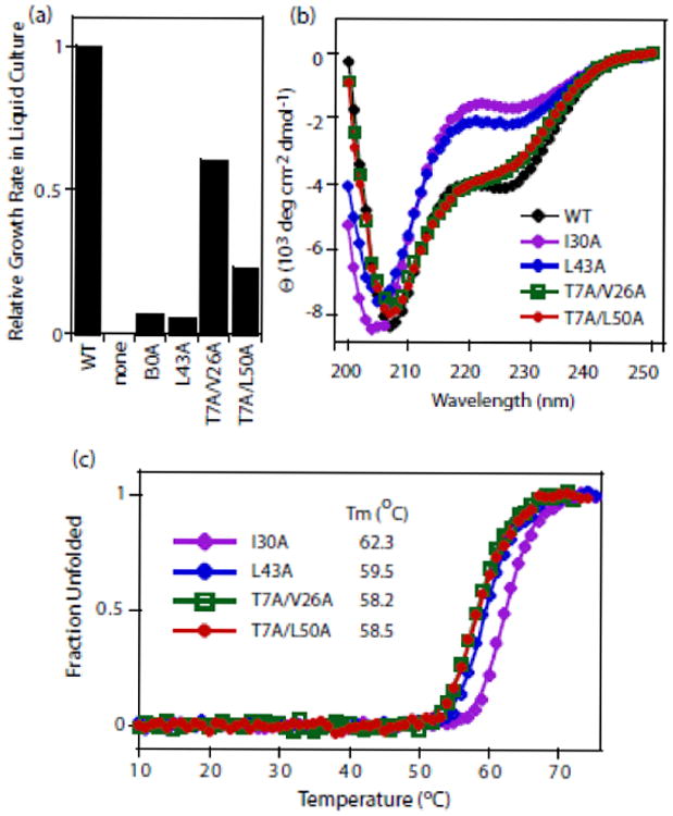
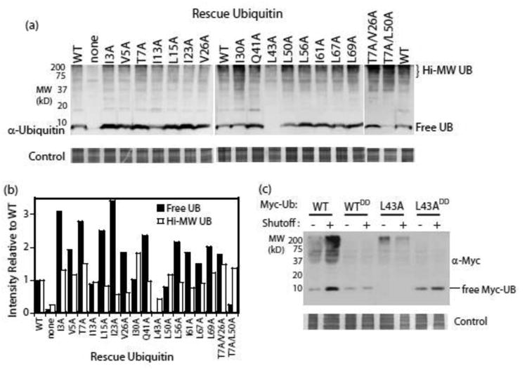
Mutations at solvent-inaccessible core positions in proteins can impact function through many biophysical mechanisms including alterations to thermodynamic stability and protein dynamics. As these properties of proteins are difficult to investigate, the impacts of core mutations on protein function are poorly understood for most systems. Here, we determined the effects of alanine mutations at all 15 core positions in ubiquitin on function in yeast. The majority (13 of 15) of alanine substitutions supported yeast growth as the sole ubiquitin. Both the two null mutants (I30A and L43A) were less stable to temperature-induced unfolding in vitro than wild type (WT) but were well folded at physiological temperatures. Heteronuclear NMR studies indicated that the L43A mutation reduces temperature stability while retaining a ground-state structure similar to WT. This structure enables L43A to bind to common ubiquitin receptors in vitro. Many of the core alanine ubiquitin mutants, including one of the null variants (I30A), exhibited an increased accumulation of high-molecular-weight species, suggesting that these mutants caused a defect in the processing of ubiquitin-substrate conjugates. In contrast, L43A exhibited a unique accumulation pattern with reduced levels of high-molecular-weight species and undetectable levels of free ubiquitin. When conjugation to other proteins was blocked, L43A ubiquitin accumulated as free ubiquitin in yeast. Based on these findings, we speculate that ubiquitin's stability to unfolding may be required for efficient recycling during proteasome-mediated substrate degradation.

Keywords: proteasome; protein function; structural dynamics; thermodynamic stability; ubiquitin recycling.


Figures

Figure 1
Figure 1. Effects of alanine mutations in the core of ubiquitin on yeast growth
(a) Illustrations of the structure of ubiquitin highlighting the locations of core positions that are shielded from solvent. (b&c) Capability of mutants to function as the sole ubiquitin. Constitutively expressed mutants were introduced into the SUB328 shutoff strain, and growth under selective conditions was examined on plates (b) and in liquid culture (c). As a negative control, cells harboring a plasmid lacking the ubiquitin open reading frame (labeled as “no UB”) were analyzed.
Figure 2
Figure 2. Protein properties of ubiquitin mutants with severe growth defects
(a) Relative growth rates of yeast with I30A or L43A ubiquitin are slower than yeast with T7A/V26A or T7A/L50A ubiquitin. (b) Circular dichroism spectra of purified ubiquitin proteins at 23 °C. (c) Stability of purified ubiquitin mutants to temperature denaturation.
Figure 3
Figure 3. Accumulation of ubiquitin mutants in yeast
(a) Western blot analyses of yeast whole cell lysates. Cells were grown in dextrose for 24 hours in order to shut off expression of WT ubiquitin. (b) The intensity of the free ubiquitin band as well as of high molecular weight species in panel (a) were quantified and compared to a WT control present on each blot. (c) Western blot analyses of lysates from yeast expressing epitope-tagged ubiquitin variants. The G75D/G76D mutations (annotated as DD) were used to block activation by E1 and hence eliminate all covalent attachment reactions. Under shutoff conditions, the epitope-tagged version is the only ubiquitin protein in the cells.
Figure 4
Figure 4. Structure and backbone dynamics of L43A ubiquitin by NMR
(a) Overlay of 1H-15N NMR spectra of L43A (red) and WT (black) ubiquitin and (b) quantification of the spectral differences between the two proteins in terms of amide chemical shift perturbations (CSPs) plotted as a function of the residue number. (c) Mapping the CSPs due to the L43A mutation on ubiquitin structure. L43 is shown in spheres, and the coloring represents CSPs in decreasing order: Red> Orange > Yellow > Green. (d) The 1H-15N residual dipolar couplings (RDCs) measured for L43A and the back-calculated RDCs using solution structure of WT ubiquitin (1D3Z.PDB) are in close agreement, indicating that the 3D structure of ubiquitin is not grossly affected by the mutation. The Pearson's correlation coefficient is r=0.976, the quality factor is 0.146 (62 residues included). (e) Conformational exchange (Rex) contributions detected in L43A (red) and WT ubiquitin (blue). (f) Backbone order parameters for L43A (red) and WT ubiquitin (blue) indicate similarity in the backbone motions on a sub-nanosecond time scale.
Figure 5
Figure 5. Recognition of L43A and WT ubiquitins by the ubiquitin-associated domain (UBA) of the proteasomal shuttle protein ubiquilin-1 (UQ1)
Comparison of spectral perturbations induced by UQ1 UBA binding to (a) L43A ubiquitin and (b) WT ubiquitin. Light-gray bars mark residues that show significant attenuation (disappearance) of the NMR signals in the course of titration with UQ1 UBA. Horizontal bars at the top of the plots indicate elements of ubiquitin's secondary structure. The spectral perturbation profiles indicate that a hydrophobic surface patch including L8, I44, and V70 mediates binding of both WT and L43A ubiquitin with this UBA domain. (c) Map of the UBA-induced spectral perturbations on the surface of ubiquitin. Shown is the structure of WT- ubiquitin/UQ1 UBA complex (2JY6.PDB) facing the hydrophobic patch surface, with ubiquitin residues showing perturbations (CSPs>0.15 ppm and/or signal attenuations) colored red for L43A (left) and WT ubiquitin (right). (d) Overlay of 1H-15N HSQC spectra of L43A (red) and WT (black) at the endpoint of titrations reveals a striking similarity of the UQ1 UBA-bound states of the two ubiquitin variants. Titration curves for L43A (e) and WT ubiquitin (f) plotted as CSPs versus the UBA: ubiquitin molar ratio. Titration data were fit with a 1:1 binding model. The average Kd values (over ∼10 residues) are 28±11 μM (L43A) and 4.2±4.9 μM (WT).
Figure 6
Figure 6. Proposed mechanistic model of ubiquitin recycling by the proteasome
In this speculative model, the stability of wild-type ubiquitin forms a strong obstruction to processing and degradation, increasing the probability that the Rpn11 subunit will remove ubiquitin before it can be unfolded and translocated into the core particle for degradation.

Comment on

Similar articles

Cited by

References

    1. Ward JJ, Sodhi JS, McGuffin LJ, Buxton BF, Jones DT. Prediction and functional analysis of native disorder in proteins from the three kingdoms of life. J Mol Biol. 2004;337:635–45. - PubMed
    1. Karplus M, Kuriyan J. Molecular dynamics and protein function. Proc Natl Acad Sci U S A. 2005;102:6679–85. - PMC - PubMed
    1. Mittermaier AK, Kay LE. Observing biological dynamics at atomic resolution using NMR. Trends Biochem Sci. 2009;34:601–11. - PubMed
    1. Cunningham BC, Wells JA. High-resolution epitope mapping of hGH-receptor interactions by alanine-scanning mutagenesis. Science. 1989;244:1081–5. - PubMed
    1. Dill KA. Dominant forces in protein folding. Biochemistry. 1990;29:7133–55. - PubMed

Publication types

MeSH terms

LinkOut - more resources