. 2014 Feb;184(2):483-93.
doi: 10.1016/j.ajpath.2013.10.027. Epub 2013 Dec 21.

CD4+ T cells and IFN-γ are required for the development of Pneumocystis-associated pulmonary hypertension

Affiliations

CD4+ T cells and IFN-γ are required for the development of Pneumocystis-associated pulmonary hypertension

Steve D Swain et al. Am J Pathol. 2014 Feb.

Abstract

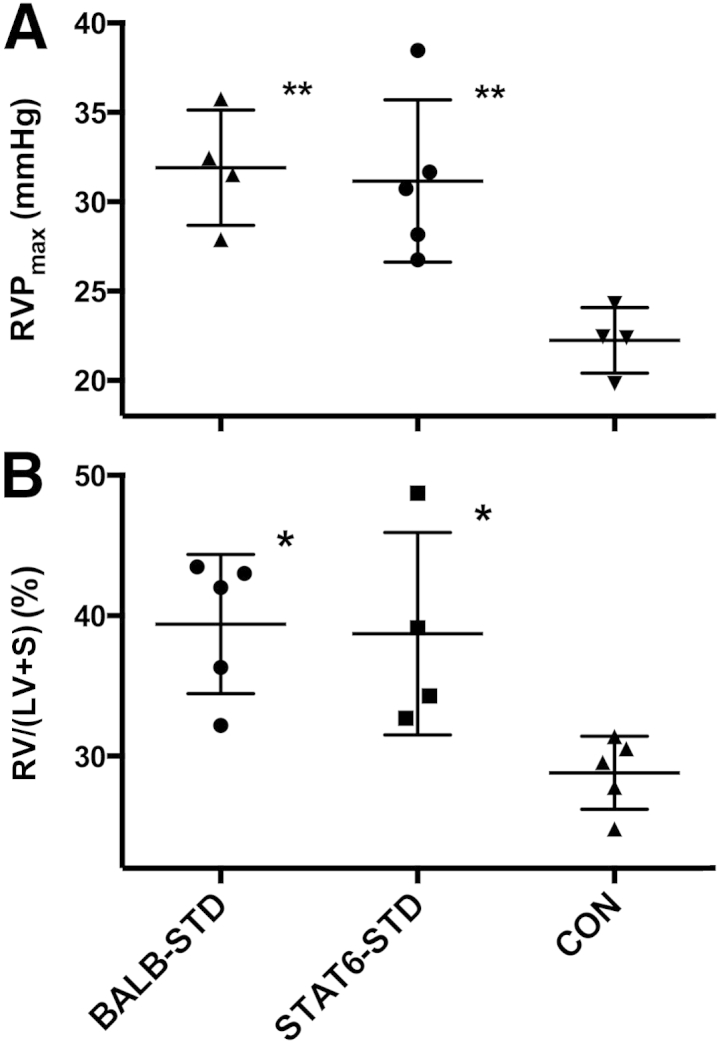
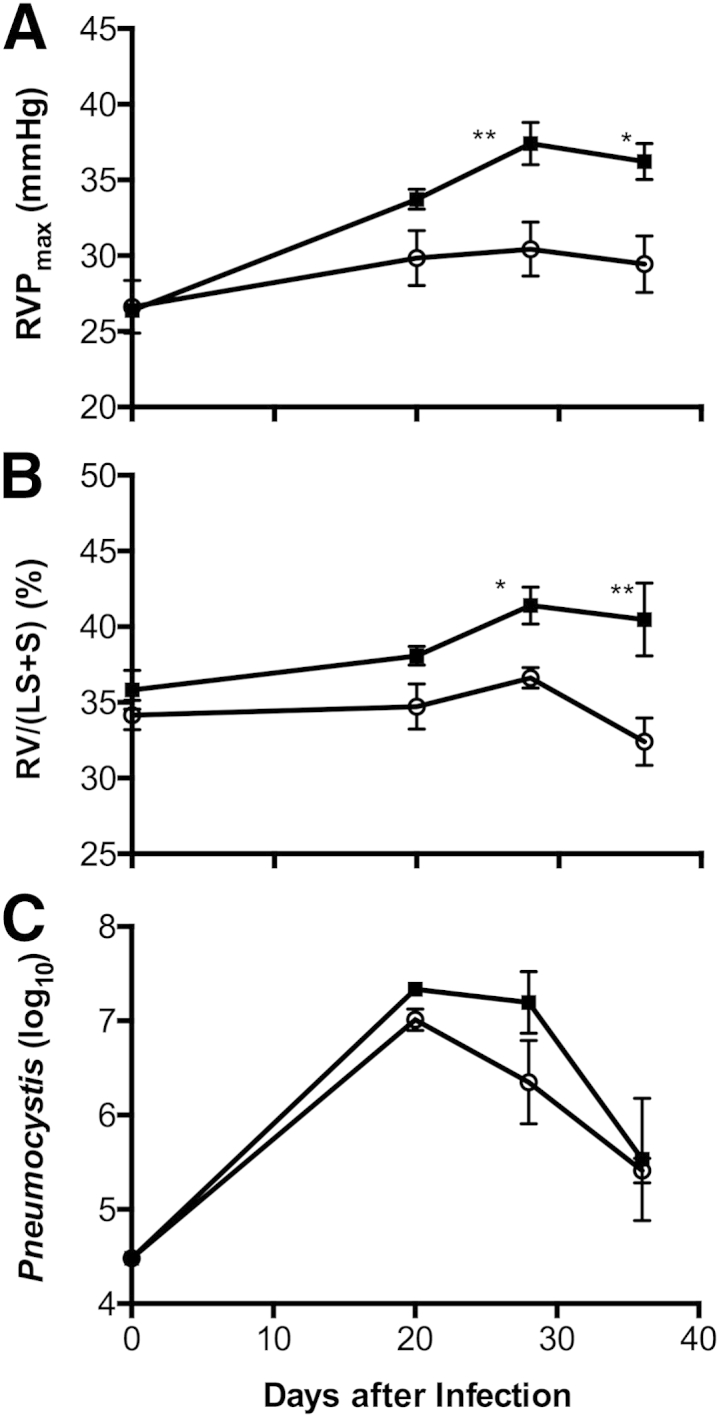
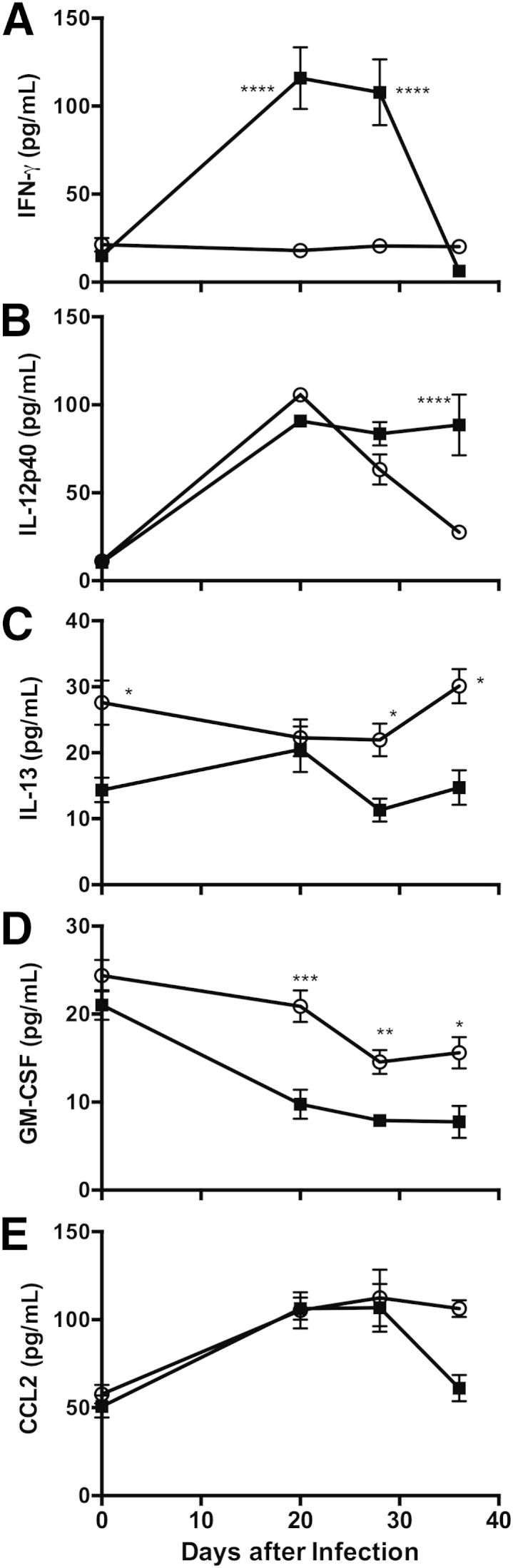
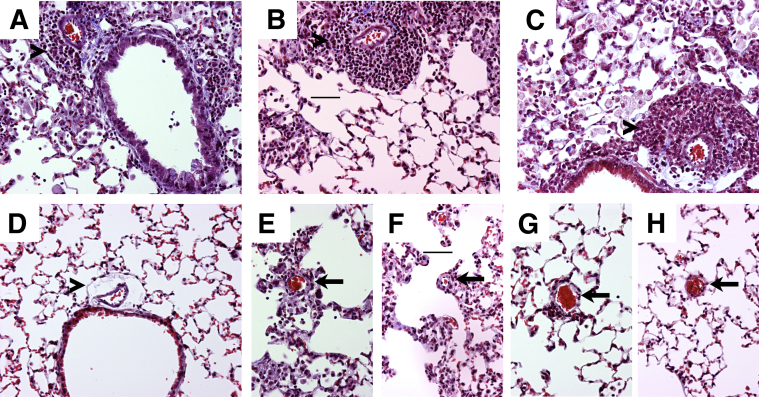
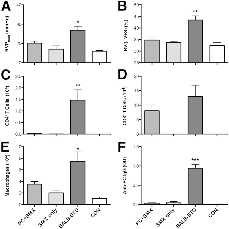
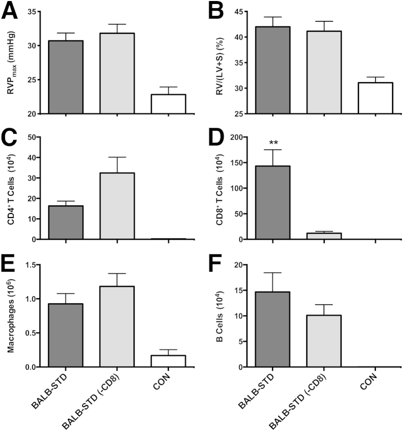
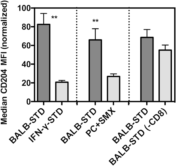
Pulmonary hypertension (PH) is a disease of diverse etiology. Although primary PH can develop in the absence of prior disease, PH more commonly develops in conjunction with other pulmonary pathologies. We previously reported a mouse model in which PH occurs as a sequela of Pneumocystis infection in the context of transient CD4 depletion. Here, we report that instead of the expected Th2 pathways, the Th1 cytokine IFN-γ is essential for the development of PH, as wild-type mice developed PH but IFN-γ knockout mice did not. Because gene expression analysis showed few strain differences that were not immune-function related, we focused on those responses as potential pathologic mechanisms. In addition to dependence on IFN-γ, we found that when CD4 cells were continuously depleted, but infection was limited by antibiotic treatment, PH did not occur, confirming that CD4 T cells are required for PH development. Also, although CD8 T-cells are implicated in the pathology of Pneumocystis pneumonia, they did not have a role in the onset of PH. Finally, we found differences in immune cell phenotypes that correlated with PH, including elevated CD204 expression in lung CD11c(+) cells, but their role remains unclear. Overall, we demonstrate that a transient, localized, immune response requiring IFN-γ and CD4-T cells can disrupt pulmonary vascular function and promote lingering PH.


Figures

Figure 1
Figure 1
Under short-term CD4 depletion and P. murina infection, STAT6 knockout mice (STAT6-STD) are not protected from developing PH. A: RVP did not differ between BALB-STD and STAT6-STD mice. B: RV hypertrophy was present in both CD4-depleted strains. Data are representative of two independent experiments and are expressed as means ± SEM. n = 4 or 5. P ≤ 0.05, ∗∗P ≤ 0.01 versus control (CON).
Figure 2
Figure 2
Under short-term CD4 depletion and P. murina infection, PH develops in wild-type BALB/c mice (BALB-STD, squares) but not in IFN-γ knockout mice (IFN-γ–STD, circles). RVP became elevated (A), and persistent RV hypertrophy developed (B), even as the P. murina infection was cleared (C). A Pneumocystis count of approximately 25,000 (4.4 on a log10 scale) is the limit of detection for this procedure. Data are representative of three independent experiments and are expressed as means ± SEM. n = 5. P ≤ 0.05, ∗∗P ≤ 0.01.
Figure 3
Figure 3
Under short-term CD4 depletion and P. murina infection, the influx of pulmonary inflammatory cells differs distinctly between BALB-STD mice (which develop PH) and IFN-γ–STD mice (which do not develop PH). Cell types counted included total lung cells; macrophages (Macs), polymorphonuclear cells (PMN), eosinophils (EOS), natural killer (NK) cells, and γδ T cells as percentage in BALF; and B cells, CD4+ T cells, CD8+ T cells, and CD4+CD8+ cells. Important differences are described in the Results section. Data are representative of three independent experiments and are expressed as means ± SEM. n = 5. P ≤ 0.05, ∗∗P ≤ 0.01, ∗∗∗P ≤ 0.001, and ∗∗∗∗P ≤ 0.0001 versus control.
Figure 4
Figure 4
IFN-γ (A) and IL-12p40 (B) levels were elevated at times in BALF from BALB-STD mice, compared with IFN-γ–STD mice (which do not develop PH), whereas levels of IL-13 (C) and GM-CSF (D) were significantly lower. E: Levels of CCL2 did not differ significantly between the two mouse strains. Data are representative of three independent experiments and are expressed as means ± SEM. n = 5. P ≤ 0.05, ∗∗P ≤ 0.01, ∗∗∗P ≤ 0.001, and ∗∗∗∗P ≤ 0.0001 versus control.
Figure 5
Figure 5
In representative images, perivascular inflammation lingers around larger pulmonary arteries adjacent to airways (arrowheads), and collagen deposition (blue) in these areas is similar in BALB-STD (A), IFN-γ–STD (B), and STAT6-STD (C) mice but is less than that in the control (D). Although slight inflammation without collagen deposition remains around smaller arterioles near alveolar ducts (arrows) in P. murina–infected mice, it is similar in BALB-STD (E), IFN-γ–STD (F), and STAT6-STD (G) mice but is absent in the control (H). Original magnification, ×200. Scale bar = 50 μm (B and F).
Figure 6
Figure 6
Mice continuously depleted of CD4+ cells but treated with SMX/TMP do not develop PH. Antibiotic treatment commenced at 20 days after infection in P. murina–infected (PC+SMX) or uninfected (SMX only) mice. At 40 days after infection, only the BALB-STD mice exhibited PH (A) and RV hypertrophy (B). Numbers of CD4+ cells (C), CD8+ cells (D), macrophages (E), and anti-Pneumocystis antibody (F) were measured in BALF. Data are representative of two independent experiments and are expressed as means ± SEM. n = 5. P ≤ 0.05, ∗∗P ≤ 0.01, and ∗∗∗P ≤ 0.001 as compared to PC+SMX. OD, optical density 405 nm.
Figure 7
Figure 7
CD8+ cells do not contribute to the development of PH. Two groups of mice were transiently depleted of CD4+ cells, and one group was also continuously depleted of CD8+ cells [BALB-STD (−CD8)]. There was no significant difference in PH (A), RV mass (B). Numbers of CD4+ cells (C), CD8+ cells (D), macrophages (E), and B cells (F) were measured in BALF. Data are representative of two independent experiments and are expressed as means ± SEM. n = 5. ∗∗P ≤ 0.01 as compared to the BALB-STD (−CD8) group.
Figure 8
Figure 8
Up-regulated CD204 expression on lung CD11c+ cells correlates with PH. Flow cytometric assessment of median fluorescence intensity (MFI) of Alexa Fluor 647–conjugated anti-mouse CD204 on gated CD11c+ BALF cells. Shown are comparisons of BALB-STD with IFN-γ–STD (Figure 3), PC+SMX (Figure 6), and BALB-STD (−CD8) (Figure 7). Statistical tests were performed on raw MFI values within each experiment, but values were normalized (100% is the highest MFI of BALB-STD group, and 0% is the mean CD204 MFI of control uninfected mice) for the composite graph. Data are representative of two independent experiments and are expressed as means ± SEM. ∗∗P ≤ 0.01.

References

    1. Simonneau G., Robbins I.M., Beghetti M., Channick R.N., Delcroix M., Denton C.P., Elliott C.G., Gaine S.P., Gladwin M.T., Jing Z.C., Krowka M.J., Langleben D., Nakanishi N., Souza R. Updated clinical classification of pulmonary hypertension. J Am Coll Cardiol. 2009;54(1 Suppl):S43–S54. - PubMed
    1. Pullamsetti S.S., Savai R., Janssen W., Dahal B.K., Seeger W., Grimminger F., Ghofrani H.A., Weissmann N., Schermuly R.T. Inflammation, immunological reaction and role of infection in pulmonary hypertension. Clin Microbiol Infect. 2011;17:7–14. - PubMed
    1. Voelkel N.F., Tuder R.M. Cellular and molecular mechanisms in the pathogenesis of severe pulmonary hypertension. Eur Respir J. 1995;8:2129–2138. - PubMed
    1. Tuder R.M., Voelkel N.F. Pulmonary hypertension and inflammation. J Lab Clin Med. 1998;132:16–24. - PubMed
    1. Larsen K.O., Yndestad A., Sjaastad I., Løberg E.M., Goverud I.L., Halvorsen B., Jia J., Andreassen A.K., Husberg C., Jonasson S., Lipp M., Christensen G., Aukrust P., Skjønsberg O.H. Lack of CCR7 induces pulmonary hypertension involving perivascular leukocyte infiltration and inflammation. Am J Physiol Lung Cell Mol Physiol. 2011;301:L50–L59. - PubMed

Publication types

MeSH terms