Review
. 2014 Apr:35:60-72.
doi: 10.1016/j.matbio.2013.12.001. Epub 2013 Dec 19.

Heparan sulfate 3-O-sulfation: a rare modification in search of a function

Affiliations
Review

Heparan sulfate 3-O-sulfation: a rare modification in search of a function

Bryan E Thacker et al. Matrix Biol. 2014 Apr.

Abstract

Many protein ligands bind to heparan sulfate, which results in their presentation, protection, oligomerization or conformational activation. Binding depends on the pattern of sulfation and arrangement of uronic acid epimers along the chains. Sulfation at the C3 position of glucosamine is a relatively rare, yet biologically significant modification, initially described as a key determinant for binding and activation of antithrombin and later for infection by type I herpes simplex virus. In mammals, a family of seven heparan sulfate 3-O-sulfotransferases installs sulfate groups at this position and constitutes the largest group of sulfotransferases involved in heparan sulfate formation. However, to date very few proteins or biological systems have been described that are influenced by 3-O-sulfation. This review describes our current understanding of the prevalence and structure of 3-O-sulfation sites, expression and substrate specificity of the 3-O-sulfotransferase family and the emerging roles of 3-O-sulfation in biology.

Keywords: 3-O-Sulfotransferases; Antithrombin; Growth factors; Heparan sulfate; Sulfation.


Figures

Figure 1
Figure 1. Structure of the antithrombin-binding pentasaccharide found in heparin
The individual residues of the pentasaccharide are numbered relative to the N-sulfo-glucosamine-3-sulfate residue at position 0. The central 3-O-sulfate group generated by Hs3st-1 (shown in red) and the 6-O-sulfate group at residue −2 (shown in blue) account for the majority of the binding energy of antithrombin to heparin. Note that the 6-O-sulfate group on the 3-O-sulfated glucosamine residue is dispensable and that residue −2 can be N-sulfated.
Figure 2
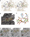
Figure 2. Structural interactions of 3-O-sulfotransferases with oligosaccharide substrates
(A) Stereo diagram of the interaction between Hs3st-3 and the tetrasaccharide D2S6-I2S6 (PDB code 1T8U). Side chains of critical amino acid residues that mediate substrate binding are shown as thick grey sticks. Possible hydrogen bonds between side chains and tetrasaccharide are pictured as black broken lines. The C3-OH group of the glucosamine residue to be sulfated is shown as a red sphere. The sulfate donor mimic, 3’-phosphoadenosine 5’-phosphate (PAP) is shown, and the protein backbone is shown as a cartoon in the background. (B) Superposition of Hs3st-3 bound tetrasaccharide (D2S6-I2S6; green, PDB code 1T8U) and heptasaccharide (A6-G0S6-I2S6-G0M0; white, PDB code 3UAN). The surface of Hs3st-1 is rendered in gray. Residues in tetrasaccharides are labeled as T-1 to T-4. Residues in the heptasaccharide are labeled as H-1 to H-6 (the anhydromannose residue is not shown). (C) Role of K259 in Hs3st-3 substrate recognition. The tetrasaccharide and heptasaccharide are colored and numbered as in (B). Putative hydrogen bonds between K259 and the 2-O-sulfate group of T-1 and the carboxylate of T-3 are shown in black dashed lines. The distance between the 2-O-sulfate of T-1 and carboxylate of T-3 are shown as a yellow broken line. The distance between the 2-O-sulfate of T-1 and the carboxylate of H-4 are shown as a red broken line. (D-F) Role of gate residues in substrate recognition. The gate residues are shown as large spheres and labeled in white, and the tetrasaccharide is shown with the C3-OH group shown as a red sphere. The molecular surface of each enzyme is shown in grey. The distance between gates residues is pictured as black broken lines. The tetrasaccharide is modeled into Hs3st-1 (PDB code 1VKJ) and Hs3st-5 (PDB code 3BD9) by superimposing their structures on that of the Hs3st-3/tetrasaccharide complex (PDB code 1T8U) using PyMol (Schrödinger).
Figure 3
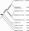
Figure 3. Phylogenetic analysis of Hs3sts
The number of Hs3st isozymes identified in each species was deduced from existing publications, NCBI protein database searches and comparison of key amino acids that define substrate and catalytic sites. The phylogenetic tree depicts their evolutionary relationship (adapted from (Medeiros et al., 2000)). The proposed origin of Hs3st is indicated with an arrow.
Figure 4
Figure 4. Stereo diagram of pentasaccharide-antithrombin interaction
(PDB code: 2GD4). Heparan sulfate binding residues of antithrombin (salmon) and the pentasaccharide (grey) are shown in sticks, and the antithrombin protein backbone is shown in cartoon (salmon). Putative salt bridges and hydrogen bonds stabilized by the 3- O-sulfate group and K114 are shown as broken lines. Sugar residues in the pentasaccharide are labeled as in Figure 1.

References

    1. Allain F, Vanpouille C, Carpentier M, Slomianny MC, Durieux S, Spik G. Interaction with glycosaminoglycans is required for cyclophilin B to trigger integrin-mediated adhesion of peripheral blood T lymphocytes to extracellular matrix. Proc Natl Acad Sci USA. 2002;99:2714–2719. - PMC - PubMed
    1. Arungundram S, Al-Mafraji K, Asong J, Leach FE, 3rd, Amster IJ, Venot A, Turnbull JE, Boons GJ. Modular synthesis of heparan sulfate oligosaccharides for structure-activity relationship studies. J Am Chem Soc. 2009;131:17394–17405. - PMC - PubMed
    1. Atha DH, Lormeau JC, Petitou M, Rosenberg RD, Choay J. Contribution of monosaccharide residues in heparin binding to antithrombin III. Biochemistry. 1985;24:6723–6729. - PubMed
    1. Atha DH, Lormeau JC, Petitou M, Rosenberg RD, Choay J. Contribution of 3-O- and 6-O-sulfated glucosamine residues in the heparin-induced conformational change in antithrombin III. Biochemistry. 1987;26:6454–6461. - PubMed
    1. Atkinson A, Garnier S, Afridi S, Fumoux F, Rihet P. Genetic variations in genes involved in heparan sulphate biosynthesis are associated with Plasmodium falciparum parasitaemia: a familial study in Burkina Faso. Malaria journal. 2012;11:108. - PMC - PubMed

Publication types