. 2014 Mar;1840(3):1214-24.
doi: 10.1016/j.bbagen.2013.12.018. Epub 2013 Dec 21.

A thermodynamic assay to test pharmacological chaperones for Fabry disease

Affiliations

A thermodynamic assay to test pharmacological chaperones for Fabry disease

Giuseppina Andreotti et al. Biochim Biophys Acta. 2014 Mar.

Abstract

Background: The majority of the disease-causing mutations affect protein stability, but not functional sites and are amenable, in principle, to be treated with pharmacological chaperones. These drugs enhance the thermodynamic stability of their targets. Fabry disease, a disorder caused by mutations in the gene encoding lysosomal alpha-galactosidase, represents an excellent model system to develop experimental protocols to test the efficiency of such drugs.

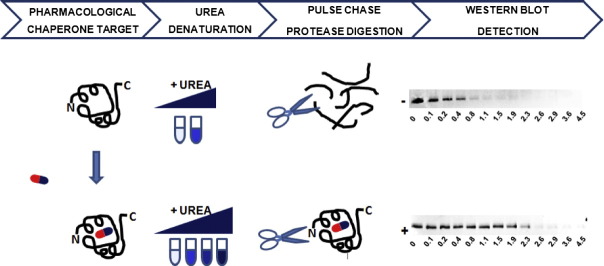
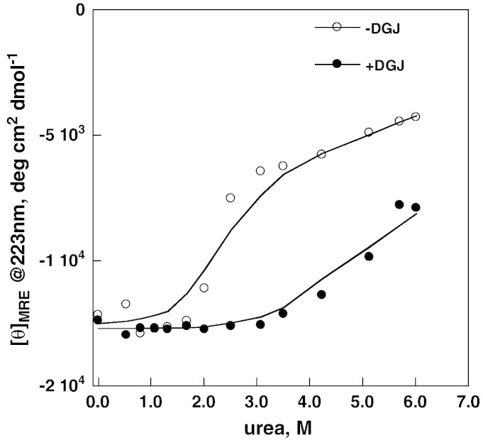
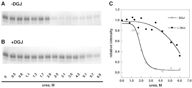
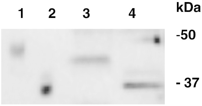
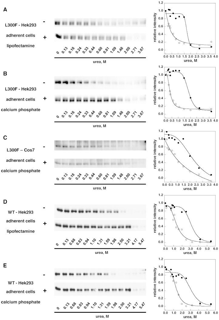
Methods: The stability of lysosomal alpha-galactosidase under different conditions was studied by urea-induced unfolding followed by limited proteolysis and Western blotting.

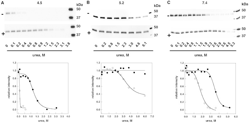
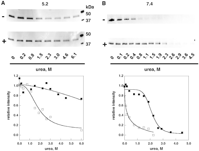
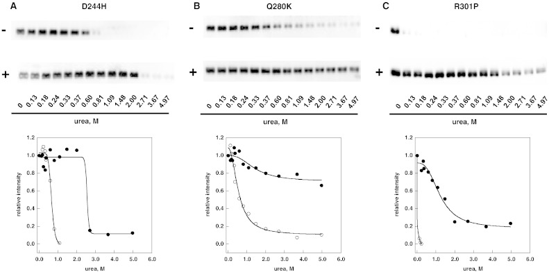
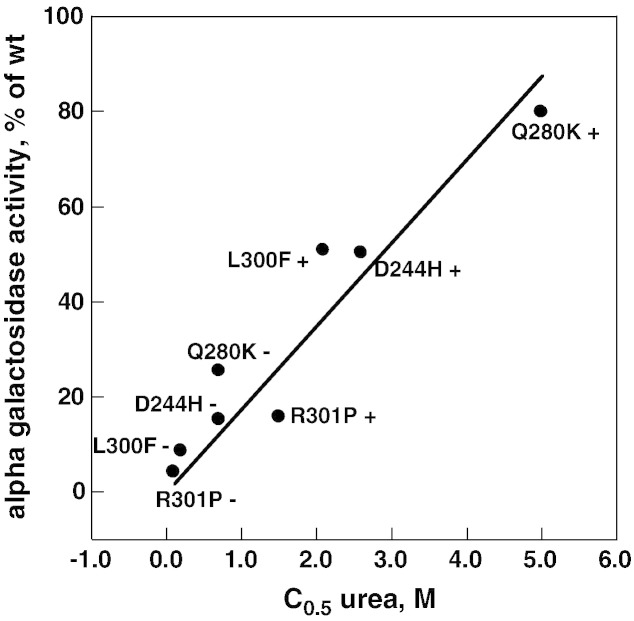
Results: We measured the concentration of urea needed to obtain half-maximal unfolding because this parameter represents an objective indicator of protein stability.

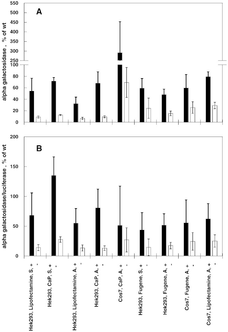
Conclusions: Urea-induced unfolding is a versatile technique that can be adapted to cell extracts containing tiny amounts of wild-type or mutant proteins. It allows testing of protein stability as a function of pH, in the presence or in the absence of drugs. Results are not influenced by the method used to express the protein in transfected cells.

General significance: Scarce and dispersed populations pose a problem for the clinical trial of drugs for rare diseases. This is particularly true for pharmacological chaperones that must be tested on each mutation associated with a given disease. Diverse in vitro tests are needed. We used a method based on chemically induced unfolding as a tool to assess whether a particular Fabry mutation is responsive to pharmacological chaperones, but, by no means is our protocol limited to this disease.

Keywords: 1-deoxy-galactonojirimycin; AGAL; CD; Cell lysate; DGJ; FD; Fabry disease; Limited proteolysis; Lysosomal storage disorder; PC; Pharmacological chaperone; Urea-induced unfolding; circular dichroism; lysosomal alpha-galactosidase; pharmacological chaperones.


Figures

None
Graphical abstract
Fig. 1
Fig. 1
Urea-induced melting profile of wild-type lysosomal alpha-galactosidase (Fabrazyme®) recorded by circular dichroism. The protein (0.3 mg/ml in McIlvaine buffer at pH 7.4) was equilibrated with urea (from 0 to 6 M) in the presence of 1-deoxy-galactonojirimycin (DGJ) 40 μM or not, then ellipticity at 223 nm was recorded. Data were expressed as mean residue ellipticity.
Fig. 2
Fig. 2
Urea-induced melting profile of wild-type lysosomal alpha-galactosidase (Fabrazyme®) recorded by pulse-proteolysis and SDS-PAGE analysis. The protein (0.3 mg/ml in McIlvaine buffer at pH 7.4) was equilibrated with urea (from 0 to 6 M) in the presence of 1-deoxy-galactonojirimycin (DGJ) 40 μM or not, then an aliquot of each sample was subjected to pulse proteolysis with thermolysin (1 min at 37 °C, 1:5 protease to substrate ratio) and then analyzed by SDS-PAGE. The protein was visualized by Coomassie Blue Staining (− DGJ, panel A; + DGJ, panel B). The intensity of the bands was quantified and expressed as relative intensity (panel C).
Fig. 3
Fig. 3
Urea-induced unfolding profiles of wild-type lysosomal alpha-galactosidase present in raw cell extracts recorded by pulse-proteolysis and western-blot. Lysates of COS7 cells expressing wild-type lysosomal alpha-galactosidase were mixed with the denaturant to obtain final urea concentrations ranging from 0 to 6 M. The experiment was conducted in McIlvaine buffer at pH 4.5 (panel A), 5.2 (panel B) or 7.4 (panel C) either in the absence or in the presence of 1-deoxy-galactonojirimycin (DGJ) 40 μM. Pulse proteolysis (1 min at 37 °C, 1:5 protease to substrate ratio) was performed after the equilibrium was reached to digest unfolded protein and then analyzed by western-blot. Thermolysin was used when operating at pH 5.2 or 7.4, pepsin when operating at pH 4.5. The intensity of the bands was quantified and data were expressed as fraction of the zero urea sample.
Fig. 4
Fig. 4
Comparison between glycosylation states of wild-type lysosomal alpha-galactosidase produced in COS7 cells and Fabrazyme®. Fabrazyme® and lysates of COS7 cells expressing wild-type lysosomal alpha-galactosidase were incubated overnight at 37 °C in the presence of N-Glycosidase F. The samples were pre-treated with SDS 0.1% and EDTA 20 mM, boiled and cooled and NP-40 was added to 0.7%. Samples were compared by SDS-PAGE and western blot: Fabrazyme® not treated (lane 1) or treated with N-Glycosidase F (lane 2); wild-type lysosomal alpha-galactosidase expressed in COS7 non-treated (lane 3) or treated with N-Glycosidase F (lane 4).
Fig. 5
Fig. 5
Urea-induced unfolding profiles of L300F lysosomal alpha-galactosidase present in raw extracts recorded by pulse-proteolysis and western-blot. Lysates of COS7 cells expressing L300F lysosomal alpha-galactosidase were mixed with the denaturant to obtain final urea concentrations ranging from 0 to 6 M. The experiment was conducted in McIlvaine buffer at 5.2 (panel A) or 7.4 (panel B) either in the absence or in the presence of DGJ 40 μM. Pulse proteolysis (1 min at 37 °C, 1:5 thermolysin to substrate ratio) was performed after the equilibrium was reached to digest unfolded protein and then analyzed by western-blot. The intensity of the bands was quantified and data were expressed as fraction of the zero urea sample.
Fig. 6
Fig. 6
Urea-induced unfolding profiles of D244H, Q280K and R301P lysosomal alpha-galactosidase mutants present in raw cell extracts recorded by pulse-proteolysis and western-blot. Lysates of COS7 cells expressing D244H (panel A), Q280K (panel B) or R301P (panel C) lysosomal alpha-galactosidase were mixed with the denaturant to obtain final urea concentrations ranging from 0 to 5 M. The experiment was conducted in McIlvaine buffer at pH 7.4 either in the absence or in the presence of DGJ 40 μM. Pulse proteolysis (1 min at 37 °C, 1:5 thermolysin to substrate ratio) was performed after the equilibrium was reached to digest unfolded protein and then analyzed by western-blot. The intensity of the bands was quantified and data were expressed as fraction of the zero urea sample.
Fig. 7
Fig. 7
Correlation between urea C0.5 and alpha galactosidase increase. Urea concentration was from Fig. 5, Fig. 6 (this paper); alpha galactosidase activity is expressed as mut_activity + DGJ / WT_activity − DGJ × 100. The Pearson correlation coefficient is 0.94.
Fig. 8
Fig. 8
Alpha-galactosidase activity resulting from 1-deoxy-galactonojirimycin administration in transfected cells. Comparison between different cell hosts and transfection methods. COS7 or HEK293 cells were co-transfected with pCMV6-AC harboring L300F-AGAL gene and with pMIR vector harboring the luciferase reporter gene. Transfections were performed with Lipofectamine® ltx, Fugene® HD or CalPhos™ Mammalian Transfection Kit, either using cell in adhesion (A) or in suspension (S). Cells were than cultivated with (+) or without (−) 0.02 mM DGJ. After cell lysis AGAL activity and luciferase activity were measured. Four independent experiments were performed in the presence of DGJ, two without DGJ. Alpha galactosidase activity (expressed as mut_activity / WT_activity − DGJ × 100) was normalized by protein concentration in panel A and by luciferase activity in panel B.
Fig. 9
Fig. 9
Stability enhancement of lysosomal alpha galactosidase by 1-deoxy-galactonojirimycin. Comparison between different cell hosts and transfection methods. Lysates of COS7 or HEK293 cells transfected with different methods and expressing L300F (panels A, B, and C) or wild type (panels D and E) lysosomal alpha-galactosidase were mixed with the denaturant to obtain final urea concentrations ranging from 0 to 6 M. The experiment was conducted in McIlvaine buffer at pH 7.4 either in the absence or in the presence of DGJ 40 μM. Pulse proteolysis (1 min at 37 °C, 1:5 thermolysin to substrate ratio) was performed after the equilibrium was reached to digest unfolded protein and then analyzed by western-blot. The intensity of the bands was quantified and data were expressed as fraction of the zero urea sample.

References

    1. Yue P., Li Z., Moult J. Loss of protein structure stability as a major causative factor in monogenic disease. J. Mol. Biol. 2005;353:459–473. - PubMed
    1. Boyd R.E., Lee G., Rybczynski P., Benjamin E.R., Khanna R., Wustman B.A., Valenzano K.J. Pharmacological chaperones as therapeutics for lysosomal storage diseases. J. Med. Chem. 2013;56:2705–2725. - PubMed
    1. Ishii S. Pharmacological chaperone therapy for Fabry disease. Proc. Jpn. Acad. Ser. B Phys. Biol. Sci. 2012;88:18–30. - PMC - PubMed
    1. Young-Gqamana B., Brignol N., Chang H.H., Khanna R., Soska R., Fuller M., Sitaraman S.A., Germain D.P., Giugliani R., Hughes D.A., Mehta A., Nicholls K., Boudes P., Lockhart D.J., Valenzano K.J., Benjamin E.R. Migalastat HCl reduces globotriaosylsphingosine (lyso-Gb3) in Fabry transgenic mice and in the plasma of Fabry patients. PLoS One. 2013;8:e57631. - PMC - PubMed
    1. Benito J.M., Garcia Fernandez J.M., Ortiz Mellet C. Pharmacological chaperone therapy for Gaucher disease: a patent review. Expert. Opin. Ther. Pat. 2011;21:885–903. - PubMed

Publication types