. 2014 Feb;118(2):464-472.
doi: 10.1213/ANE.0000000000000047.

Spinal cord stimulation reduces mechanical hyperalgesia and glial cell activation in animals with neuropathic pain

Affiliations

Spinal cord stimulation reduces mechanical hyperalgesia and glial cell activation in animals with neuropathic pain

Karina L Sato et al. Anesth Analg. 2014 Feb.

Abstract

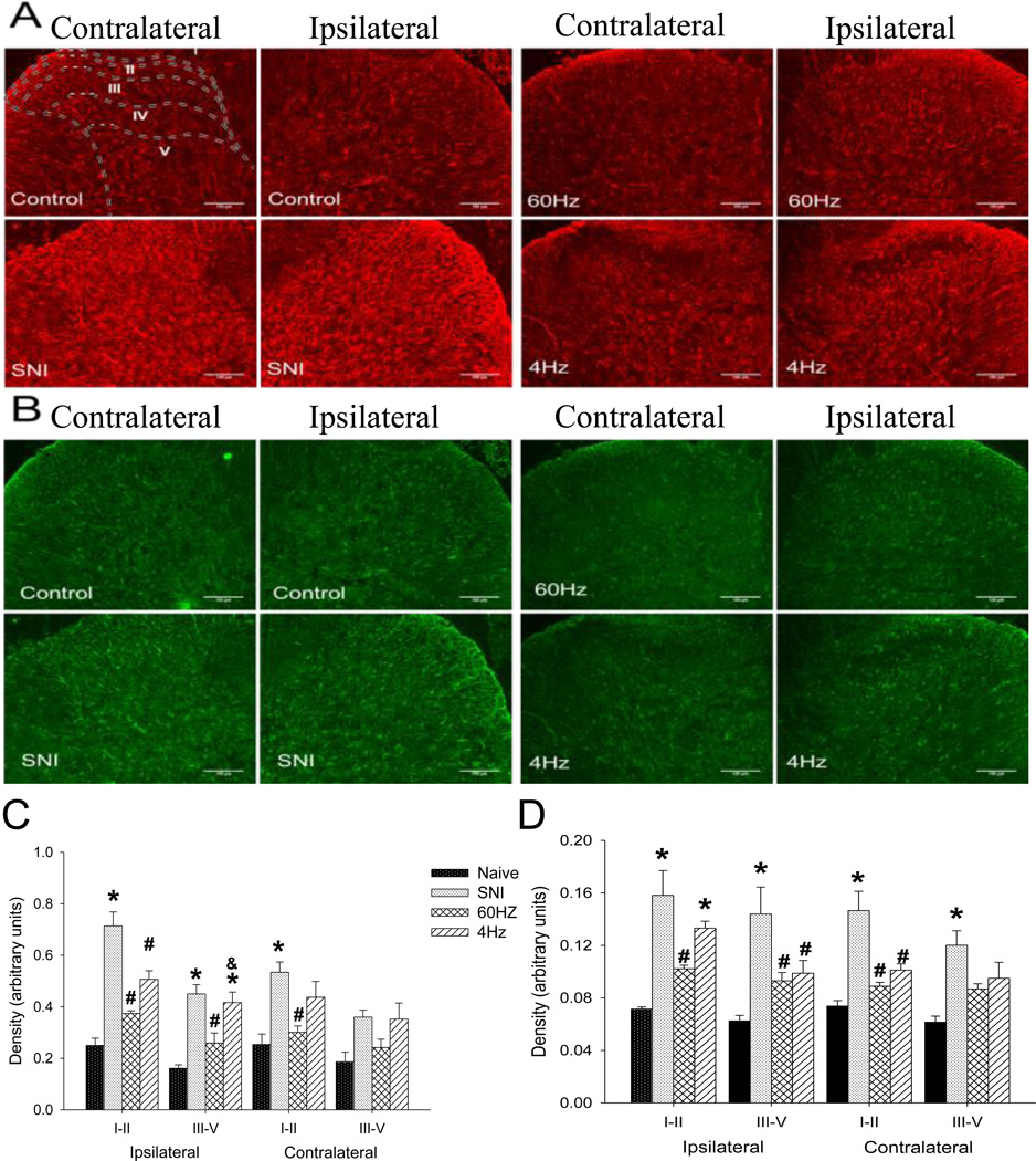
Background: Spinal cord stimulation (SCS) is commonly used for neuropathic pain; the optimal variables and mechanisms of action are unclear. We tested whether modulation of SCS variables improved analgesia in animals with neuropathic pain by comparing 6-hour vs 30-minute duration and 50%, 75%, or 90% motor threshold (MT) intensity (amplitude). Furthermore, we examined whether maximally effective SCS reduced glial activation in the spinal cord in neuropathic animals.

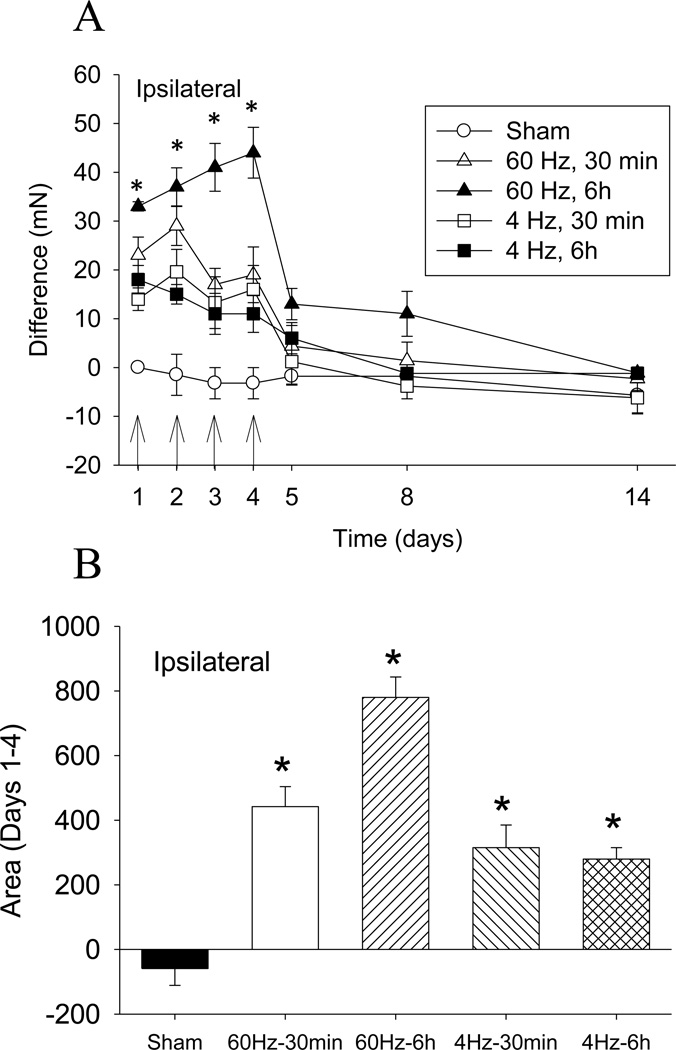
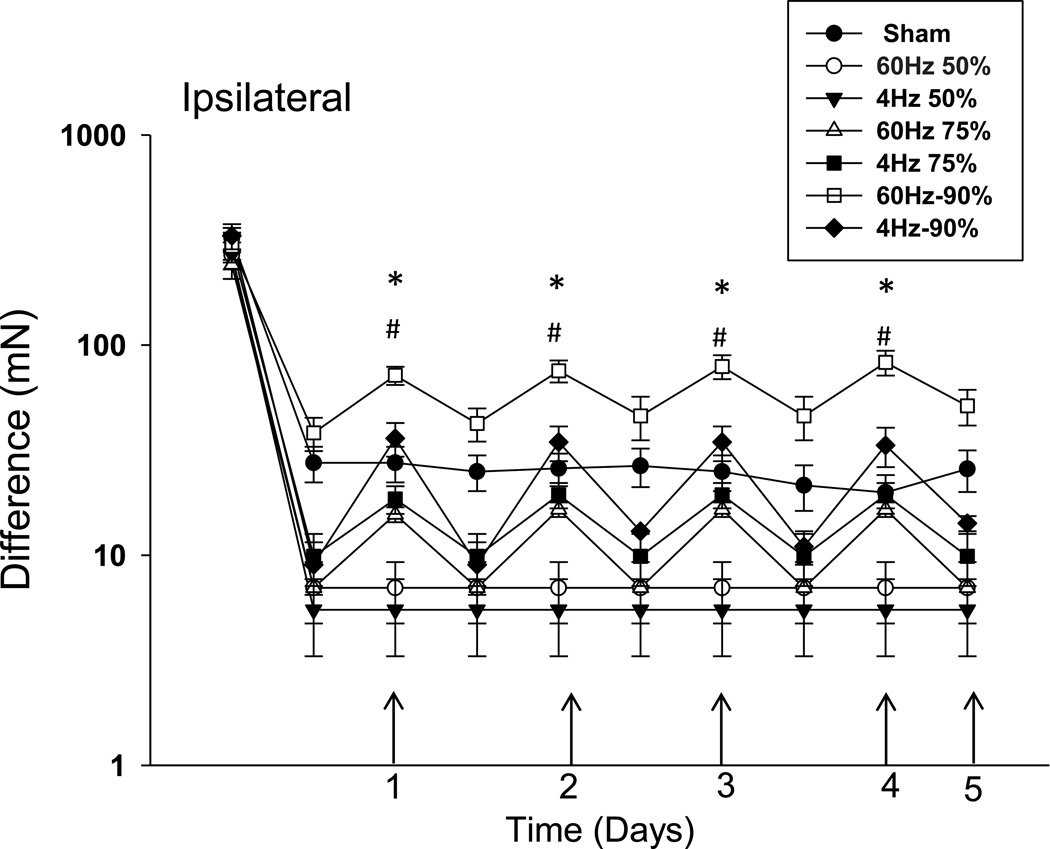
Methods: Sprague-Dawley rats received the spared nerve injury model and were implanted with an epidural SCS lead. Animals were tested for mechanical withdrawal threshold of the paw before and 2 weeks after spared nerve injury, before and after SCS daily for 4 days, and 1, 4, and 9 days after SCS. Spinal cords were examined for the effects of SCS on glial cell activation.

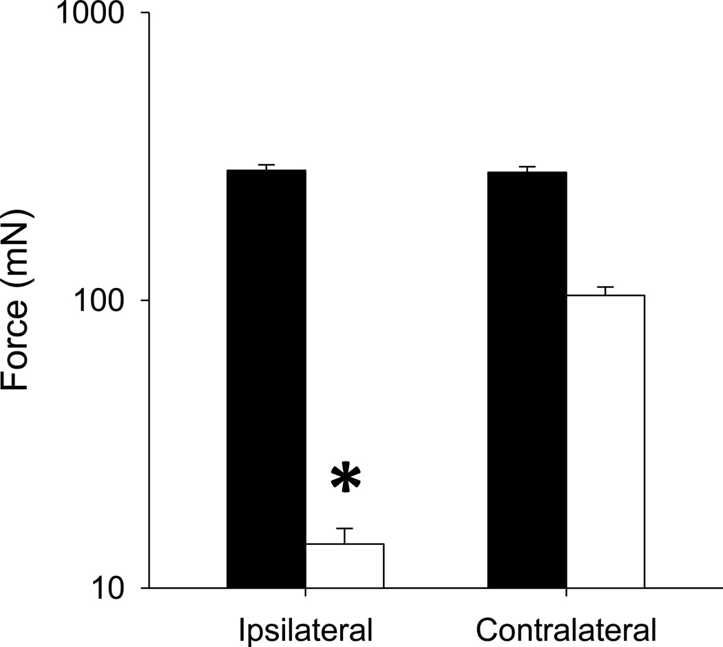
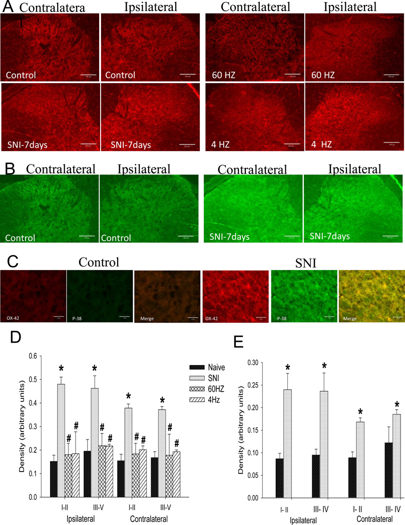
Results: The mechanical withdrawal threshold decreased, and glial immunoreactivity increased 2 weeks after spared nerve injury. For duration, 6-hour SCS significantly increased the mechanical withdrawal threshold when compared with 30-minute SCS or sham SCS; 30-minute SCS was greater than sham SCS. For intensity (amplitude), 90% MT SCS significantly increased the withdrawal threshold when compared with 75% MT SCS, 50% MT SCS, and sham SCS. Both 4 and 60 Hz SCS decreased glial activation (GFAP, MCP-1, and OX-42) in the spinal cord dorsal horn when compared with sham.

Conclusions: Six-hour duration SCS with 90% MT showed the largest increase in mechanical withdrawal threshold, suggesting that the variables of stimulation are important for clinical effectiveness. One potential mechanism for SCS may be to reduce glial activation at the level of the spinal cord.


Conflict of interest statement

Conflict of Interest Statement: There are no financial or other relationships that might lead to conflicts of interest.

Figures

Figure 1
Figure 1
Average withdrawal thresholds before (baseline) and 2 weeks SNI for the ipsilateral and contralateral sides. Data are man + S.E.M. *, p<0.05
Figure 2
Figure 2
A. Time course for changes in withdrawal thresholds after SNI and after SCS for up to 14 days. SCS significantly increased the mechanical withdrawal threshold bilaterally when compared to sham SCS. A greater effect was observed with 6h of SCS compared 30min SCS. The arrow shows the time of SCS treatment. B. Averege area under the curve for the changes in withdrawal thresholds during SCS compared to prior to SCS for all groups averaged over the first 4 days of SNI. Data are mean difference scores between after SCS compared to before SCS with S.E.M. * p<0.05, different from sham group.
Figure 3
Figure 3
SCS at 90% MT and 75% MT significantly increased withdrawal thresholds of the paw. Notice a dose-response effect ipsilaterally with increasing intensity of SCS. The difference in paw withdrawal threshold after SCS when compared to pre-SCS values 2 weeks after SNI. * diference between 90% MT and others groups, # difference between 75% and 50% and sham.
Figure 4
Figure 4
A. Representative tissue sections for OX-42 immunostaining in the dorsal horn of naive control rats, SNI-7days, 60 Hz SCS and 4 Hz SCS. Bar =100µm. B. Representative tissue sections for p-p-38 immunostaining in the dorsal horn of naive control rats compared to 7 days after SNI. Bar =100µm. C. Representative tissue sections with high magnification (40X) for OX-42 in red, p-p-38 in green and merge in yellow of control rats and 7 days after SNI. Bar =50µm. D. There is a significant increase in the density of OX-42 staining bilaterally in the dorsal horn after SNI. Both 60 Hz SCS and 4 Hz SCS significantly decrease this staining. E. There is a significant increase in the density of staining bilaterally in the dorsal horn 7 days after SNI in p-p-38 staining. (*) compared with the naieve controls, (#) compared with the SNI, Data represent mean ≤ p=0.005.
Figure 5
Figure 5
A. Representative tissue sections for GFAP immunostaining in the dorsal horn of control rats, SNI, 60 Hz SCS and 4 Hz SCS. Sketches delineating boundaries of different laminae are superimposed over the spinal sections of control rats. B. Representative tissue sections for MCP-1 immunostaining in the dorsal horn of native control rats, SNI, 60 Hz SCS and 4 Hz SCS. C. There is a significant increase in the density of GFAP staining bilaterally in the dorsal horn after SNI. Both 60 Hz SCS and 4 Hz SCS significantly decrease this staining. D. There is a significant increase in the density of staining bilaterally in the dorsal horn after SNI. Both 60 Hz SCS and 4 Hz SCS significantly decrease MCP-1 staining. (*) compared with naive controls, (#) compared with the SNI, (&) compared with 60Hz SCS. Data represent mean ≤ p=0.005. Bar =100µm.

References

    1. Barchini J, Tchachaghian S, Shamaa F, Jabbur SJ, Meyerson BA, Song Z, Linderoth B, Saade NE. Spinal segmental and supraspinal mechanisms underlying the pain-relieving effects of spinal cord stimulation: an experimental study in a rat model of neuropathy. Neuroscience. 2012;215:196–208. - PubMed
    1. Campbell JN, Meyer RA. Mechanisms of neuropathic pain. Neuron. 2006;52:77–92. - PMC - PubMed
    1. Casamenti F, Prosperi C, Scali C, Giovannelli L, Colivicchi MA, Faussone-Pellegrini MS, Pepeu G. Interleukin-1beta activates forebrain glial cells and increases nitric oxide production and cortical glutamate and GABA release in vivo: implications for Alzheimer’s disease. Neuroscience. 1999;91:831–842. - PubMed
    1. Chen T, Koga K, Li XY, Zhuo M. Spinal microglial motility is independent of neuronal activity and plasticity in adult mice. Mol Pain. 2010;6:19. - PMC - PubMed
    1. Chen T, Willoughby KA, Ellis EF. Group I metabotropic receptor antagonism blocks depletion of calcium stores and reduces potentiated capacitative calcium entry in strain-injured neurons and astrocytes. J Neurotrauma. 2004;21:271–281. - PubMed

Publication types