. 2014 Feb 7;289(6):3724-35.
doi: 10.1074/jbc.M113.529032. Epub 2013 Dec 20.

Structure of the TbBILBO1 protein N-terminal domain from Trypanosoma brucei reveals an essential requirement for a conserved surface patch

Affiliations

Structure of the TbBILBO1 protein N-terminal domain from Trypanosoma brucei reveals an essential requirement for a conserved surface patch

Keni Vidilaseris et al. J Biol Chem. .

Abstract

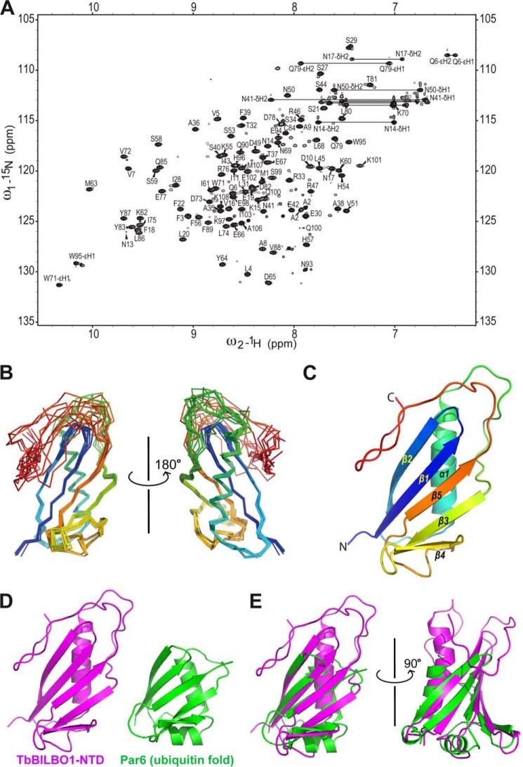
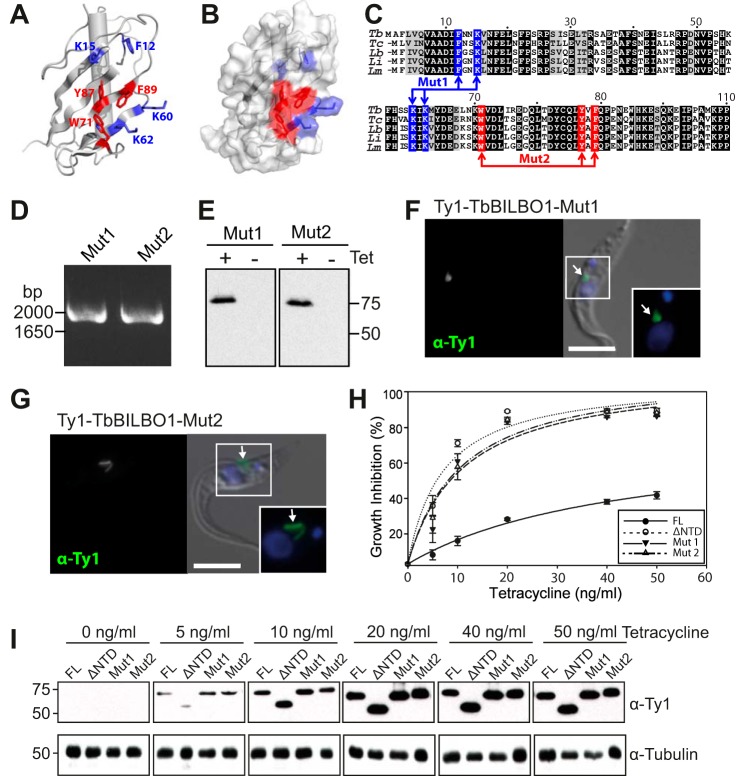
TbBILBO1 is the only known component of the flagellar pocket collar, a cytoskeletal barrier element found in trypanosomes. The N-terminal domain (NTD) of TbBILBO1 was found to be dispensable for targeting of the protein in vivo. However, overexpression of constructs lacking the NTD caused complete growth inhibition, implying an essential requirement for this domain. A high resolution structure of the NTD of TbBILBO1 showed that it forms a ubiquitin-like fold with a conserved surface patch. Mutagenesis of this patch recapitulated the phenotypic effects of deleting the entire domain and was found to cause cell death. The surface patch on the NTD of TbBILBO1 is therefore a potential drug target.

Keywords: Cytoskeleton; Flagellar Pocket Collar; Infectious Diseases; Molecular Cell Biology; Nuclear Magnetic Resonance; Parasite; Protein Structure; Structural Biology; TbBILBO1; Trypanosoma brucei.


Figures

FIGURE 1.
FIGURE 1.
The TbBILBO1-NTD is not required for localization. A, schematic depicting the arrangement of the three domains of TbBILBO1: N-terminal domain (NTD), EF hands (EF), and coiled coil domain. Amino acid numbers are indicated above the schematic. B, TbBILBO1 truncation constructs used for the localization experiments. Abbreviations are defined in the text. C–E, localizations of YFP-tagged TbBILBO1 full-length and truncation constructs in transiently transfected T. brucei cells. Intact cells were analyzed by immunofluorescence microscopy. Anti-GFP antibodies were used to visualize YFP-TbBILBO1-NTD as low expression levels precluded direct observation of YFP. DAPI was used to stain DNA (blue). Regions enlarged in insets are indicated with white boxes. Scale bars, 5 μm. F, immunoblot of whole cell lysates from transiently transfected T. brucei cells, probed using anti-GFP antibodies. Arrows indicate the expected size of the YFP-tagged TbBILBO1 constructs.
FIGURE 2.
FIGURE 2.
The TbBILBO1-NTD is required for normal cellular growth. A, PCR amplification of genomic DNA confirms the presence of the indicated TbBILBO1 transgenes. Primers annealing to sequences encoding the Ty1 epitope tag (5′-end) and ORF (3′-end) were used. B, immunoblots of whole cell lysates from cells inducibly expressed the indicated Ty1-tagged TbBILBO1 transgenes in the presence or absence of 20 ng/ml tetracycline. Probing with anti-Ty1 antibodies confirmed that both transgenes were inducibly and tightly expressed. C–E, immunofluorescence microscopy using anti-Ty1 antibodies confirmed correct and inducible localization of the transgenic proteins. DAPI was used to stain DNA. Scale bars, 5 μm. C, no labeling was seen in the absence of tetracycline. D and E, both Ty1-tagged TbBILBO1 constructs localized correctly. Arrows indicate the point of flagellum entry into the cell. Boxed areas are enlarged in insets. F, growth inhibition curves from stably transfected cells inducibly expressed Ty1-tagged TbBILBO1-FL or TbBILBO1-ΔNTD at different concentrations of tetracycline after a 4-day time course. Values were calculated from the mean of at least three independent experiments. Error bars show S.E. G, immunoblot analysis shows whole cell lysates taken from cells expressing Ty1-tagged TbBILBO1-FL and -ΔNTD at different concentrations of tetracycline after 4 days of induction. TbBILBO1 constructs were detected using anti-Ty1 antibodies (upper panel). An immunoblot using anti-tubulin antibodies was used as a loading control (lower panel).
FIGURE 3.
FIGURE 3.
NMR structure of the TbBILBO1-NTD reveals a ubiquitin-like fold. A, 15N-1H correlated HSQC spectrum of the TbBILBO1-NTD. The spectrum was collected from a sample of TbBILBO1-NTD at 25 °C in a 20 mm sodium phosphate buffer (pH 7.8) containing 100 mm NaCl, 10% (v/v) 2H2O, and 0.2% (w/v) NaN3. Resonance assignments for all resolved cross-peaks are labeled. B, two views of superimposed 10 energy-minimized NMR conformers of the TbBILBO1-NTD. C, ribbon diagram of the TbBILBO1-NTD structure, color-ramped from blue at the N terminus to red at the C terminus. The five β strands (β1–β5) and the single α helix (α1) are labeled. D, structural comparison between the TbBILBO1-NTD and the ubiquitin-like PB1 domain of Par6 (Protein Data Bank ID code 1WMH). E, two different views of the TbBILBO1-NTD structure superimposed on the Par6-PB1.
FIGURE 4.
FIGURE 4.
Essential requirement for a conserved surface patch on the TbBILBO1-NTD. A, ribbon diagram of the TbBILBO1-NTD with the seven conserved residues shown as sticks. The three aromatic residues at the bottom of the conserved crater are colored in red; the four flanking residues at the rim are shown in blue. B, surface plot with the same orientation and color scheme as in A. C, primary sequence alignment of the NTDs of TbBILBO1 and homologs. Residues altered by site-directed mutagenesis in the Mut1 (rim residues) and Mut2 (aromatic residues at the crater bottom) constructs are indicated. Tb, T. brucei; Tc, T. cruzi; Lb, Leishmania braziliensis; Li, Leishmania infantum; Lm, Leishmania major. D, PCR amplification of genomic DNA to confirm the presence of the indicated TbBILBO1 transgenes. E, anti-Ty1 immunoblots of whole cell lysates from cells inducibly expressing Ty1-tagged TbBILBO1-Mut1 or -Mut2 transgenes in the presence or absence of 20 ng/ml tetracycline. F and G, Ty1-tagged TbBILBO1-Mut1 and -Mut2 constructs localizing correctly to the FPC. Arrows indicate the point of flagellum entry into the cell. DNA is labeled with DAPI. Scale bars, 5 μm. H, mutagenesis of the TbBILBO1-NTD causing cell growth inhibition. Growth inhibition curves are from stably transfected cells inducibly expressing Ty1-tagged TbBILBO1 constructs at different concentrations of tetracycline after 4 days of induction. I, immunoblots of whole cell lysates from the four cell lines taken at various concentrations of tetracycline and probed using anti-Ty1 antibodies (upper panels). The expression of all four constructs was approximately the same at each tetracycline concentration and saturated at around 20 ng/ml. Immunoblotting with anti-tubulin antibodies was used as a loading control (lower panels).
FIGURE 5.
FIGURE 5.
TbBILBO1-NTD deletion or mutagenesis causes dominant negative effects. All cells were assayed by immunofluorescence microscopy after a 2-day induction using 20 ng/ml tetracycline. Cells were labeled using anti-Ty1 antibodies; DAPI was used to stain DNA. Boxed regions are enlarged in insets. A, Ty1-TbBILBO1-FL does not cause gross morphological abnormalities. In replicating cells, Ty1-TbBILBO1-FL was associated with both old and new flagella (arrows). B–D, overexpression of Ty1-TbBILBO1-ΔNTD, -Mut1, and -Mut2 causes gross morphological effects, notably a detached new flagellum in replicating cells (arrowheads). Note the absence of TbBILBO1 labeling at the detached new flagella. Scale bars, 5 μm.
FIGURE 6.
FIGURE 6.
The detached flagellum in overexpressing cells has an associated basal body. Expression of the various Ty1-tagged TbBILBO1 constructs was induced for 2 days using 20 ng/ml tetracycline. Detergent-extracted cells were labeled with anti-Ty1 and anti-TbCentrin4 antibodies. DAPI was used to stain DNA. Boxed areas are enlarged in insets. A, in replicating cells overexpressing Ty1-TbBILBO1-FL, both Ty1-TbBILBO1-FL and TbCentrin4 had normal localizations and were associated with both old and new flagella. B–D, the detached new flagella in replicating cells overexpressing the other TbBILBO1 constructs are associated with TbCentrin4-positive basal bodies (arrowheads). Scale bars, 5 μm.
FIGURE 7.
FIGURE 7.
Loss or mutagenesis of the TbBILBO1-NTD is lethal. Stably transfected cells conditionally expressing the indicated constructs were induced using 20 ng/ml tetracycline for 4 days. They were then treated with fluorescein isothiocyanate-labeled anti-Annexin V antibodies, stained with propidium iodide, and analyzed by FACS. A, diagram showing the different cell populations detected by staining with Annexin V and propidium iodide. B, negative control using uninduced cells. C, positive control for programmed cell death using 4 mm DTT for 24 h. D–G, FACS traces for cells overexpressing the indicated constructs. H, quantitation of the data shown in B–G. Values shown are the mean from three independent experiments. Error bars show S.E.
FIGURE 8.
FIGURE 8.
Structural comparison of the TbBILBO1-NTD with the PB1 domain of Par6 in the Par6-aPKC complex. A, ribbon diagram of the TbBILBO1-NTD NMR structure. Residues in the essential surface patch are shown as red/blue sticks. The magenta arrow indicates the crater-like area mutated in this study. The orange oval encircles the flexible loop at the C terminus of the NTD. B, ribbon diagram of the Par6-aPKC complex (Protein Data Bank ID code 1WMH). The magenta oval indicates the interaction interface between the two proteins. C, superimposition of the TbBILBO1-NTD onto the Par6-PB1 in the Par6-aPKC complex. The C-terminal loop of the TbBILBO1-NTD obscures the surface corresponding to the aPKC binding site on Par6. The conserved surface patch of the TbBILBO1-NTD is also distant from the corresponding aPKC binding site on Par6. D, two views of the superimposed structures with the TbBILBO1-NTD shown in white surface plot.

Similar articles

Cited by

References

    1. Simpson A. G., Stevens J. R., Lukes J. (2006) The evolution and diversity of kinetoplastid flagellates. Trends Parasitol. 22, 168–174 - PubMed
    1. Welburn S. C., Maudlin I. (2012) Priorities for the elimination of sleeping sickness. Adv. Parasitol. 79, 299–337 - PubMed
    1. Schmunis G. A., Yadon Z. E. (2010) Chagas disease: a Latin American health problem becoming a world health problem. Acta Trop. 115, 14–21 - PubMed
    1. Alvar J., Vélez I. D., Bern C., Herrero M., Desjeux P., Cano J., Jannin J., den Boer M., and WHO Leishmaniasis Control Team (2012) Leishmaniasis worldwide and global estimates of its incidence. PLoS One 7, e35671. - PMC - PubMed
    1. Zucca M., Scutera S., Savoia D. (2013) New chemotherapeutic strategies against malaria, leishmaniasis and trypanosomiases. Curr. Med. Chem. 20, 502–526 - PubMed

Publication types

MeSH terms

Substances

Associated data

LinkOut - more resources