. 2014 Feb 7;289(6):3405-15.
doi: 10.1074/jbc.M113.531251. Epub 2013 Dec 20.

Structural insights into the epimerization of β-1,4-linked oligosaccharides catalyzed by cellobiose 2-epimerase, the sole enzyme epimerizing non-anomeric hydroxyl groups of unmodified sugars

Affiliations

Structural insights into the epimerization of β-1,4-linked oligosaccharides catalyzed by cellobiose 2-epimerase, the sole enzyme epimerizing non-anomeric hydroxyl groups of unmodified sugars

Takaaki Fujiwara et al. J Biol Chem. .

Abstract

Cellobiose 2-epimerase (CE) reversibly converts d-glucose residues into d-mannose residues at the reducing end of unmodified β1,4-linked oligosaccharides, including β-1,4-mannobiose, cellobiose, and lactose. CE is responsible for conversion of β1,4-mannobiose to 4-O-β-d-mannosyl-d-glucose in mannan metabolism. However, the detailed catalytic mechanism of CE is unclear due to the lack of structural data in complex with ligands. We determined the crystal structures of halothermophile Rhodothermus marinus CE (RmCE) in complex with substrates/products or intermediate analogs, and its apo form. The structures in complex with the substrates/products indicated that the residues in the β5-β6 loop as well as those in the inner six helices form the catalytic site. Trp-322 and Trp-385 interact with reducing and non-reducing end parts of these ligands, respectively, by stacking interactions. The architecture of the catalytic site also provided insights into the mechanism of reversible epimerization. His-259 abstracts the H2 proton of the d-mannose residue at the reducing end, and consistently forms the cis-enediol intermediate by facilitated depolarization of the 2-OH group mediated by hydrogen bonding interaction with His-200. His-390 subsequently donates the proton to the C2 atom of the intermediate to form a d-glucose residue. The reverse reaction is mediated by these three histidines with the inverse roles of acid/base catalysts. The conformation of cellobiitol demonstrated that the deprotonation/reprotonation step is coupled with rotation of the C2-C3 bond of the open form of the ligand. Moreover, it is postulated that His-390 is closely related to ring opening/closure by transferring a proton between the O5 and O1 atoms of the ligand.

Keywords: AGE Superfamily; Bacterial Metabolism; Carbohydrate-binding Protein; Cellobiitol; Cellobiose 2-Epimerase; Crystal Structure; Enzyme Mechanisms; Epimerization; Glycobiology.


Figures
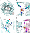
FIGURE 1.
FIGURE 1.
A, overall structure of apo-RmCE displayed as a ribbon diagram. B, chloride ion binding site. The residues around the bound chloride ion are represented as sticks. Chloride ion and water molecule are represented as spheres in red and green, respectively. Polar interactions are shown by dotted lines. Electron density of the bound chloride ion and water in the omit |Fo| − |Fc| map (gray) are calculated without the ligand and contoured at 3.0 σ. C and D, phosphate ion binding site. The residues around the bound phosphate ions are represented as sticks. Polar interactions are shown by dotted lines. Electron density of the bound phosphate in the omit |Fo| − |Fc| map (gray) are calculated without the ligand and contoured at 3.0 σ.
FIGURE 2.
FIGURE 2.
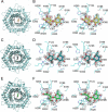
A, C, and D, the overall structures of RmCE-Glc-Man, RmCE-epilactose, and RmCE-cellobiitol, respectively, displayed as ribbon diagrams. B, D, and F, stereo views of catalytic site architecture of RmCE-Glc-Man, RmCE-epilactose, and RmCE-cellobiitol, respectively. The residues involved in substrate recognition are represented as lines. Water molecules are represented as spheres (red). Electron densities of the bound Glc-Man, epilactose, and cellobiitol in the omit |Fo| − |Fc]bar] map (gray) were calculated without the substrate and contoured at 3.0 σ.
FIGURE 3.
FIGURE 3.
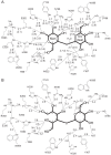
A, contact between Glc-Man or epilactose and RmCE. The distances between specific atoms are shown in Å as dotted lines. Numbers in parentheses are for contact between epilactose and RmCE. B, contact between cellobiitol and RmCE. The distances between specific atoms are shown in Å as dotted lines.
FIGURE 4.
FIGURE 4.
A, the structure of apo-form of RmCE is superposed on that of Glc-Man-bound RmCE (upper panel), and that of epilactose (lower panel). Apo structures in both panels are shown in light green. The β5-β6 loops in RmCE in complex with Glc-Man (blue) and epilactose (dark green) are represented and the residues close to the substrate are depicted as sticks. B, multiple sequence alignment of several CEs. Serine and aspartic acid residues related to substrate binding are indicated by stars and solid circles, respectively. The multiple sequence alignment was performed using the programs ClustalW and ESPript. The sequences are as follows (GenBankTM accession numbers in parentheses): RmCE (BAK61777.1), RaCE (BAF81108.1), BfCE (BAH23773.1), EcCE (BAG68451.1), CsCE (WP_011915904.1), and DtCE (BAM66298.1). C, the ribbon diagrams of β11-α8 loop in SeYihS with or without d-mannose are shown in green and blue, respectively. The residues of the orientations of which are changed to make contact with the ligand are depicted by sticks.
FIGURE 5.
FIGURE 5.
A, relative orientations of three catalytic histidine residues to cellobiitol and Glc-Man. Residues in RmCE-cellobiitol and RmCE-Glc-Man are represented as sticks in yellow and blue, respectively. B, the closed form sugars at the reducing end of cellobiose (left) and Glc-Man (right), and the sugar alcohol part of cellobiitol (middle) viewed along with the C2-C3 bond. C, projection of the closed form sugars at the reducing end of cellobiose (left) and Glc-Man (right), and the sugar alcohol part of cellobiitol (middle) viewed along with the C2-C3 bond.
FIGURE 6.
FIGURE 6.
Schematic diagram of the proposed epimerization catalyzed by RmCE. The electron transfer processes to convert glucose into mannose and its reverse conversion are represented by black and gray arrows, respectively.
FIGURE 7.
FIGURE 7.
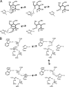
A, schematic diagram of the proposed ring opening mechanism involving His-390. The electron transfer reaction is represented by arrows (black). B, schematic diagram of the proposed proton-relay system. The electron transfer processes of forward and reverse reactions are represented by black and gray arrows, respectively.
FIGURE 8.
FIGURE 8.
Relationships between structural features to thermal stability of RmCE. The structure displayed is the Glc-Man-bound form of RmCE. A, hydrophobic core formed by helices α1, α10, α11, and α12. Ribbon diagrams of RmCE (upper panel) and RaCE (lower panel) are shown, and hydrophobic residues are represented as sticks. Extended parts of helices in RmCE are shown in dark blue. B, positions of Pro and Arg residues in RmCE (upper left and upper right, respectively) and RaCE (lower left and lower right, respectively) are indicated. Surface representations of Pro and Arg residues are shown in orange and blue, respectively.

References

    1. Tyler T. R., Leatherwood J. M. (1967) Epimerization of disaccharides by enzyme preparations from Ruminococcus albus. Arch. Biochem. Biophys. 119, 363–367 - PubMed
    1. Senoura T., Ito S., Taguchi H., Higa M., Hamada S., Matsui H., Ozawa T., Jin S., Watanabe J., Wasaki J., Ito S. (2011) New microbial mannan catabolic pathway that involves a novel mannosylglucose phosphorylase. Biochem. Biophys. Res. Commun. 408, 701–706 - PubMed
    1. Kawahara R., Saburi W., Odaka R., Taguchi H., Ito S., Mori H., Matsui H. (2012) Metabolic mechanism of mannan in ruminal bacterium, Ruminococcuss albus, involving two mannoside phosphorylases and cellobiose 2-epimerase. Discovery of a new carbohydrate phosphorylase, β-1,4-mannooligosaccharide phosphorylase. J. Biol. Chem. 287, 42389–42399 - PMC - PubMed
    1. Ito S., Taguchi H., Hamada S., Kawauchi S., Ito H., Senoura T., Watanabe J., Nishimukai M., Ito S., Matsui H. (2008) Enzymatic properties of cellobiose 2-epimerase from Ruminococcus albus and the synthesis of rare oligosaccharides by the enzyme. Appl. Microbiol. Biotechnol. 79, 433–441 - PubMed
    1. Watanabe J., Nishimukai M., Taguchi H., Senoura T., Hamada S., Matsui H., Yamamoto T., Wasaki J., Hara H., Ito S. (2008) Prebiotic properties of epilactose. J. Dairy Sci. 91, 4518–4526 - PubMed

Publication types

MeSH terms

LinkOut - more resources