. 2013:372:3-38.
doi: 10.1007/978-3-642-38919-1_1.

Respiratory syncytial virus: virology, reverse genetics, and pathogenesis of disease

Affiliations

Respiratory syncytial virus: virology, reverse genetics, and pathogenesis of disease

Peter L Collins et al. Curr Top Microbiol Immunol. 2013.

Abstract

Human respiratory syncytial virus (RSV) is an enveloped, nonsegmented negative-strand RNA virus of family Paramyxoviridae. RSV is the most complex member of the family in terms of the number of genes and proteins. It is also relatively divergent and distinct from the prototype members of the family. In the past 30 years, we have seen a tremendous increase in our understanding of the molecular biology of RSV based on a succession of advances involving molecular cloning, reverse genetics, and detailed studies of protein function and structure. Much remains to be learned. RSV disease is complex and variable, and the host and viral factors that determine tropism and disease are poorly understood. RSV is notable for a historic vaccine failure in the 1960s involving a formalin-inactivated vaccine that primed for enhanced disease in RSV naïve recipients. Live vaccine candidates have been shown to be free of this complication. However, development of subunit or other protein-based vaccines for pediatric use is hampered by the possibility of enhanced disease and the difficulty of reliably demonstrating its absence in preclinical studies.


Figures

Fig. 1
Fig. 1
Photomicrographs (a and b) and electron micrographs (ce) of RSV-infected cells and associated viral structures. a is a photomicrograph of a syncytium in an RSV-infected cell monolayer (several nuclei are indicated with arrows; courtesy of Dr. Alexander Bukreyev). b is a fluorescence photomicrograph of a syncytium in an RSV-infected cell monolayer (not the same one as in a) stained with an antibody specific to the F protein, showing filamentous viral projections (courtesy of Dr. Ursula J. Buchholz). c is an electron micrograph of negatively stained budding RSV virions: V indicates a budding virion and F indicates filamentous cytoplasmic structures that likely are nucleocapsids (courtesy of Dr. Robert M. Chanock) (Kalica et al. 1973). d and e are field emission scanning electron micrographs of the surface of uninfected (d) and RSV-infected (e) cells, illustrating viral filamentous structures (VF in e) that are thought to form at sites of virus budding and may yield filamentous particles; also shown are microvilli (mv in d) that are found in uninfected cells (courtesy of Dr. Richard Sugrue) (Jeffree et al. 2003)
Fig. 2
Fig. 2
RSV proteins and their functions and location in the virion, shown in reference strained electron micrographs of negatively stained budding (a) and free (b) virions (courtesy of Dr. Robert M. Chanock)
Fig. 3
Fig. 3
Diagram of the 3′ to 5′ negative-sense RSV genome (approximately to scale, strain A2), and sequences of the leader region, gene junctions, overlap, and trailer region. a Genome diagram: each box represents a gene encoding a separate mRNA. The first row of numbers over the diagram indicates the nucleotide lengths of the genes, and the second, upper row of numbers (italicized) indicates the amino acid lengths of the primary, unmodified proteins. The overlapping ORFs of the M2 mRNA are illustrated over the M2 gene. The numbers under the diagram indicate the lengths of the leader, intergenic, and trailer regions (underlined) and the gene overlap (parentheses). b Sequences of the leader region, gene junctions, overlap, and trailer region (3′ to 5′, negative-sense). This shows the leader region, followed by the NS1, NS2, N, P, M, SH, G, and F genes and their intergenic regions, followed by the overlapping M2 and L genes and the trailer region. The main body of each gene is deleted and is represented by a box with the gene name. Nucleotide assignments that are conserved between the 3′ ends of the genome and the antigenome (represented here as the reverse complement in the trailer region) are in bold capitals. The gene-start and gene-end signals of the gene are underlined, and conserved asignments are in capitals
Fig. 4
Fig. 4
Overview of RSV transcription and RNA replication. The polymerase enters the negative-sense genome at its 3′ end executes transcription to yield positive-sense subgenomic mRNAs (in a polar gradient) or executes the first step in RNA replication to yield full-length positive-sense antigenome. The polymerase enters the antigenome at its 3′ end and executes the second step of RNA replication to yield full-length progeny genomes. Note that the L gene yields two polyadenylated mRNAs: a very short species due to termination in the gene overlap, and full-length L mRNA
Fig. 5
Fig. 5
Comparison of the genes and gene order of RSV with those of selected members of Paramyxoviridae: HMPV, human parainfluenza virus (HPIV) serotypes 1, 2, and 3, and measles virus (MeV). The genes are shown in their 3′ to 5′ order in genomic RNA, which is the direction of transcription. Genes are not to scale, and orthologous genes are aligned vertically as much as possible (the only genes that could not be appropriately aligned are SH and G of RSV and SH of HMPV), with gaps introduced to maximize the alignments. Genes encoding major protective antigens are in dark shading. Asterisks indicate proteins that can be deleted from RSV without loss of replication, although this may be reduced. Proteins that have no direct ortholog in RSV include: C small accessory protein, V cysteine-rich accessory protein, HN hemagglutinin-neuraminidase glycoprotein, H hemagglutinin glycoprotein. The Henipavirus and Avulavirus genera and a number of unclassified viruses within Paramyxovirinae are not represented
Fig. 6
Fig. 6
Nucleotide positions in the leader region of genomic RNA that are important for transcription (top) and RNA replication (bottom). Important residues present in positions 1–11 are indicated with open boxes; note that those that are important for transcription are a subset of those important for RNA replication. A region that increases the efficiency of transcription is indicated with a dashed box. The GS signal of the first gene, necessary for transcriptional initiation but not involved in RNA replication, is underlined in the diagram for transcription. A region that contains an apparent encapsidation signal necessary to produce full-length replication products is indicated with a shaded box. Sequences are in negative-sense
Fig. 7
Fig. 7
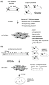
Reverse genetic systems. A Helper-dependent mini-replicon system, illustrated with a dicistronic mini-replicon (shown here as an antigenome-sense replicon) containing chloramphenicol acetyl transferase (CAT) and luciferase (LUC) marker genes under the control of RSV GS and GE signals (not shown) and flanked by the complements of the RSV leader and trailer regions (gray boxes, lec and trc, respectively). The mini-replicon cDNA is flanked by a T7 RNA polymerase promoter (arrow, T7 pr) and a self-cleaving ribozyme (black box, rbz). This is complemented by support plasmids encoding various RSV proteins. b Recovery of complete infectious virus from a full-length antigenome expressed from a transfected plasmid in the presence of support plasmids encoding the N, P, M2-1, and L proteins. The complements of the leader and trailer regions are shown (gray boxes, lec and trc, respectively), as are the T7 RNA polymerase promoter (arrow, T7pr) and ribozyme (black box)

References

    1. Asenjo A, Rodriguez L, Villanueva N. Determination of phosphorylated residues from human respiratory syncytial virus P protein that are dynamically dephosphorylated by cellular phosphatases: a possible role for serine 54. J Gen Virol. 2005;86(Pt 4):1109–1120. doi: 10.1099/vir.0.80692-086/4/1109. [pii] - DOI - PubMed
    1. Asenjo A, Calvo E, Villanueva N. Phosphorylation of human respiratory syncytial virus P protein at threonine 108 controls its interaction with the M2-1 protein in the viral RNA polymerase complex. J Gen Virol. 2006;87(Pt 12):3637–3642. doi: 10.1099/vir.0.82165-087/12/3637. [pii] - DOI - PubMed
    1. Asenjo A, Gonzalez-Armas JC, Villanueva N. Phosphorylation of human respiratory syncytial virus P protein at serine 54 regulates viral uncoating. Virology. 2008;380(1):26–33. doi: 10.1016/j.virol.2008.06.045. S0042-6822(08)00427-3 [pii] - DOI - PubMed
    1. Atreya PL, Peeples ME, Collins PL. The NS1 protein of human respiratory syncytial virus is a potent inhibitor of minigenome transcription and RNA replication. J Virol. 1998;72(2):1452–1461. - PMC - PubMed
    1. Ausar SF, Espina M, Brock J, Thyagarayapuran N, Repetto R, Khandke L, Middaugh CR. High-throughput screening of stabilizers for respiratory syncytial virus: identification of stabilizers and their effects on the conformational thermostability of viral particles. Hum Vaccin. 2007;3(3):94–103. doi: 10.1099/vir.0.82165-04149. [pii] - DOI - PubMed

Publication types

MeSH terms

Substances