. 2014 Feb;17(2):207-14.
doi: 10.1038/nn.3610. Epub 2013 Dec 22.

Adult neural stem cells in distinct microdomains generate previously unknown interneuron types

Affiliations

Adult neural stem cells in distinct microdomains generate previously unknown interneuron types

Florian T Merkle et al. Nat Neurosci. 2014 Feb.

Abstract

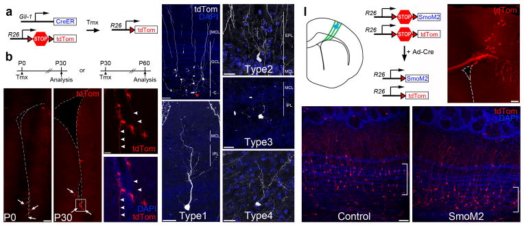
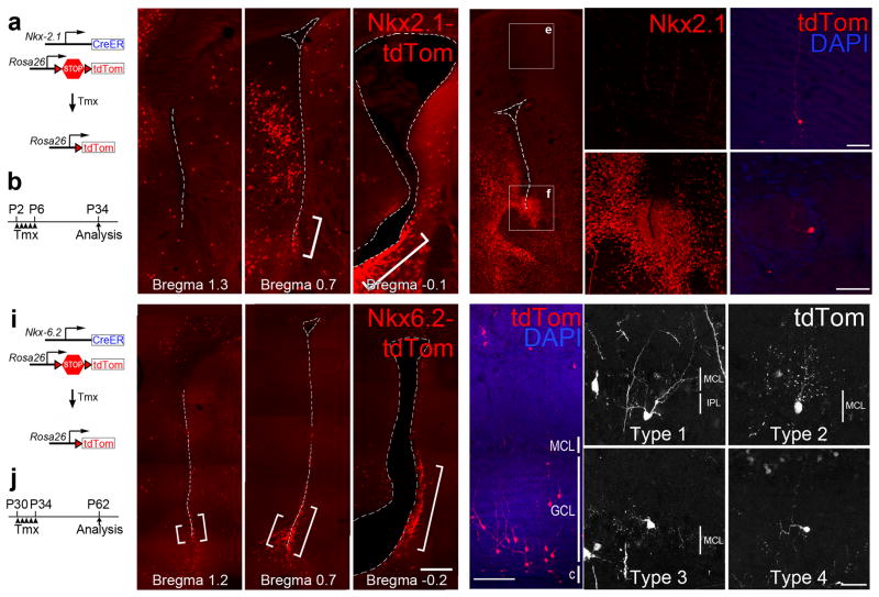
Throughout life, neural stem cells (NSCs) in different domains of the ventricular-subventricular zone (V-SVZ) of the adult rodent brain generate several subtypes of interneurons that regulate the function of the olfactory bulb. The full extent of diversity among adult NSCs and their progeny is not known. Here, we report the generation of at least four previously unknown olfactory bulb interneuron subtypes that are produced in finely patterned progenitor domains in the anterior ventral V-SVZ of both the neonatal and adult mouse brain. Progenitors of these interneurons are responsive to sonic hedgehog and are organized into microdomains that correlate with the expression domains of the Nkx6.2 and Zic family of transcription factors. This work reveals an unexpected degree of complexity in the specification and patterning of NSCs in the postnatal mouse brain.


Figures

Figure 1
Figure 1. Production of novel OB cell types by specifically labeled NSCs
a) Schematic diagram of radial glial cell targeting. Stereotaxic injections of small volumes of adenovirus infect cells locally at the injection site and needle tract (blue) as well as the long basal processes of radial glia (green), which transform into adult NSCs. b–d) DAB-stained photomicrographs of labeled cells in the P28 brain on the medial (b,c) and lateral (d) walls of the lateral ventricle. e–h) Labeled NSCs in this region of the V-SVZ generated novel labeled OB cell types (Type 1-4), stained here by DAB. i) Schematic diagram showing camera lucida traces of known adult-born OB interneurons (black) and novel cell types (red, blue, magenta, green) superimposed over a photomicrograph of a hematoxylin-stained OB to show their relative positions. Cells are shown to scale, and the pial surface is up. Scale bars are 75 μm for b–d and 25 μm for E-H. EPL, external plexiform layer; GC, granule cell; GL, glomerular layer; GRL, granule cell layer, IPL, internal plexiform layer; MCL, mitral cell layer; PCG, periglomerular cell.
Figure 2
Figure 2. Type 1-4 cells continue to be produced by GFAP+ NSCs in the adult anterior ventral V-SVZ
a) Diagram showing the injection tract (blue line) and injection site (blue circle) of Ad:GFAP-Cre targeting the anterior-ventral V-SVZ in a coronal schematic diagram of the adult (P60) brain hemisphere (midline to the left, dorsal is up). b) DAB-stained photomicrograph of targeted V-SVZ and labeled striatal astrocytes 28 days after Ad:GFAP-cre injection. c–f) Type 1-4 cells can be clearly identified by their distinctive morphology in the OB 28 days after adult neural stem cell labeling. Scale bar is 150 μm for B, 42 μm for c and f, and 25 μm for d and e.
Figure 3
Figure 3. Molecular and morphological characterization of Type 1-4 cells
a) Cell body diameters measured along the minor and major axes are shown for Type 1-4 cells alongside granule and periglomerular cells. b) Quantification of soma position in the OB relative to the MCL normalized to the thickness of the GRL and EPL. c) Maximum reach of the dendritic arbor into the EPL for each cell type. Note that although Type 1 and 2 cells had superficially located cell bodies, their dendrites were restricted to the lower EPL, whereas superficial granule cells (GIII, GIIIM) consistently reached the most superficial EPL. d) Analysis of the number of secondary and tertiary branches extending from the primary dendrite reveals a unique branching pattern that, along with other morphological characteristics, distinguishes Type 1-4 cells from granule and periglomerular cells. e) A subset of Type 1-4 cells expressed CalR. Data are expressed as mean±s.d. for a–d and mean±s.e.m for e. At least five mice were examined in each experimental group. Abbreviations for cell types and OB regions are given in Figure 1. CalR, calretinin.
Figure 4
Figure 4. Type 1-4 cells are produced by Gli1-expressing progenitors
a) Diagram showing the Gli1-CreERT2 and Rosa26-Ai14 alleles. b) Gli1::CreERT2;R26R-Ai14 neonates (P0) and adult (P30) mice were treated with tamoxifen and analyzed 30 days later. c–f) The majority of Gli1-expressing progenitors are located in the ventral V-SVZ in P0 (c) or P30 (d) animals (arrows) and frequently have B1 cell-like morphology with long basal processes (arrowheads, e). g) Gli1+-progenitors generate mainly deep GCs located close to the core of the OB. Arrowhead (red) denotes a migrating neuroblast in the OB core (c, core). h–k) Type 1-4 cells are produced by Gli1-expressing progenitors. In this image, the tdTomato staining has been pseudocolored to improve its contrast. These are representative examples of type 1-4 observed from n=13 mice. l) Schematic diagram showing the targeting of radial glia (green) in the cortical V-SVZ with Ad:Cre (blue) of a neonatal (P0) R26R-LSL-SmoM2;R26R-Ai14 mouse brain. Upon Cre recombination, a constitutive active form of the Smo receptor (SmoM2) and tdTomato are expressed in cortical radial glia and their progeny. m) Photomicrograph of a targeted R26R-Ai14 brain section 28 days after Ad:Cre injection. n–o) Cortical Ad:Cre injections in control R26R-Ai14 neonates generated mostly superficial GCs (in brackets, m), while SmoM2;R26R-Ai14 neonates produced deep granule cells (in brackets, n), but no Type 1-4 cells. These images are representative for each genotype of at least three independent experiments. Scale bar is 120 μm for c, 20 μm for e–j, 200 μm for l, and 60 μm for e.
Figure 5
Figure 5. Type 1-4 cells are produced by postnatal progenitors expressing Nkx6.2, but not in Nkx2.1
a,b) Diagram showing the Nkx2.1::CreERT2 and Rosa26-Ai14 alleles (a) and timing of tamoxifen (Tmx) administration and analysis (b) c) Nkx2.1 lineage-traced (tdTomato-expressing) cells are present in the ventral V-SVZ (brackets), shown here in coronal sections at different caudal-rostral levels. d–f) Immunofluorescence for Nkx2.1 protein in the neonatal mouse brain showing strong Nkx2.1 expression in the ventral V-SVZ (near the bed nucleus of the stria terminalis in d and f) but no expression in the cortex (d and e). g–h) Nkx2.1-expressing progenitors generate granule (g) and periglomerular (h) cells but not Type 1-4 cells. i,j) Diagram showing the Nkx6.2::CreERT2 and Rosa26-Ai14 alleles (i) and timing of Tmx administration and analysis (j). k) Nkx6.2 lineage-traced cells with B1 cell-like morphology are present in the anterior ventral V-SVZ (brackets). l) Nkx6.2+-progenitors generate mainly deep GCs located close to the core of the OB. m–p) Type 1-4 cells are produced by Nkx6.2-expressing progenitors. In this image, the tdTomato staining has been pseudocolored to improve its contrast. Scale bar is 60 μm for g–h, 200 μm for k–l, and 25 μm for m–p.
Figure 6
Figure 6. Zic1/2/3 expression partitions Type 1-4 production domains
a–c) Photomicrographs of coronal adult brain sections at three rostrocaudal levels showing Zic immunopositive cells in the medial wall of the adult V-SVZ. d) Zic expression in the OB, including the glomerular layer (GL), external plexiform layer (EPL), mitral cells layer (MCL), internal plexiform layer (IPL), and superficial granule cell layer (GCL). e) Co-localization of Zic and the OB interneuronal marker calretinin (CalR). Data are expressed as mean±s.e.m. At least three mice per group were examined. f) Quantification of CalR, CalB and TH expression in Zic+ periglomerular cells. g–h) Zic expression in GFP-positive V-SVZ cell types derived from Ad:Cre targeted radial glia in the medial V-SVZ. Zic is barely detectable in a small subpopulation of GFAP+ B1 cells (closed arrow, B1 cell body; open arrows, basal process, in g). Zic is expressed in cells with morphologies characteristics of transit-amplifying cells (arrowheads in g) and migrating neuroblasts (in the RMS) (arrow in h). i–j) Zic+ proliferating progenitors in the medial V-SVZ incorporate EdU (1 hr survival) (i), and express Ki67 (j). k) Within chains of migrating neuroblasts, Zic is expressed by a subpopulation of doublecortin (Dcx)+ neuroblasts (arrowheads). l–o) The majority of Type 1 (l) and Type 3 (n) cells express Zic protein, while Type 2 (m) and Type 4 (o) cells are mostly Zic negative. p) Quantification of Zic expression in Type 1-4 cells. Data are presented as mean±s.e.m., n=13 mice. Scale bar is 60 μm for a–c, and e and 20 μm for g–o.
Figure 7
Figure 7. Type 1-4 cells are produced in distinct subregions of the anterior ventral V-SVZ
a) Schematic lateral view of an adult mouse brain indicating the approximate spatial extent of the V-SVZ (light blue) including the subdomains that give rise to Type 1-4 cells (darker blue). Vertical lines indicate the approximate regions from which the outline of the lateral ventricle is shown in coronal section in (b–f). b) Labeled V-SVZ regions (blue) of brains containing labeled Type 1-4 cells were traced and overlaid onto a common template to reveal the origin of Type 1-4 cells (darker blue). c–f) This analysis was repeated for those brain regions giving rise to the highest percentage of Type 1, 2, 3, or 4 cells, revealing a unique spatial origin for each of these cell types. g–i) Approximate spatial extent of the Gli1 (g) and Nkx2.1 (h), and Nkx6.2 (i) expression domains, based on CreERT2 lineage tracing. Dashed line in (g) indicates lower expression. Nkx2.1 data in (h) are supported by immunostaining. j) Approximate spatial extent of the Zic1/2/3 expression domain based on immunostaining (see also Fig. 6 and S4).

References

    1. Alvarez-Buylla A, Garcia-Verdugo JM. Neurogenesis in adult subventricular zone. Journal of Neuroscience. 2002;22:629–634. - PMC - PubMed
    1. Sakamoto M, et al. Continuous neurogenesis in the adult forebrain is required for innate olfactory responses. Proceedings of the National Academy of Sciences. 2011;108:8479–8484. - PMC - PubMed
    1. Imayoshi I, et al. Roles of continuous neurogenesis in the structural and functional integrity of the adult forebrain. Nature Neuroscience. 2008;11:1153–1161. - PubMed
    1. Kosaka K, et al. Chemically defined neuron groups and their subpopulations in the glomerular layer of the rat main olfactory bulb. Neurosci Res. 1995;23:73–88. - PubMed
    1. Price JL, Powell TP. The mitral and short axon cells of the olfactory bulb. J Cell Sci. 1970;7:631–651. - PubMed

Publication types

MeSH terms