. 2014 Feb;46(2):176-181.
doi: 10.1038/ng.2856. Epub 2013 Dec 22.

Integrated genomic analysis identifies recurrent mutations and evolution patterns driving the initiation and progression of follicular lymphoma

Affiliations

Integrated genomic analysis identifies recurrent mutations and evolution patterns driving the initiation and progression of follicular lymphoma

Jessica Okosun et al. Nat Genet. 2014 Feb.

Abstract

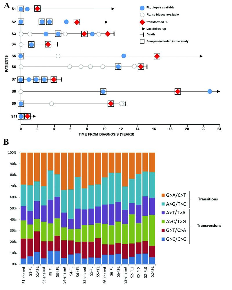
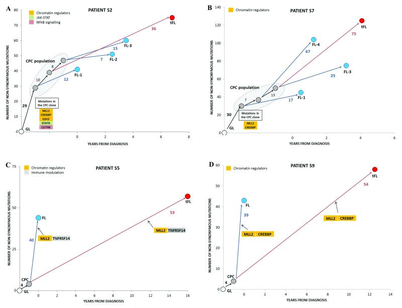
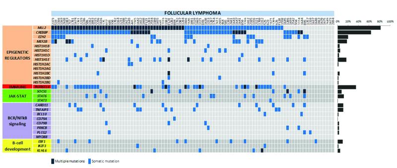
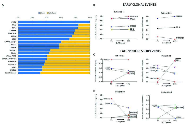
Follicular lymphoma is an incurable malignancy, with transformation to an aggressive subtype representing a critical event during disease progression. Here we performed whole-genome or whole-exome sequencing on 10 follicular lymphoma-transformed follicular lymphoma pairs followed by deep sequencing of 28 genes in an extension cohort, and we report the key events and evolutionary processes governing tumor initiation and transformation. Tumor evolution occurred through either a 'rich' or 'sparse' ancestral common progenitor clone (CPC). We identified recurrent mutations in linker histone, JAK-STAT signaling, NF-κB signaling and B cell developmental genes. Longitudinal analyses identified early driver mutations in chromatin regulator genes (CREBBP, EZH2 and KMT2D (MLL2)), whereas mutations in EBF1 and regulators of NF-κB signaling (MYD88 and TNFAIP3) were gained at transformation. Collectively, this study provides new insights into the genetic basis of follicular lymphoma and the clonal dynamics of transformation and suggests that personalizing therapies to target key genetic alterations in the CPC represents an attractive therapeutic strategy.


Figures

Figure 1
Figure 1. Clinical timelines and somatic mutation profiles for the discovery cases.
(a) Biopsy information with disease event timelines for the 10 WGS and WES cases. (b) Distribution of base substitution patterns of all somatic single nucleotide variants (SNVs) identified in the 6 paired FL-tFL genomes. The somatic SNVs for each case were sorted into three categories: shared (variants present in both FL and tFL samples), FL-specific and tFL-specific. Aberrant somatic hypermutation (aSHM) did not contribute to the majority of variants detected, with the exception of previously described gene targets (Supplementary Table 13).
Figure 2
Figure 2. Clonal evolution patterns of FL to tFL for four cases.
In each case, a phylogenetic tree was constructed using non-synonymous mutations and illustrated alongside the corresponding event timeline. All trees have shared mutations (‘trunk’), which represents a common ancestral origin, the CPC, and unique ‘branches’ depicting the mutations present in that biopsy alone. The length of individual branches denotes the number of mutations separating the two disease events. S2 (a) and S7 (b), both with 3 FL (blue) and a single tFL (red) biopsy show divergent clonal evolution with a ‘rich’ CPC pool from which subsequent events arise. S5 (c) and S9 (d) illustrating a ‘sparse’ CPC (4 shared mutations between FL and tFL in both cases) and convergent evolution, in which similar genetic alterations occurs independently in separate branches of the tree. All paired tumors were confirmed to be clonally-related with BCL2-IGH rearrangement analyses. The pattern of clonal evolution was identical with all somatic variants included (Supplementary Fig. 6). Functional annotation of the mutated genes was based on previously reported functions-.
Figure 3
Figure 3. Distribution of mutations in 28 genes in FL.
The heat map indicates the case-specific, concurrent and mutually exclusive mutation patterns in 100 FL cases. Each column represents an individual affected case and each row denotes a specific gene assigned into one of five functional categories as labeled on the left. The bar graph on the right shows the frequency of mutations found per gene across all samples. A heat map corresponding to the paired FL-tFL series is included in Supplementary Fig. 7b.
Figure 4
Figure 4. Functional analyses of linker histone, HIST1H1C, and EBF1 mutations.
(a) Reversed phase HPLC (RP-HPLC) profiles of histones extracted from chromatin isolated from hH1cWT or hH1cS102F expressing H1 TKO mouse ESC cells. The hH1cS102F mutant demonstrated a higher hydrophobicity compared to WT. (b) The ratio of individual H1 variants (and total H1) to the nucleosome of the indicated ESC cells. The ratio is calculated, from the HPLC analysis shown in (a) and demonstrates that the total H1 levels in H1 TKO/hH1cS102F ESCs were reduced compared to H1 TKO/hH1cWT, as a result of a poorer association of FLAG-hH1cS102F histones with chromatin (only 35% of FLAG-hH1c). (c) Schematics of the domains and location of mutations in EBF1 detected by targeted resequencing (NCBI protein reference sequence: NP_076870.1). DBD, DNA-binding domain; IPT, immunoglobulin domain; HLH, helix-loop-helix. Coding mutations were colored green (missense), yellow (nonsense) or red (frameshift). Mutations at the same amino acid residues in different cases are depicted. (d) Quantitative reverse transcription PCR (Q-RT-PCR)-based gene expression from Ebf1-deficient primary murine pre-pro-B-cells transduced with wild-type or mutant Ebf1. All 3 Ebf1 mutants demonstrate similar expression levels. Both DBD-mutants (G171D and S238T) exhibit abrogation of target gene activation. Igll1 and Cd79b transcripts were normalized to the control (pMYs) vector whilst Ebf1 transcripts of mutant Ebf1 constructs were normalized to wt Ebf1.
Figure 5
Figure 5. Early and late mutations in FL and tFL.
(a) Clonality profiles of 28 recurrent gene mutations. In cases with genes harboring multiple mutations, the mutation with the highest VAF was considered. (b) Longitudinal analyses of paired FL-tFLs show early initiating mutations with corrected variant allele frequencies that were unchanged with disease progression in two representative cases. (c) Acquisition of clonal EBF1 mutations at transformation illustrated with two examples. (d) Acquisition of NF-κB genes at transformation, particularly MYD88 which demonstrated no mutations in the extension cohort of 100 FLs, suggesting these are predominantly late genetic events during the disease course.

References

    1. Swenson WT, et al. Improved survival of follicular lymphoma patients in the United States. J Clin Oncol. 2005;23:5019–26. - PubMed
    1. Al-Tourah AJ, et al. Population-based analysis of incidence and outcome of transformed non-Hodgkin’s lymphoma. J Clin Oncol. 2008;26:5165–9. - PubMed
    1. Montoto S, Fitzgibbon J. Transformation of indolent B-cell lymphomas. J Clin Oncol. 2011;29:1827–34. - PubMed
    1. Montoto S, et al. Risk and clinical implications of transformation of follicular lymphoma to diffuse large B-cell lymphoma. J Clin Oncol. 2007;25:2426–33. - PubMed
    1. Carlotti E, et al. Transformation of follicular lymphoma to diffuse large B-cell lymphoma may occur by divergent evolution from a common progenitor cell or by direct evolution from the follicular lymphoma clone. Blood. 2009;113:3553–7. - PubMed

Publication types

MeSH terms

Associated data