. 2014 May 1;23(9):2468-80.
doi: 10.1093/hmg/ddt642. Epub 2013 Dec 20.

Endoplasmic reticulum stress in amelogenesis imperfecta and phenotypic rescue using 4-phenylbutyrate

Affiliations

Endoplasmic reticulum stress in amelogenesis imperfecta and phenotypic rescue using 4-phenylbutyrate

Steven J Brookes et al. Hum Mol Genet. .

Abstract

Inherited diseases caused by genetic mutations can arise due to loss of protein function. Alternatively, mutated proteins may mis-fold, impairing endoplasmic reticulum (ER) trafficking, causing ER stress and triggering the unfolded protein response (UPR). The UPR attempts to restore proteostasis but if unsuccessful drives affected cells towards apoptosis. Previously, we reported that in mice, the p.Tyr64His mutation in the enamel extracellular matrix (EEM) protein amelogenin disrupts the secretory pathway in the enamel-forming ameloblasts, resulting in eruption of malformed tooth enamel that phenocopies human amelogenesis imperfecta (AI). Defective amelogenin post-secretory self-assembly and processing within the developing EEM has been suggested to underlie the pathogenesis of X chromosome-linked AI. Here, we challenge this concept by showing that AI pathogenesis associated with the p.Tyr64His amelogenin mutation involves ameloblast apoptosis induced by ER stress. Furthermore, we show that 4-phenylbutyrate can rescue the enamel phenotype in affected female mice by promoting cell survival over apoptosis such that they are able to complete enamel formation despite the presence of the mutation, offering a potential therapeutic option for patients with this form of AI and emphasizing the importance of ER stress in the pathogenesis of this inherited conformational disease.


Figures
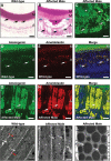
Figure 1.
Figure 1.
Secretory stage ameloblasts of affected male mice manifest a defect in the amelogenin secretory pathway. (A) Wild-type secretory stage ameloblasts (n = 3) are organized as a tall columnar epithelium with secretory Tomes' processes that inter-digitate with the enamel ECM (arrows). (B) In contrast, affected male secretory ameloblasts (n = 3) are shorter, lose their Tomes' processes (blue arrows), exhibit highly eosinophilic cytoplasm (black arrows) and secrete less enamel ECM. The secreted ECM shows a biphasic eosin staining pattern with the secreted matrix (M′) deposited later in the secretory stage being less eosinophilic than that produced at the earliest stages of amelogenesis (M). (C) The cytoplasmic vesicles observed in the secretory ameloblasts of affected male mice (n = 3) are strongly immuno-reactive for amelogenin (arrows) as is the secreted enamel ECM (asterisk). Scale bars: (A) and (B) 20 μm; (C) 10 μm. (D–F) Dual immunofluorescence for amelogenin and ameloblastin demonstrates that both proteins co-localize in the cytoplasm (arrows) of wild-type mice (n = 3). Nuclei are stained with DAPI (blue). A, ameloblasts; M, matrix. Scale bars: 10 μm. (G–I) Dual immunofluorescence for amelogenin and ameloblastin demonstrates that both proteins co-localize in the large cytoplasmic vesicles (arrows) (n = 3). Nuclei are stained with DAPI (blue). A, ameloblasts. Scale bars: 10 μm. (J–L) Transmission electron microscopy of secretory stage ameloblasts. (J) In wild-type mice (n = 3), the ER is organized longitudinally and arranged peripherally close to the plasma membrane (white arrows), while the Golgi apparatus and secretory granules are located towards the centre of the cell (red arrows). (K) In early secretory ameloblasts of affected male mice (n = 3), protein-containing inclusions form towards the periphery of the cell in the same region as the ER (white arrows) while the central area of the cell lacks a recognizable Golgi apparatus but contains dilated cisternae (asterisk). (L) The membrane-bound inclusions in affected male mouse ameloblasts enlarge progressively until they occupy the majority of the cellular volume (arrows). Scale bars: (D) and (E) 1 μm; (F) 500 nm.
Figure 2.
Figure 2.
Characterization of the cytoplasmic vesicles observed in affected male mice. (A–F) Dual immuno-staining for amelogenin (red fluorescence) or (G–I) ameloblastin (red fluorescence) and components of the secretory pathway (green fluorescence) in affected male mice (n = 4) secretory ameloblasts indicate arrest of amelogenin trafficking at a stage prior to the trans-Golgi. (A) The ER membrane protein calnexin circumscribes the amelogenin-containing vesicles (arrows), while the ER luminal molecule protein disulphide isomerase (B) co-localizes with amelogenin within the vesicles (arrows). (C) Immuno-reactivity for the ER/Golgi intermediate compartment molecule Ergic 53 also co-localizes with amelogenin in the vesicles (arrows), whereas (D) β-COP, a component of COP1 vesicles found at the cis-Golgi and ER/Golgi intermediate compartment, co-localizes with amelogenin at the periphery of the vesicles (arrows). (E) The cis-Golgi protein GM130 co-localizes with amelogenin within the vesicles (arrows). (F) Golgin 245, a trans-Golgi protein, showed a variable pattern of immuno-reactivity; in most instances, little amelogenin co-localization was observed in the vesicles (white arrows), although, less typically, partial or complete co-localization was seen (green arrows). (G) Dual immunofluorescence for ameloblastin and the ER membrane protein calnexin indicates that calnexin circumscribes the inclusions (arrows). (H) Similar analyses using antibodies directed against ameloblastin and the ER luminal molecule protein disulphide isomerase (PDI) indicate co-localization within the vesicles (arrows). (I) In contrast, dual immunofluorescence with ameloblastin and Ergic 53, a protein found in the ER/Golgi intermediate compartment, demonstrates distinct localization within the ameloblasts (arrows). Nuclei are stained with DAPI (blue). Scale bars: 5 μm.
Figure 3.
Figure 3.
The p.Tyr64His mutation in amelogenin results in ER stress increased intermolecular interactions of mutant amelogenins. (A and B) In situ hybridization indicates that the secretory stage ameloblasts of affected female mice (n = 3) (B) express Hspa5 (BiP) more strongly than wild-type littermates (n = 3) (A). Scale bar: 10 μm. (C) Real-time PCR analysis of micro-dissected secretory enamel organs indicates that the levels of ER stress response genes are elevated in affected mice (n = 4) compared with wild-type (n = 4) littermates. P = 0.014, Hspa5, Hspa90b1, Atf4 and Ddit3; P = 0.043 Xbp1.
Figure 4.
Figure 4.
Phenotypic rescue using 4-phenylbutyrate in vivo. (A) The incisor teeth of wild-type mice are smooth and opalescent while those of affected female mice exhibit a chalky-white, opaque appearance with roughened/pitted surfaces. 4-Phenylbutyrate treatment restores the appearance of affected female mice incisors to wild type. In contrast, phenotypic rescue of the molar teeth, which do not erupt continuously, is not observed. (B) Qualitative assessment of the enamel appearance by independent researchers. This difference is statistically significant (Fisher's exact test; P = 0.019). (C) Histological analysis indicates that the tall columnar secretory stage ameloblasts of wild-type mice secrete a thick, eosinophilic, enamel ECM while the maturation stage ameloblasts are shorter and the enamel ECM has been digested (asterisk). The secretory ameloblasts of untreated affected female mice are disorganized with intensely eosinophilic cytoplasm and numerous apoptotic cells (arrows). A thinner enamel matrix has been secreted. At the maturation stage, multicellular masses of ameloblasts have formed around retained enamel ECM. Many of the ameloblasts are apoptotic (arrows). By contrast, the histological appearance of the enamel organ of 4-phenylbutyrate-treated affected female mice closely resembles that of wild-type mice with only occasional secretory ameloblasts displaying a strongly eosinophilic cytoplasm (arrows). A, ameloblasts; M, enamel matrix. Scale bars: 50 μm. (D) Morphometric analysis of the enamel organ indicates that the length of the ameloblast secretory zone (P = 0.0031) and the maximum thickness of the enamel ECM (P = 0.0064) deposited is increased in affected female mice treated with 4-phenylbutyrate. PB-treated mice, n = 15; control mice, n = 12. PB, 4-phenylbutyrate. Error bars represent the standard deviation of the mean for all datasets.
Figure 5.
Figure 5.
Effect of PB treatment on the secretory stage enamel organ. (A) Effect of 4-phenylbutyrate (PB) treatment on the size of cytoplasmic vesicles in mutant mice. Ameloblastin immuno-staining was used to delineate the cytoplasmic vesicles in the secretory stage ameloblasts of untreated and PB-treated affected female and affected male mice with image analysis being used to determine their area. As indicated in the scattergram, the distribution of vesicle area shifted to lower values in PB-treated affected female and affected male mice compared with their untreated littermates. Four mice were examined, and three images analysed in each group. Comparison of the median values using Kruskal–Wallis analysis followed by a Dunn's multiple comparison test showed highly significant differences in vesicle size between untreated and PB-treated mice of both genotypes (P < 0.0001). In contrast, there was no statistically significant differences in median vesicle size between PB-treated male and PB-treated female mice (P = 0.064). (B) Western blotting of secreted matrix proteins suggests that PB does not completely relieve secretory impairment. Two wild-type, four PB-treated affected male and two untreated affected male mice were analysed. (C and D) Activated caspase 3 immuno-staining was used to identify secretory stage ameloblasts undergoing apoptosis in untreated and PB-treated affected female mice. In untreated mice (C), numerous apoptotic cells were observed (arrows) while in the PB-treated affected female mice (D), fewer apoptotic cells were detected (arrows). Three mice were examined in each group. Scale bars: 100 μm.
Figure 6.
Figure 6.
Characterization of phenotypic rescue achieved with 4-phenylbutyrate. (A) SEM of wild-type incisors showing ordered decussating enamel rod structure (E) and transverse and longitudinal CT scans (calibrated for mineral density) showing the dense enamel layer overlying dentine (D). The cartoon shows enamel being deposited by ameloblasts during the secretory stage as the tooth erupts left to right. Maturation stage ameloblasts shorten as the enamel attains its final mineral density. To accommodate the large secretory cargo transiting the ER and spontaneously mis-folded/aggregated wild-type amelogenin (depicted in red), the UPR is activated, IRE1 is activated in secretory ameloblasts (16) and cell survival is promoted. (B) The enamel of affected female mouse incisors is biphasic with an inner layer of apparently normal decussating enamel (∼35 µm thick) and an outer layer containing fewer, disorganized prisms. Outer enamel is frequently missing having fractured along the inner–outer enamel boundary (dashed line). The increase in mis-folded/aggregated mutated amelogenin accumulating in the ER is tolerated initially and enamel secretion is unaffected. However, after ∼35 µm of enamel is deposited, the increasing burden of ER stress drives the UPR towards an apoptotic endpoint. Those ameloblasts expressing p.Tyr64His amelogenin die, resulting in disruption of the ameloblast layer which compromises subsequent enamel deposition. (C) 4-Phenylbutyrate restores the decussating prism structure and appearance of the enamel layer to wild type. Western blotting (Fig. 5B) and cellular retention of eosinophilic vesicles (Fig. 4C) indicate that 4-phenylbutyrate does not greatly improve amelogenin secretion. We therefore suggest that 4-phenylbutyrate modulates the UPR response in favour of survival meaning that a functional ameloblast layer is maintained and enamel deposition proceeds undisturbed even though amelogenin secretion is presumably reduced owing to cellular retention of mutated protein. Three mice were examined in each group. PB, 4-phenylbutyrate.

Similar articles

Cited by

References

    1. Hartl F.U., Bracher A., Hayer-Hartl M. Molecular chaperones in protein folding and proteostasis. Nature. 2011;475:324–332. doi:10.1038/nature10317. - DOI - PubMed
    1. Hartl F.U., Hayer-Hartl M. Converging concepts of protein folding in vitro and in vivo. Nat. Struct. Mol. Biol. 2009;16:574–581. doi:10.1038/nsmb.1591. - DOI - PubMed
    1. Ellisdon A.M., Bottomley S.P. The role of protein misfolding in the pathogenesis of human diseases. IUBMB Life. 2004;56:119–123. - PubMed
    1. Ozcan L., Tabas I. Role of endoplasmic reticulum stress in metabolic disease and other disorders. Annu. Rev. Med. 2012;63:317–328. doi:10.1146/annurev-med-043010-144749. - DOI - PMC - PubMed
    1. Yoshida H. ER stress and diseases. FEBS J. 2007;274:630–658. doi:10.1111/j.1742-4658.2007.05639.x. - DOI - PubMed

Publication types