

Impact of inflammation on the osteoblast in rheumatic diseases
- PMID: 24363057
- PMCID: PMC3943531
- DOI: 10.1007/s11914-013-0183-y
Impact of inflammation on the osteoblast in rheumatic diseases
Abstract
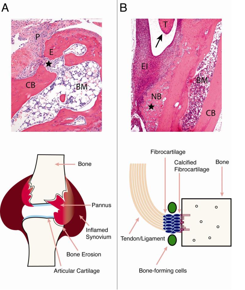
Normal bone remodeling depends upon a balance between the action of bone-resorbing cells, osteoclasts, and bone-forming cells, osteoblasts. When this balance is disrupted, as is seen in inflammatory diseases such as rheumatoid arthritis (RA) and ankylosing spondylitis (AS), abnormal bone loss or bone formation occurs. In RA, proinflammatory cytokines induce osteoclast differentiation and inhibit osteoblast maturation, leading to articular bone erosions. In contrast, the inflammatory milieu in AS leads to excessive osteoblast activation and bone formation at sites of entheses. While much information exists about the effects of proinflammatory cytokines on osteoclast differentiation and function, more recent studies have begun to elucidate the impact of inflammation on the osteoblast. This review will summarize the mechanisms by which inflammation perturbs bone homeostasis, with a specific focus on the osteoblast.
Figures
Similar articles
-
Mediators of inflammation and bone remodeling in rheumatic disease.Semin Cell Dev Biol. 2016 Jan;49:2-10. doi: 10.1016/j.semcdb.2015.10.013. Epub 2015 Oct 19. Semin Cell Dev Biol. 2016. PMID: 26481971 Free PMC article. Review.
-
Bone as a Target Organ in Rheumatic Disease: Impact on Osteoclasts and Osteoblasts.Clin Rev Allergy Immunol. 2016 Aug;51(1):1-15. doi: 10.1007/s12016-015-8515-6. Clin Rev Allergy Immunol. 2016. PMID: 26411424 Free PMC article. Review.
-
Inflammation in bone physiology and pathology.Curr Opin Rheumatol. 2018 Jan;30(1):59-64. doi: 10.1097/BOR.0000000000000449. Curr Opin Rheumatol. 2018. PMID: 29016371 Free PMC article. Review.
-
TNF and Bone Remodeling.Curr Osteoporos Rep. 2017 Jun;15(3):126-134. doi: 10.1007/s11914-017-0358-z. Curr Osteoporos Rep. 2017. PMID: 28477234 Free PMC article. Review.
-
Bone remodeling in rheumatic disease: a question of balance.Immunol Rev. 2010 Jan;233(1):301-12. doi: 10.1111/j.0105-2896.2009.00857.x. Immunol Rev. 2010. PMID: 20193007 Review.
Cited by
-
Targeting S1PRs as a Therapeutic Strategy for Inflammatory Bone Loss Diseases-Beyond Regulating S1P Signaling.Int J Mol Sci. 2021 Apr 23;22(9):4411. doi: 10.3390/ijms22094411. Int J Mol Sci. 2021. PMID: 33922596 Free PMC article. Review.
-
Animal Models of Bone Loss in Inflammatory Arthritis: from Cytokines in the Bench to Novel Treatments for Bone Loss in the Bedside-a Comprehensive Review.Clin Rev Allergy Immunol. 2016 Aug;51(1):27-47. doi: 10.1007/s12016-015-8522-7. Clin Rev Allergy Immunol. 2016. PMID: 26634933 Free PMC article. Review.
-
CD301b+ macrophage: the new booster for activating bone regeneration in periodontitis treatment.Int J Oral Sci. 2023 May 17;15(1):19. doi: 10.1038/s41368-023-00225-4. Int J Oral Sci. 2023. PMID: 37198150 Free PMC article.
-
PLCG2 and IFNAR1: The Potential Biomarkers Mediated by Immune Infiltration and Osteoclast Differentiation of Ankylosing Spondylitis in the Peripheral Blood.Mediators Inflamm. 2024 Jan 5;2024:3358184. doi: 10.1155/2024/3358184. eCollection 2024. Mediators Inflamm. 2024. PMID: 38223749 Free PMC article.
-
Moxibustion of Zusanli (ST36) and Shenshu (BL23) Alleviates Cartilage Degradation through RANKL/OPG Signaling in a Rabbit Model of Rheumatoid Arthritis.Evid Based Complement Alternat Med. 2019 Jan 3;2019:6436420. doi: 10.1155/2019/6436420. eCollection 2019. Evid Based Complement Alternat Med. 2019. PMID: 30719064 Free PMC article.
References
-
- Sims NA, Gooi JH. Bone remodeling: Multiple cellular interactions required for coupling of bone formation and resorption. Semin Cell Dev Biol. 2008;19(5):444–51. - PubMed
-
- Kular J, et al. An overview of the regulation of bone remodelling at the cellular level. Clin Biochem. 2012;45(12):863–73. - PubMed
-
- Teitelbaum SL. Bone resorption by osteoclasts. Science. 2000;289(5484):1504–8. - PubMed
-
- Matsuo K, Irie N. Osteoclast-osteoblast communication. Arch Biochem Biophys. 2008;473(2):201–9. - PubMed
-
- Hayden JM, Mohan S, Baylink DJ. The insulin-like growth factor system and the coupling of formation to resorption. Bone. 1995;17(2):93S–98S. - PubMed
Publication types
MeSH terms
Substances
Grants and funding
LinkOut - more resources
Full Text Sources
Other Literature Sources
Medical
Research Materials
