

Editorial
doi: 10.1007/s11999-013-3428-2.
Guest editorial: The Robert's view: a historical and clinical perspective
Affiliations
- PMID: 24363184
- PMCID: PMC3940739
- DOI: 10.1007/s11999-013-3428-2
Item in Clipboard
Editorial
Guest editorial: The Robert's view: a historical and clinical perspective
Clin Orthop Relat Res.
2014 Apr.
No abstract available
Figures
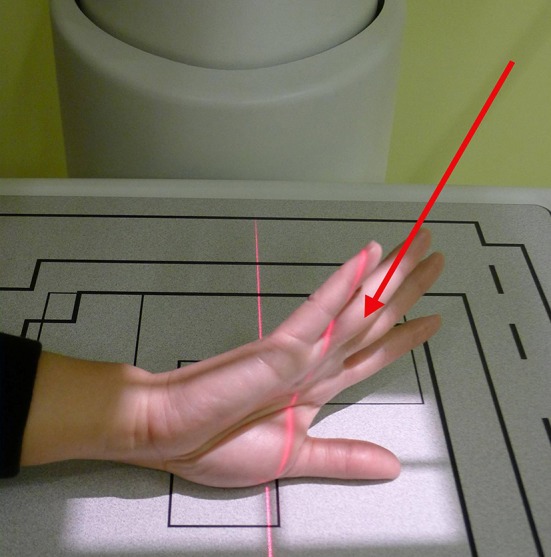
The figure shows the radiograph beam aimed 15° proximal. Published with permission of © Amy L. Ladd 2013. All Rights Reserved.
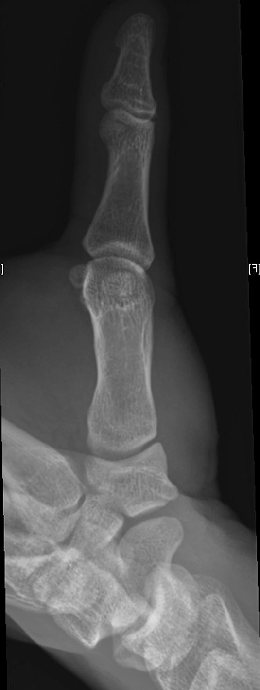
The figure shows a modified Robert’s [4] view of an asymptomatic thumb demonstrating the horizontal profile of both the trapeziometacarpal and scaphotrapezial joints. Published with permission of © Amy L. Ladd 2013. All Rights Reserved.
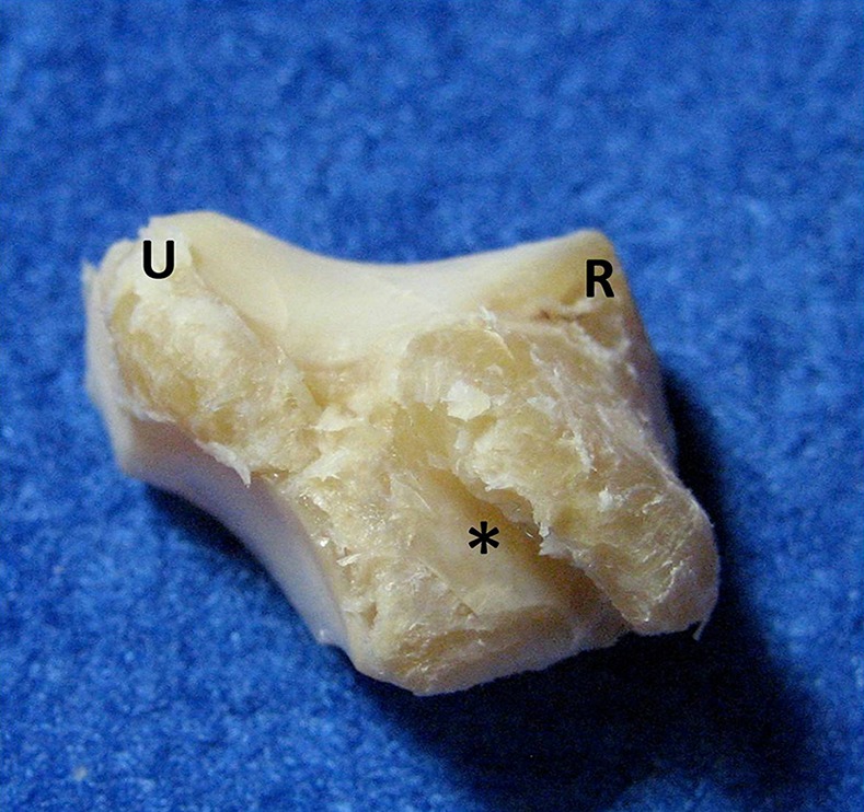
The normal right trapezium is shown from the palmar (volar) side in the same horizontal plane as the radiograph in Fig. 2. The trapezium assumes a convex shape in the ulnar (U) to radial (R) direction, and a convex shape in the volar to dorsal direction. The oblique position of the flexor carpi radialis groove (*), a relatively longitudinal structure in the palm, illustrates the tangential position of the trapezium and base of thumb relative to the rest of the hand. Published with permission of © Amy L. Ladd 2013. All Rights Reserved.
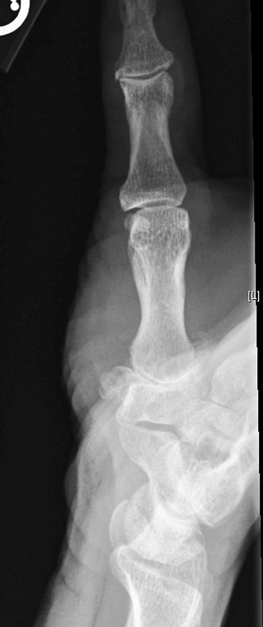
The modified Robert’s view of a patient with advanced disease is shown. The figure shows the radiograph of a patient with significant radial and ulnar osteophytes of the distal articulating trapezial surface, and loss of trapezial height and joint space degradation. Published with permission of © Amy L. Ladd 2013. All Rights Reserved.
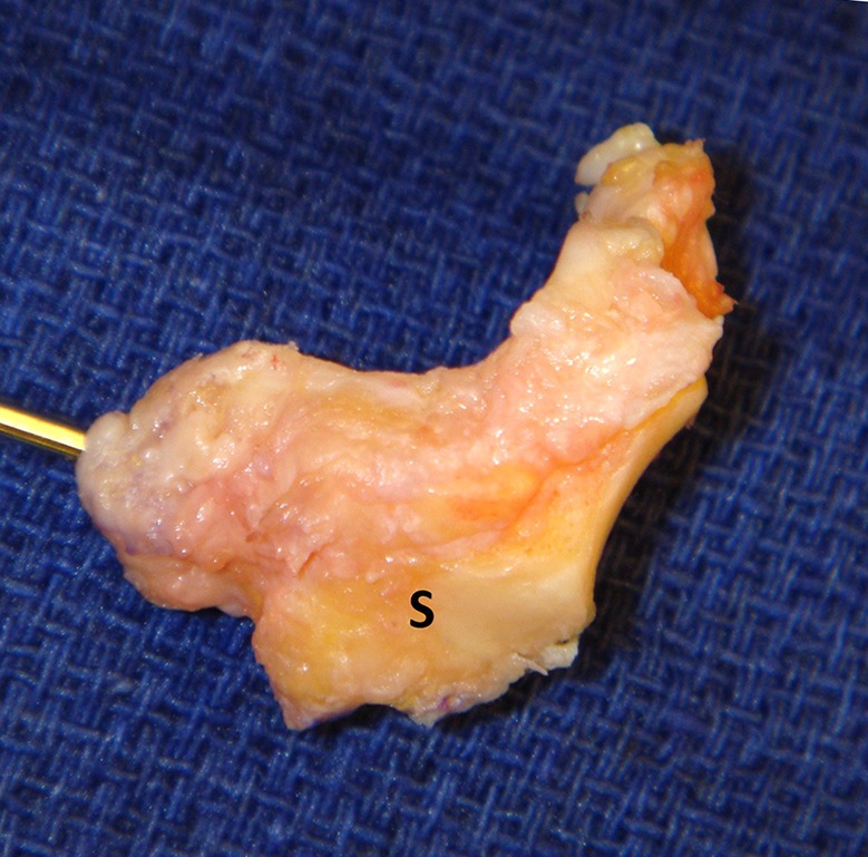
The surgically excised right trapezium of the patient shown in Fig. 4, as viewed from the dorsal side. The extensive osteophyte formation on the distal surface is seen both on the radial side (with positioning wire) and the ulnar side. Although the scaphoid articular surface (S) has a normal appearing facet with no articular wear, an adjacent nonarticular osteophyte is present. Published with permission of © Amy L. Ladd 2013. All Rights Reserved.
Comment on
-
The classic: Radiography of the trapeziometacarpal joint. Degenerative changes of this joint. 1936.Clin Orthop Relat Res. 2014 Apr;472(4):1095-6. doi: 10.1007/s11999-013-2930-x. Clin Orthop Relat Res. 2014. PMID: 23575807 Free PMC article.
References
-
- Ballinger P, Frank E, Merrill V. Merrill’s Atlas of Radiographic Positions & Radiologic Procedures. 10th ed. St. Louis, MO. Elsevier;2003:108–109.
-
- Gervis WH. Excision of the trapezium for osteoarthritis 329 of the trapezio-metacarpal joint. J Bone Joint Surg Br. 1949;31B:537–539. - PubMed
-
- Lewis S. New angles on the radiographic examination of the hand—III. Radiogr Today. 1988;54:47–48. - PubMed
Publication types
MeSH terms
LinkOut - more resources
Full Text Sources
Other Literature Sources
Medical
