. 2013 Dec;25(12):4924-40.
doi: 10.1105/tpc.113.118521. Epub 2013 Dec 20.

Sufficient amounts of functional HOP2/MND1 complex promote interhomolog DNA repair but are dispensable for intersister DNA repair during meiosis in Arabidopsis

Affiliations

Sufficient amounts of functional HOP2/MND1 complex promote interhomolog DNA repair but are dispensable for intersister DNA repair during meiosis in Arabidopsis

Clemens Uanschou et al. Plant Cell. 2013 Dec.

Abstract

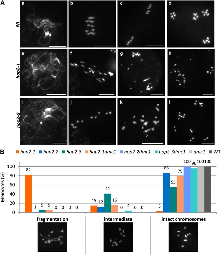
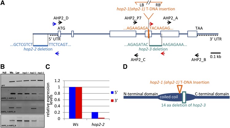
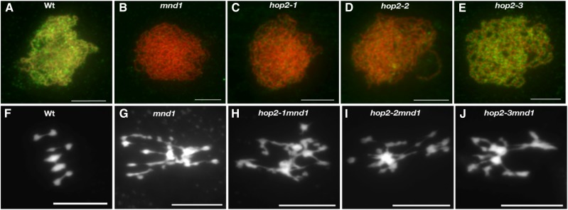
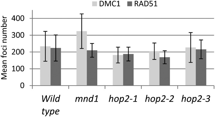
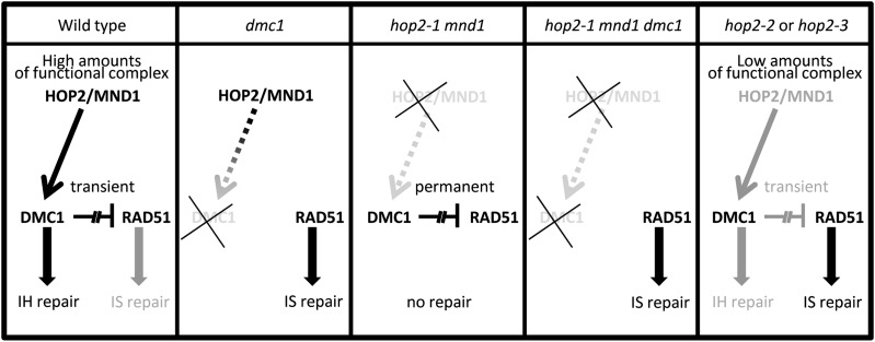
During meiosis, homologous recombination (HR) is essential to repair programmed DNA double-strand breaks (DSBs), and a dedicated protein machinery ensures that the homologous chromosome is favored over the nearby sister chromatid as a repair template. The homologous-pairing protein2/meiotic nuclear division protein1 (HOP2/MND1) protein complex has been identified as a crucial factor of meiotic HR in Arabidopsis thaliana, since loss of either MND1 or HOP2 results in failure of DNA repair. We isolated two mutant alleles of HOP2 (hop2-2 and hop2-3) that retained the capacity to repair meiotic DSBs via the sister chromatid but failed to use the homologous chromosome. We show that in these alleles, the recombinases radiation sensitive51 (RAD51) and disrupted meiotic cDNA1 (DMC1) are loaded, but only the intersister DNA repair pathway is activated. The hop2-2 phenotype is correlated with a decrease in HOP2/MND1 complex abundance. In hop2-3, a truncated HOP2 protein is produced that retains its ability to bind to DMC1 and DNA but forms less stable complexes with MND1 and fails to efficiently stimulate DMC1-driven D-loop formation. Genetic analyses demonstrated that in the absence of DMC1, HOP2/MND1 is dispensable for RAD51-mediated intersister DNA repair, while in the presence of DMC1, a minimal amount of functional HOP2/MND1 is essential to drive intersister DNA repair.


Figures

Figure 1.
Figure 1.
Meiotic Progression in hop2 Mutants. (A) 4′,6-Diamidino-2-phenylindole staining of chromosomes of wild-type (Wt; [a] to [d]), hop2-1 ([e] to [h]), and hop2-2 ([i] to [l]) pollen mother cells. (a), (e), and (i), Pachytene/pachytene-like stage; (b), (f), (j), and (k), metaphase I–anaphase I transition; (c) and (g), metaphase II; (d), (h), and (l), end of anaphase II. While full synapsis was achieved at pachytene in the wild type (a), no evidence of synapsis could be observed in any of the three hop2 mutant lines (shown here for hop2-1 in [e] and hop2-2 in [i]). At metaphase I in the wild type, five bivalents were always observed aligned on the metaphase plate (b). Homologous chromosomes segregated during anaphase I, generating two pools of five chromosomes (c). Then, during the second meiotic division, sister chromatids were separated, producing four haploid cells (d). By contrast, during hop2-1 meiosis (f), severe chromosomal defects (fragmentation and chromosome bridges) were observed at anaphase I (f) and anaphase II (g), yielding aberrant meiotic products (h). In hop2-2 ([j] to [l]) and hop2-3, hardly any chromosomal defects could be observed; instead, a mixture of intact univalents and bivalents could be seen ([j], 10 univalents; [k], six univalents and two bivalents). Random segregation of these univalents at anaphase I ([j] and [k]) and the subsequent sister chromatid segregation at meiosis II (data not shown) yielded unbalanced pools of chromosomes at the end of meiosis (l) instead of the four pools of five chromosomes observed in the wild type (d). u, univalent; b, bivalent. Bars = 10 μm. (B) dmc1 suppresses the DNA repair defects observed in hop2-1, hop2-2, and hop2-3 mutants. In hop2-1, a large majority of meiocytes showed drastic chromosomal defects (fragmentation and chromosome bridges) at metaphase I–anaphase I transition. By contrast, hop2-2 cells predominantly present intact chromosomes (86%). In hop2-3 meiocytes, the situation is intermediate with 55% of the cells showing no DNA fragmentation and 41% showing mild chromosomal defects (“intermediate” class). The severe chromosome fragmentation observed in hop2-1 mutants is largely suppressed by the dmc1 mutation. The limited chromosome fragmentation observed in hop2-2 and hop2-3 mutants is nearly completely suppressed by the dmc1 mutation.
Figure 2.
Figure 2.
Molecular Analysis of HOP2 Alleles. (A) Schematic representation of the HOP2(AHP2) gene and three mutant alleles. Open boxes represent exons, ATG and TAA encompass the open reading frame. The inverted triangle in orange shows the position of the T-DNA insertion in hop2-1 (the renamed ahp2-1 allele described in Schommer et al., 2003). Text and symbols in light blue indicate the 147-bp deletion in the promoter region of the hypomorphic hop2-2 allele. Text and symbols in turquoise indicate the 128-bp deletion in the hypomorphic hop2-3 allele. (B) RT-PCR analysis of HOP2 and hop2 mutant allele expression in young flower buds. A HOP2 transcript spanning the T-DNA insertion site was not detected in hop2-1 (primers AHP2_P7 and AHP2_B), while sequences upstream (AHP2_C and AHP2_D primers) and downstream (AHP2_A and AHP2_B primers) of the insertion could be amplified. In hop2-2, reduced levels of mRNA can be detected with all primer combinations. In hop2-3, normal levels of mRNA can be detected with all primer combinations. Nevertheless, an expected shorter transcript is amplified when using primers AHP2_P7 and AHP2_B, which span the genomic deletion of hop2-3. The mRNA/cDNA of the phosphoribosyltransferase (APT) gene was used for normalization. The corresponding wild-type controls were from ecotypes Ws and Ler. (C) Quantitative RT-PCR of HOP2 and hop2-2 mutant expression in young flower buds. Primers were designed to determine expression levels of the 5′ part of the mRNA (blue arrows in [A]; blue bar) and the 3′ part of mRNA (red arrows in [A]; red bar). All values were normalized to Actin2/7 gene expression. The mRNA levels detected in hop2-2 mutants are strongly decreased compared with the wild type: approximately fivefold decreased when evaluating the 5′ part of mRNA (green bars) and ∼30-fold decreased when evaluating the 3′ part of the mRNA. The corresponding wild-type control was from ecotype Ws. (D) Schematic representation of the anticipated HOP2 protein and its hypomorphic variant. The HOP2 wild-type protein is structured into an N-terminal domain, a putative coiled coil region, and a C-terminal domain. hop2-1 plants do not express the protein (Stronghill et al., 2010). Plants carrying the hypomorphic hop2-2 allele produce very low levels of the mRNA and anticipated very low levels of HOP2 protein. The deletion in hop2-3 leads to expression of an mRNA variant, anticipated to encode a protein variant that lacks 14 amino acids (position 123 to 136 of the wild-type protein) within the coiled coil region of the protein.
Figure 3.
Figure 3.
The Formation of MND1 Foci Is Normal in hop2-3, Reduced in hop2-2, and Abolished in hop2-1 Mutants. (A) to (E) Coimmunolocalization of ASY1 (red) and MND1 (green) in wild-type (Wt; [A]), mnd1 (B), and hop2 mutant meiocytes ([C] to [E]). In accordance with our previous studies (Vignard et al., 2007), no MND1 foci were detected in mnd1 or hop2-1 meiocytes ([B] and [C]). By contrast, MND1 signal appeared normal in hop2-3 mutant meiocytes (E) and reduced in hop2-2 meiocytes (D). Bars = 5 μm. (F) to (J) 4′,6-Diamidino-2-phenylindole staining of metaphase I pollen mother cells in the wild type (F), mnd1 (G), hop2-1 mnd1 (H), hop2-2 mnd1 (I), and hop2-3 mnd1 (J) mutants. Bars = 10 μm.
Figure 4.
Figure 4.
No Major Differences in DMC1 and RAD51 Foci Numbers in All hop2 Mutant Alleles. Chromosome spreads of Arabidopsis meiocytes from the wild type, mnd1, hop2-1, hop2-2, and hop2-3 mutants were stained with an α-ASY1 antibody and an α-RAD51 or α-DMC1 antibody (see Supplemental Figure 4 online). Foci numbers were determined and the counts blotted. While DMC1 and RAD51 foci numbers are not dramatically changed in all hop2 mutant alleles, the DMC1 foci number in mnd1 is significantly higher than in the wild type and all other mutant lines (Vignard et al., 2007). Please refer to text for more details.
Figure 5.
Figure 5.
HOP2-3/MND1 Protein Complex Is Less Stable Than the HOP2/MND1 Complex. (A) HOP2 and HOP2-3 form complexes with MND1. SDS-PAGE and Coomassie blue staining of the coexpressed and copurified recombinant HOP2/MND1 and the HOP2-3/MND1 complexes. HOP2 or HOP2-3 has been fused to a 6xHis-tag, which was used to purify HOP2 or HOP2-3 and to copurify MND1. Subsequent immunoblotting was performed to demonstrate the presence of both proteins (see Supplemental Figure 5 online). (B) Temperature-mediated unfolding of the HOP2/MND1 and HOP2-3/MND1 protein complexes followed by ECD in the far UV. Conditions: 5 mM phosphate buffer, pH 7.7. Comparison of far-UV ECD spectra of HOP2/MND1 (black) and HOP2-3/MND1 (red) at 20°C (solid lines), 85°C (dashed lines), and 20°C after heating and cooling (dotted lines). (C) Thermal unfolding of HOP2/MND1 (solid line) and HOP2-3/MND1 (dashed line) followed at 222 nm. The insets show the corresponding van’t Hoff plots. (D) DNA binding activities of the recombinant HOP2/MND1 and HOP2-3/MND1 protein complexes. Linearized PhiX174 Replication form I dsDNA and circular PhiX174 virion ssDNA were incubated with the following amounts of protein: 0, 1, 2.5, 5, 10 µM HOP2/MND1 for lanes 1 to 5 and 1, 2.5, 5, and 10 µM HOP2-3/MND1 for lanes 6 to 9. Reactions were loaded and separated on a 0.8% agarose gel and stained with ethidium bromide. Diagrams below the gel pictures ([A] and [D]) indicate the recombinant proteins: red, MND1; blue, HOP2 or HOP2-3; ellipsoids represent the putative coiled coil domains; the turquoise rectangle indicates the 14–amino acid deletion in the second half of the putative coiled coil region in HOP2-3. M, Fermentas prestained protein ladder (numbers indicate molecular mass in kilodaltons). The calculated molecular weight for MND1 is 26.5 kD, for 6xHIS/HOP2-3 is 26.3 kD, and for 6xHIS/HOP2 is 28 kD.
Figure 6.
Figure 6.
HOP2-3/MND1 Fails to Activate DMC1-Mediated D-Loop Formation Efficiently. Principle of the D-loop assay (A). A labeled ssDNA is first incubated with DMC1, followed by addition of HOP2/MND1 and then the supercoiled plasmid DNA to allow formation of D-loops. Reactions are stopped, proteins are removed, and DNA structures are separated by gel electrophoresis. Gel pictures showing D-loop assays employing rice DMC1A (B) or DMC1B (C) proteins with either the addition of wild-type HOP2/MND1 complex or HOP2-3/MND1 complex in increasing concentrations. All reactions were performed in triplicate and quantifications are shown below the gel pictures as percentages of ssDNA incorporated into D-loops.
Figure 7.
Figure 7.
Model of the Functional Relationship between HOP2/MND1, DMC1, and RAD51. In a wild-type context, high levels of HOP2/MND1 stimulate DMC1 activity, thereby promoting IH DNA repair. We anticipate that RAD51-mediated IS DNA repair is transiently repressed, limiting IS repair to a backup function. In the absence of DMC1, RAD51 efficiently repairs meiotic DSBs via sister chromatids. We anticipate that this repair is independent of HOP2/MND1, since in hop2 mnd1 dmc1 triple mutants, IS DNA repair is in place. Importantly, in the absence of HOP2 and/or MND1, DMC1 is not stimulated and permanently inhibits RAD51-mediated IS DNA repair. Therefore, in the absence of HOP2/MND1, RAD51-mediated DNA repair can only take place if DMC1 is removed. Last, in conditions with only limited amounts of functional HOP2/MND1 complex available (hop2-2 and hop2-3 mutant alleles), DMC1 is not optimally supported and only limited IH DNA repair takes place. The limited stimulation of DMC1 is sufficient to indirectly promote RAD51-mediated IS repair. Possible mechanisms are discussed in the text.

Comment in

Similar articles

Cited by

References

    1. Allers T., Lichten M. (2001). Differential timing and control of noncrossover and crossover recombination during meiosis. Cell 106: 47–57 - PubMed
    1. Armstrong S.J., Caryl A.P., Jones G.H., Franklin F.C. (2002). Asy1, a protein required for meiotic chromosome synapsis, localizes to axis-associated chromatin in Arabidopsis and Brassica. J. Cell Sci. 115: 3645–3655 - PubMed
    1. Baudat F., de Massy B. (2007). Regulating double-stranded DNA break repair towards crossover or non-crossover during mammalian meiosis. Chromosome Res. 15: 565–577 - PubMed
    1. Baumann P., Benson F.E., West S.C. (1996). Human Rad51 protein promotes ATP-dependent homologous pairing and strand transfer reactions in vitro. Cell 87: 757–766 - PubMed
    1. Bechtold N., Ellis J., Pelletier G. (1993). In planta Agrobacterium-mediated gene transfer by infiltration of adult Arabidopsis thaliana plants. Comptes Rendus Acad. Sci. Ser. Iii-Sciences De La Vie. Life Sciences. 316: 1194–1199

Publication types

MeSH terms

LinkOut - more resources