

Blood feeding induces hemocyte proliferation and activation in the African malaria mosquito, Anopheles gambiae Giles
- PMID: 24363411
- PMCID: PMC3990356
- DOI: 10.1242/jeb.094573
Blood feeding induces hemocyte proliferation and activation in the African malaria mosquito, Anopheles gambiae Giles
Abstract
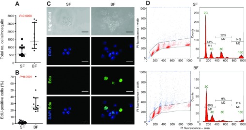
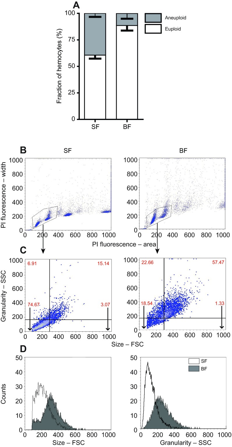
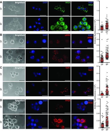
Malaria is a global public health problem, especially in sub-Saharan Africa, where the mosquito Anopheles gambiae Giles serves as the major vector for the protozoan Plasmodium falciparum Welch. One determinant of malaria vector competence is the mosquito's immune system. Hemocytes are a critical component as they produce soluble immune factors that either support or prevent malaria parasite development. However, despite their importance in vector competence, understanding of their basic biology is just developing. Applying novel technologies to the study of mosquito hemocytes, we investigated the effect of blood meal on hemocyte population dynamics, DNA replication and cell cycle progression. In contrast to prevailing published work, the data presented here demonstrate that hemocytes in adult mosquitoes continue to undergo low basal levels of replication. In addition, blood ingestion caused significant changes in hemocytes within 24 h. Hemocytes displayed an increase in cell number, size, granularity and Ras-MAPK signaling as well as altered cell surface moieties. As these changes are well-known markers of immune cell activation in mammals and Drosophila melanogaster Meigen, we further investigated whether a blood meal changes the expression of hemocyte-derived immune factors. Indeed, hemocytes 24 h post-blood meal displayed higher levels of critical components of the complement and melanization immune reactions in mosquitoes. Taken together, this study demonstrates that the normal physiological process of a blood meal activates the innate immune response in mosquitoes. This process is likely in part regulated by Ras-MAPK signaling, highlighting a novel mechanistic link between blood feeding and immunity.
Keywords: Blood meal; Immunology; Infectious disease; Innate immunity; Phenoloxidase.
Figures
Similar articles
-
Anopheles gambiae phagocytic hemocytes promote Plasmodium falciparum infection by regulating midgut epithelial integrity.Nat Commun. 2025 Feb 8;16(1):1465. doi: 10.1038/s41467-025-56313-y. Nat Commun. 2025. PMID: 39920122 Free PMC article.
-
Molecular Profiling of Phagocytic Immune Cells in Anopheles gambiae Reveals Integral Roles for Hemocytes in Mosquito Innate Immunity.Mol Cell Proteomics. 2016 Nov;15(11):3373-3387. doi: 10.1074/mcp.M116.060723. Epub 2016 Sep 13. Mol Cell Proteomics. 2016. PMID: 27624304 Free PMC article.
-
Anopheles gambiae hemocytes exhibit transient states of activation.Dev Comp Immunol. 2016 Feb;55:119-29. doi: 10.1016/j.dci.2015.10.020. Epub 2015 Oct 26. Dev Comp Immunol. 2016. PMID: 26515540 Free PMC article.
-
The Anopheles innate immune system in the defense against malaria infection.J Innate Immun. 2014;6(2):169-81. doi: 10.1159/000353602. Epub 2013 Aug 28. J Innate Immun. 2014. PMID: 23988482 Free PMC article. Review.
-
NF-κB-Like Signaling Pathway REL2 in Immune Defenses of the Malaria Vector Anopheles gambiae.Front Cell Infect Microbiol. 2017 Jun 21;7:258. doi: 10.3389/fcimb.2017.00258. eCollection 2017. Front Cell Infect Microbiol. 2017. PMID: 28680852 Free PMC article. Review.
Cited by
-
Warmer environmental temperature accelerates aging in mosquitoes, decreasing longevity and worsening infection outcomes.Immun Ageing. 2024 Sep 11;21(1):61. doi: 10.1186/s12979-024-00465-w. Immun Ageing. 2024. PMID: 39261928 Free PMC article.
-
Anopheles gambiae larvae mount stronger immune responses against bacterial infection than adults: evidence of adaptive decoupling in mosquitoes.Parasit Vectors. 2017 Aug 1;10(1):367. doi: 10.1186/s13071-017-2302-6. Parasit Vectors. 2017. PMID: 28764812 Free PMC article.
-
Anopheles gambiae phagocytic hemocytes promote Plasmodium falciparum infection by regulating midgut epithelial integrity.Nat Commun. 2025 Feb 8;16(1):1465. doi: 10.1038/s41467-025-56313-y. Nat Commun. 2025. PMID: 39920122 Free PMC article.
-
Fluctuating Warm and Humid Conditions Differentially Impact Immunity and Development in the Malaria Vector Anopheles stephensi.Glob Chang Biol. 2025 Aug;31(8):e70382. doi: 10.1111/gcb.70382. Glob Chang Biol. 2025. PMID: 40762096 Free PMC article.
-
Cell-specific responses of Anopheles gambiae fat body to blood feeding and infection at a single nuclei resolution.bioRxiv [Preprint]. 2025 Jul 29:2025.07.29.667437. doi: 10.1101/2025.07.29.667437. bioRxiv. 2025. PMID: 40766566 Free PMC article. Preprint.
References
-
- Bellingan G. (1999). Inflammatory cell activation in sepsis. Br. Med. Bull. 55, 12-29 - PubMed
Publication types
MeSH terms
Grants and funding
LinkOut - more resources
Full Text Sources
Other Literature Sources
Research Materials
