. 2013:2013:357014.
doi: 10.1155/2013/357014. Epub 2013 Dec 10.

Conditioned media from adipose-tissue-derived mesenchymal stem cells downregulate degradative mediators induced by interleukin-1β in osteoarthritic chondrocytes

Affiliations

Conditioned media from adipose-tissue-derived mesenchymal stem cells downregulate degradative mediators induced by interleukin-1β in osteoarthritic chondrocytes

Julia Platas et al. Mediators Inflamm. 2013.

Abstract

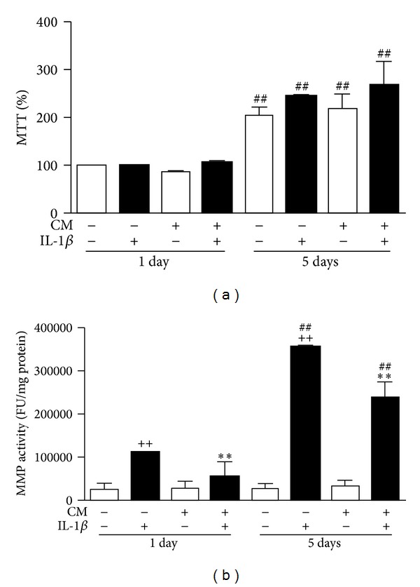
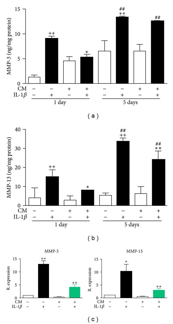
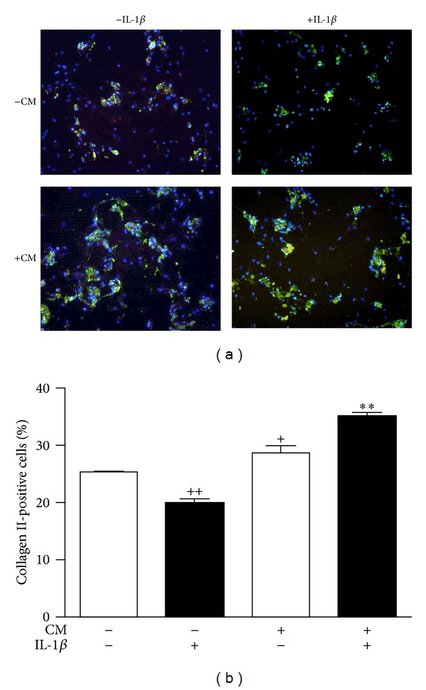
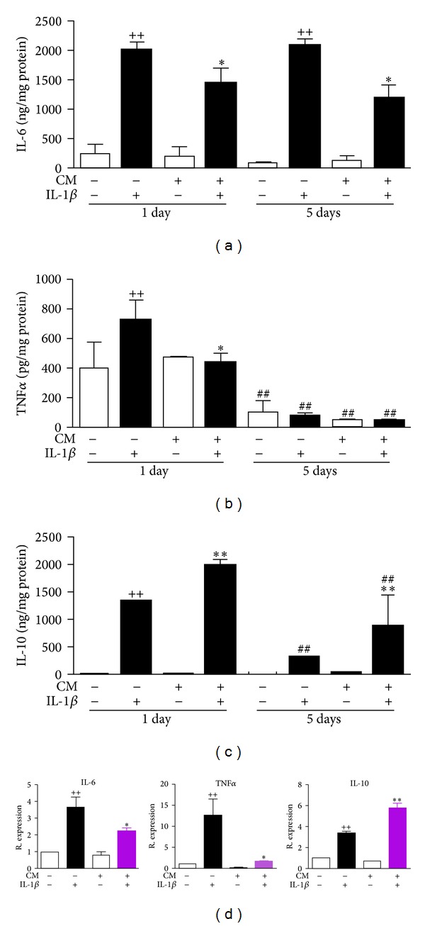
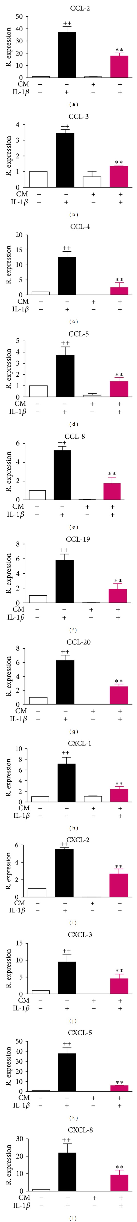
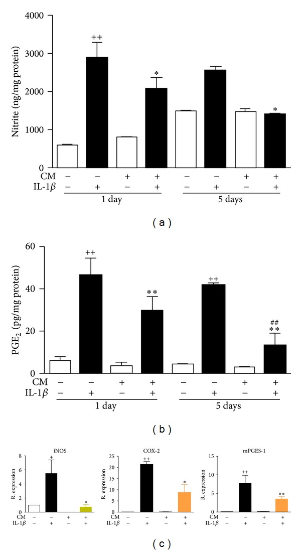
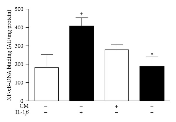
Osteoarthritis (OA) is the most frequent joint disorder and an important cause of disability. Recent studies have shown the potential of adipose-tissue-derived mesenchymal stem cells (AD-MSC) for cartilage repair. We have investigated whether conditioned medium from AD-MSC (CM) may regulate in OA chondrocytes a number of key mediators involved in cartilage degeneration. CM enhanced type II collagen expression in OA chondrocytes while decreasing matrix metalloproteinase (MMP) activity in cell supernatants as well as the levels of MMP-3 and MMP-13 proteins and mRNA in OA chondrocytes stimulated with interleukin- (IL-) 1β. In addition, CM increased IL-10 levels and counteracted the stimulating effects of IL-1β on the production of tumor necrosis factor-α, IL-6, prostaglandin E2, and NO measured as nitrite and the mRNA expression of these cytokines, CCL-2, CCL-3, CCL-4, CCL-5, CCL-8, CCL-19, CCL-20, CXCL-1, CXCL-2, CXCL-3, CXCL-5, CXCL-8, cyclooxygenase-2, microsomal prostaglandin E synthase-1, and inducible NO synthase. These effects may be dependent on the inhibition of nuclear factor-κB activation by CM. Our data demonstrate the chondroprotective actions of CM and provide support for further studies of this approach in joint disease.


Figures

Figure 1
Figure 1
Effects of CM on (a) cell proliferation and (b) MMP activity in OA chondrocytes. (a) Cell proliferation was measured by the MTT method. Results are expressed as % MTT with respect to nonstimulated cells at 24 h. (b) MMP activity was measured by fluorometry in cell culture supernatants after 24 h or 5 days of incubation of chondrocytes in the presence or absence of IL-1β and CM. Activity is expressed as fluorescence units (FU) per mg of protein. Data represent mean ± S.E.M. of independent cultures with chondrocytes from 4 different donors. ++ P < 0.01 with respect to nonstimulated cells; **P < 0.01 with respect to IL-1β; ## P < 0.01 with respect to the corresponding group after 24 h incubation.
Figure 2
Figure 2
Effects of CM on MMP-3 (a) and MMP-13 (b) proteins and mRNA (c) expression in OA chondrocytes. (a, b) Protein levels were measured by ELISA in cell supernatants after 24 h or 5 days of incubation of chondrocytes in the presence or absence of IL-1β and CM. (c) mRNA expression was determined by real-time PCR analysis in OA chondrocytes after 24 h of incubation in the presence or absence of IL-1β and CM. Results indicate relative expression with respect to nonstimulated OA chondrocytes. Data represent mean ± S.E.M. of independent cultures with chondrocytes from 4 different donors. + P < 0.05, ++ P < 0.01 with respect to nonstimulated cells; *P < 0.05, **P < 0.01 with respect to IL-1β; ## P < 0.01 with respect to the corresponding group after 24 h incubation.
Figure 3
Figure 3
Immunocytochemical analysis of collagen II expression. Chondrocytes were treated with CM and/or IL-1β for 5 days. (a) Representative image showing the expression of collagen II (FITC-immunofluorescence, green) and DAPI (blue) for cellular nuclei. Original magnification ×200. (b) Percentages of collagen II-positive cells with respect to total cell numbers. Data represent mean ± S.E.M. of independent cultures with chondrocytes from 4 different donors. + P < 0.05, ++ P < 0.01 with respect to nonstimulated cells; **P < 0.01 with respect to IL-1β.
Figure 4
Figure 4
Effects of CM on the protein levels of IL-6 (a), TNFα (b), and IL-10 (c) in supernatants and mRNA expression in OA chondrocytes (d). (a–c) Cytokines were measured by ELISA in cell culture supernatants after 24 h or 5 days of incubation of chondrocytes with CM in the presence or absence of IL-1β. (d) mRNA expression was determined by real-time PCR analysis in OA chondrocytes after 24 h of incubation and results indicate relative expression with respect to nonstimulated OA chondrocytes. Data represent mean ± S.E.M. of independent cultures with chondrocytes from 6 different donors. ++ P < 0.01 with respect to nonstimulated cells; *P < 0.05, **P < 0.01 with respect to IL-1β; ## P < 0.01 with respect to the corresponding group after 24 h incubation.
Figure 5
Figure 5
Effects of CM on chemokine mRNA expression. mRNA expression was determined by real-time PCR analysis in OA chondrocytes after 24 h of incubation in the presence or absence of IL-1β and/or CM. Results indicate relative expression with respect to nonstimulated OA chondrocytes. Data represent mean ± S.E.M. of independent cultures with chondrocytes from 4 different donors. ++ P < 0.01 with respect to nonstimulated cells; **P < 0.01 with respect to IL-1β.
Figure 6
Figure 6
Effects of CM on NO (a) and PGE2 (b) production and iNOS, COX-2, and mPGES-1 mRNA (c) expression in OA chondrocytes. (a) NO was measured by fluorometry as nitrite. (b) PGE2 was measured by radioimmunoassay. Mediators were determined in cell culture supernatants after 24 h or 5 days of incubation of OA chondrocytes with CM in the presence or absence of IL-1β. (c) mRNA expression was determined by real-time PCR analysis in OA chondrocytes after 24 h of incubation and results indicate relative expression with respect to nonstimulated OA chondrocytes. Data represent mean ± S.E.M of independent cultures with chondrocytes from 6 (NO, PGE2) or 4 (mRNA) different donors. ++ P < 0.01 with respect to nonstimulated cells; *P < 0.05, **P < 0.01 with respect to IL-1β; ## P < 0.01 with respect to the corresponding group after 24 h incubation.
Figure 7
Figure 7
Effects of CM on NF-κB activation in OA chondrocytes. Cells were stimulated with IL-1β for 1 h and p65-NF-κB binding to DNA was determined by ELISA in nuclear fractions. Data are expressed as mean ± S.E.M. of independent cultures with chondrocytes from 3 different donors. AU: arbitrary units. + P < 0.05 with respect to nonstimulated cells; *P < 0.05 with respect to IL-1β.

References

    1. Goldring MB, Goldring SR. Osteoarthritis. Journal of Cellular Physiology. 2007;213(3):626–634. - PubMed
    1. Fernandes JC, Martel-Pelletier J, Pelletier J-P. The role of cytokines in osteoarthritis pathophysiology. Biorheology. 2002;39(1-2):237–246. - PubMed
    1. Goldring MB, Birkhead JR, Suen L-F, et al. Interleukin-1β-modulated gene expression in immortalized human chondrocytes. Journal of Clinical Investigation. 1994;94(6):2307–2316. - PMC - PubMed
    1. Mengshol JA, Vincenti MP, Coon CI, et al. Interleukin-1 induction of collagenase 3 (matrix metalloproteinase 13) gene expression in chondrocytes requires p38, c-Jun N-terminal kinase, and nuclear factor kappaB: differential regulation of collagenase 1 and collagenase 3. Arthritis and Rheumatism. 2000;43:801–811. - PubMed
    1. Tetlow LC, Adlam DJ, Woolley DE. Matrix metalloproteinase and proinflammatory cytokine production by chondrocytes of human osteoarthritic cartilage: associations with degenerative changes. Arthritis and Rheumatism. 2001;44:585–594. - PubMed

Publication types

MeSH terms