. 2013;9(11):e1003334.
doi: 10.1371/journal.pcbi.1003334. Epub 2013 Nov 21.

A Petri net model of granulomatous inflammation: implications for IL-10 mediated control of Leishmania donovani infection

Affiliations

A Petri net model of granulomatous inflammation: implications for IL-10 mediated control of Leishmania donovani infection

Luca Albergante et al. PLoS Comput Biol. 2013.

Abstract

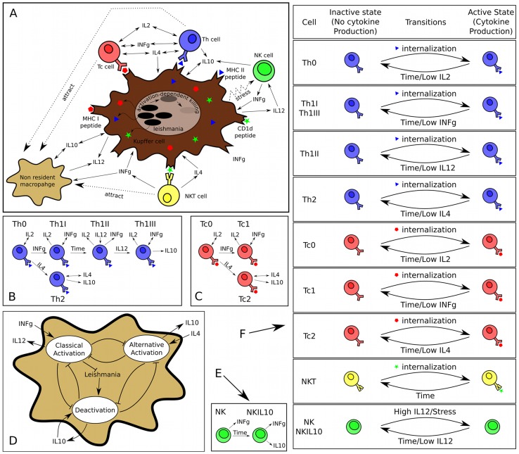
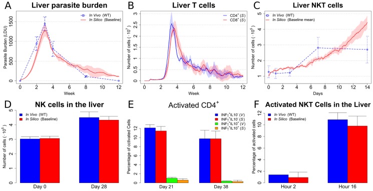
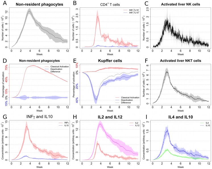
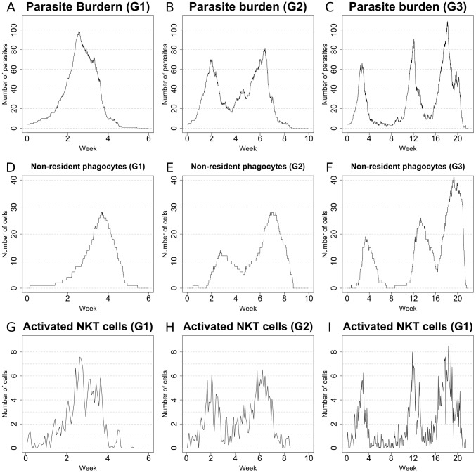
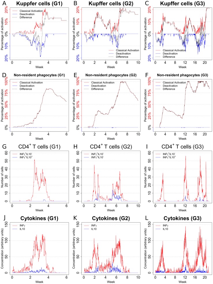
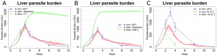
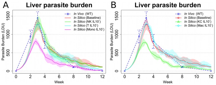
Experimental visceral leishmaniasis, caused by infection of mice with the protozoan parasite Leishmania donovani, is characterized by focal accumulation of inflammatory cells in the liver, forming discrete "granulomas" within which the parasite is eventually eliminated. To shed new light on fundamental aspects of granuloma formation and function, we have developed an in silico Petri net model that simulates hepatic granuloma development throughout the course of infection. The model was extensively validated by comparison with data derived from experimental studies in mice, and the model robustness was assessed by a sensitivity analysis. The model recapitulated the progression of disease as seen during experimental infection and also faithfully predicted many of the changes in cellular composition seen within granulomas over time. By conducting in silico experiments, we have identified a previously unappreciated level of inter-granuloma diversity in terms of the development of anti-leishmanial activity. Furthermore, by simulating the impact of IL-10 gene deficiency in a variety of lymphocyte and myeloid cell populations, our data suggest a dominant local regulatory role for IL-10 produced by infected Kupffer cells at the core of the granuloma.


Conflict of interest statement

The authors have declared that no competing interests exist.

Figures

Figure 1
Figure 1. Schematics of the model dynamics.
(A) High-level depiction of the interactions among the entities modeled. (B) Differentiation of helper T cells. Labels on arrows indicate the conditions for differentiation. Arrows pointing to/originating from a cytokine name indicate that the cytokine is produced/consumed by the cell. (C) Differentiation of cytotoxic T cells. Arrow conventions as in panel B. (D) Dynamics of activation types in macrophages. Leishmania interactions are restricted to Kupffer cells only. Note how different cytokines promote different types of activation and how different types of activation result in the production of different cytokines. (E) Differentiation of NK cells. Arrow conventions as in panel B. (F) Transitions from/to inactive to/from active states for the modeled leukocytes. This representation stresses the complexity of the model and the degree of interaction among the different cell populations; see Section 1 of Supplementary Information for a more detailed description.
Figure 2
Figure 2. Baseline model reproduces many biological features of EVL.
In all the panels, means and standard deviation are reported. Standard deviation is indicated by error bars or shaded areas (A) Organ level parasite burden (compared with [58]). (B) Number of CD4+ and CD8+ T cells over the course of infection of in silico data. The same plotting convention as panel A is used. (C) Number of NKT cells (compared with [59]). The same plotting conventions are used as in panel A. (D) NK cell number (compared with [29]). (E) Percentage of activated CD4+ T cells (compared with unpublished data). (V) and (S) indicate in vivo and in silico, respectively. (F) Percentage of activated NKT cells (compared with and [60]).
Figure 3
Figure 3. Baseline model allows the exploration of biological quantities difficult to access experimentally.
In all the panels, means and standard deviation (indicated by shaded area around the mean) are reported. All numbers are relative to cells in the liver associated with a granuloma microenvironment. (A) Number of granuloma-associated non-resident macrophages. (B) Number of differentiated Th1 cells. (C) Number of activated NK cells. (D) Level of activation and deactivation of non-resident macrophages. (E) Level of activation and deactivation of Kupffer cells. (F) Number of activated NKT cells. (G) Concentration of IFNγ and IL-10. (H) Concentration of IL-2and IL-12. (I) Concentration of IL-4 and IL-10.
Figure 4
Figure 4. Sample behaviours of different in silico granulomas: 1.
Number of parasites (AC), number of non-resident phagocytes (DF) and number of activated NKT cells (GI) in sample granulomas. Note how remarkable diversity is observed among the different granulomas, with rather simple dynamics (G1) coexisting with more complex ones (G2, G3) in the simulations. Total organ parasite load can be reflected by the aggregate results from 50 granulomas.
Figure 5
Figure 5. Sample behaviours of different in silico granulomas: 2.
Level of activation in Kupffer cells (AC; classical activation, red; deactivation, blue; difference, dotted), level of activation in non-resident phagocytes (DF; classical activation, red; deactivation, blue; difference, dotted), number of activated effector Th1 cells (GI; IFNγ+IL-10, red; IFNγ+IL-10+, blue), and cytokine concentration (JL; IFNγ, red; IL-10, blue) in samples granulomas.
Figure 6
Figure 6. Simulations reflecting gene KO qualitatively reproduce expected changes in disease outcome.
(A and B) Parasite burden after in silico knock out of T cells (A) or IFNγ (B), compared to the results from the baseline model (baseline) and in vivo (; WT). (C) In silico knock out of IL-10 compared with and data adapted from [61]). In all the panels, means and standard deviation are reported. Standard deviation is indicated by error bars or shaded areas.
Figure 7
Figure 7. In silico cell-specific knock out of IL-10 implicates Kupffer cell IL-10 production as a major determinant of leishmanicidal activity within granulomas.
(A) In silico knockout of IL-10 in mononuclear phagocytes (Mono IL-10), T cells (T IL10), and NK cells (NK IL10) compared with baseline in silico model and in vivo (; WT). (B) In silico knockout of IL-10 from Kupffer cells (KC IL10) and non-resident macrophages/monocytes/DC (Mac IL10), compared with baseline in silico model and . In all the panels, means and standard deviation are reported. Standard deviation is indicated by error bars or shaded areas.

References

    1. Desjeux P (2004) Leishmaniasis: current situation and new perspectives. Comp Immunol Microbiol Infect Dis 27: 305–318. - PubMed
    1. Alvar J, Velez ID, Bern C, Herrero M, Desjeux P, et al. (2012) Leishmaniasis worldwide and global estimates of its incidence. PLoS One 7: e35671. - PMC - PubMed
    1. Kaye PM, Aebischer T (2011) Visceral leishmaniasis: immunology and prospects for a vaccine. Clin Microbiol Infect 17: 1462–1470. - PubMed
    1. Kaye PM, Svensson M, Ato M, Maroof A, Polley R, et al. (2004) The immunopathology of experimental visceral leishmaniasis. Immunol Rev 201: 239–253. - PubMed
    1. Murray HW (2001) Tissue granuloma structure-function in experimental visceral leishmaniasis. Int J Exp Pathol 82: 249–267. - PMC - PubMed

Publication types