

Reappraisal of field dynamics of motor cortex during self-paced finger movements
- PMID: 24363977
- PMCID: PMC3868179
- DOI: 10.1002/brb3.186
Reappraisal of field dynamics of motor cortex during self-paced finger movements
Abstract
Background: The exact origin of neuronal responses in the human sensorimotor cortex subserving the generation of voluntary movements remains unclear, despite the presence of characteristic but robust waveforms in the records of electroencephalography or magnetoencephalography (MEG).
Aims: To clarify this fundamental and important problem, we analyzed MEG in more detail using a multidipole model during pulsatile extension of the index finger, and made some important new findings.
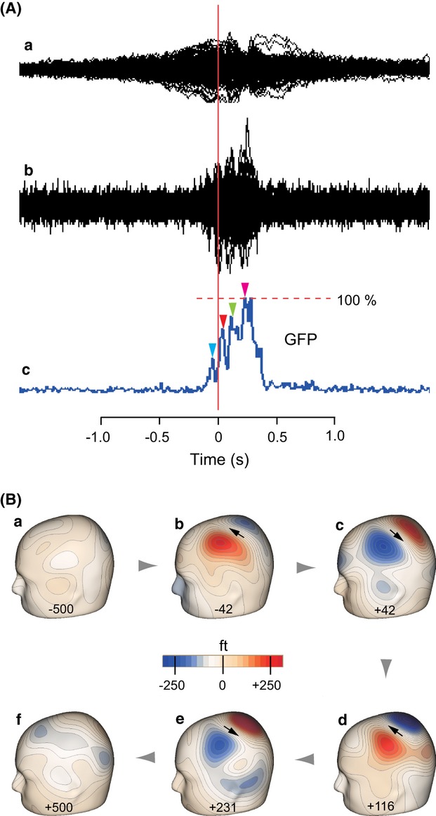
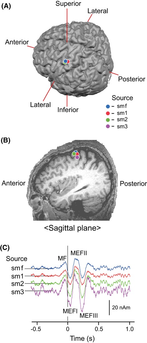
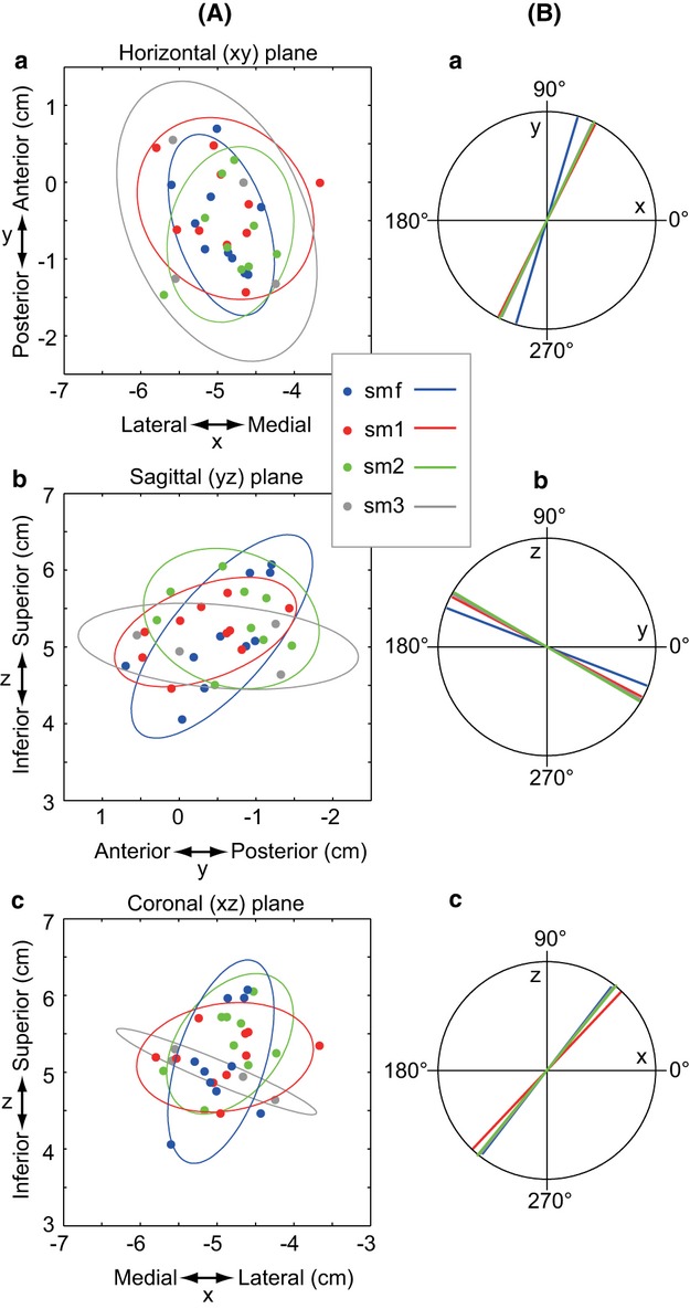
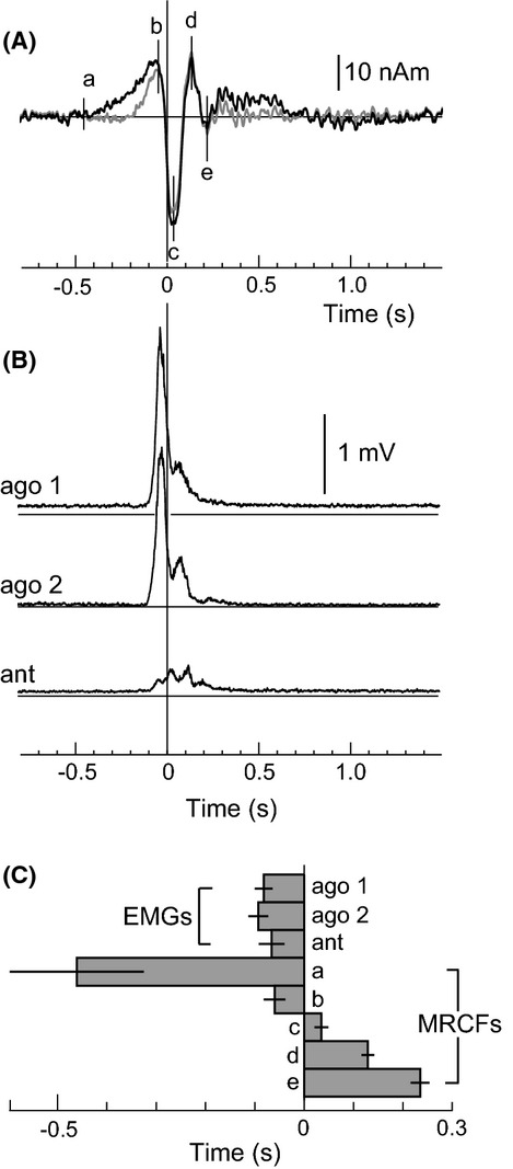
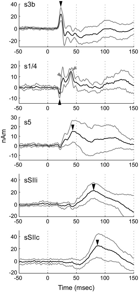
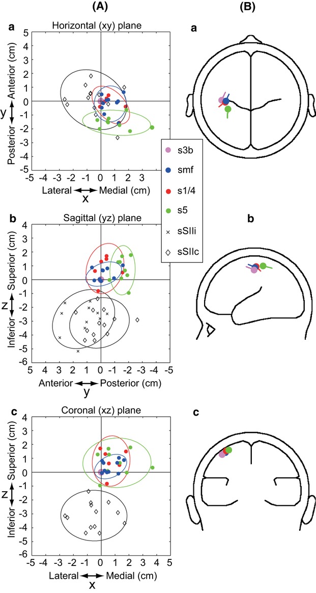
Results: Movement-related cerebral fields (MRCFs) were confirmed over the sensorimotor region contralateral to the movement, consisting of a temporal succession of the first premovement component termed motor field, followed by two or three postmovement components termed movement evoked fields. A source analysis was applied to separately model each of these field components. Equivalent current diploes of all components of MRCFs were estimated to be located in the same precentral motor region, and did not differ with respect to their locations and orientations. The somatosensory evoked fields following median nerve stimulation were used to validate these findings through comparisons of the location and orientation of composite sources with those specified in MRCFs. The sources for the earliest components were evoked in Brodmann's area 3b located lateral to the sources of MRCFs, and those for subsequent components in area 5 and the secondary somatosensory area were located posterior to and inferior to the sources of MRCFs, respectively. Another component peaking at a comparable latency with the area 3b source was identified in the precentral motor region where all sources of MRCFs were located.
Conclusion: These results suggest that the MRCF waveform reflects a series of responses originating in the precentral motor area.
Keywords: Diploe sources; magnetoencephalography; motor cortex; movement-related cerebral fields; somatosensory evoked fields.
Figures
Similar articles
-
Magnetoencephalographic representation of the sensorimotor hand area in cases of intracerebral tumour.J Neurol Neurosurg Psychiatry. 2003 Dec;74(12):1649-54. doi: 10.1136/jnnp.74.12.1649. J Neurol Neurosurg Psychiatry. 2003. PMID: 14638884 Free PMC article.
-
[Presurgical functional mapping of the sensorimotor area using evoked magnetic fields].No Shinkei Geka. 2002 Apr;30(4):391-7. No Shinkei Geka. 2002. PMID: 11968825 Japanese.
-
Identification of motor and sensory brain activities during unilateral finger movement: spatiotemporal source analysis of movement-associated magnetic fields.Exp Brain Res. 1997 Jun;115(1):6-14. doi: 10.1007/pl00005685. Exp Brain Res. 1997. PMID: 9224829 Clinical Trial.
-
Electroencephalographic and magnetoencephalographic studies of motor function.Adv Neurol. 1990;54:193-205. Adv Neurol. 1990. PMID: 2270804 Review.
-
[Functional localization of the somatomotor area by magnetoencephalography].Rinsho Shinkeigaku. 1999 Jan;39(1):42-3. Rinsho Shinkeigaku. 1999. PMID: 10377797 Review. Japanese.
Cited by
-
A touch of hierarchy: population receptive fields reveal fingertip integration in Brodmann areas in human primary somatosensory cortex.Brain Struct Funct. 2021 Sep;226(7):2099-2112. doi: 10.1007/s00429-021-02309-5. Epub 2021 Jun 5. Brain Struct Funct. 2021. PMID: 34091731 Free PMC article.
-
A Dynamical Systems Approach to Characterizing Brain-Body Interactions during Movement: Challenges, Interpretations, and Recommendations.Sensors (Basel). 2023 Jul 11;23(14):6296. doi: 10.3390/s23146296. Sensors (Basel). 2023. PMID: 37514591 Free PMC article. Review.
-
Contrasting MEG effects of anodal and cathodal high-definition TDCS on sensorimotor activity during voluntary finger movements.Front Neuroimaging. 2024 Feb 5;3:1341732. doi: 10.3389/fnimg.2024.1341732. eCollection 2024. Front Neuroimaging. 2024. PMID: 38379832 Free PMC article.
References
-
- Allison T, McCarthy G, Wood CC, Darcey TM, Spencer DD, Williamson PD. Human cortical potentials evoked by stimulation of the median nerve. II. Cytoarchitectonic areas generating short-latency activity. J. Neurophysiol. 1989;62:694–710. - PubMed
-
- Asanuma H, Larsen K, Yumiya H. Peripheral input pathways to the monkey motor cortex. Exp. Brain Res. 1980;38:349–355. - PubMed
-
- Avendano C, Isla AJ, Rausell E. Area 3a in the cat II. Projections to the motor cortex and their relations to other corticocortical connections. J. Comp. Neurol. 1992;321:373–386. - PubMed
-
- Ball T, Schreiber A, Feige B, Wagner M, Lucking CH, Kristeva-Feige R. The role of higher-order motor areas in voluntary movement as revealed by high-resolution EEG and fMRI. Neuroimage. 1999;10:682–694. - PubMed
-
- Barrett G, Shibasaki H, Neshige R. Cortical potentials preceding voluntary movement: evidence for three periods of preparation in man. Electroencephalogr. Clin. Neurophysiol. 1986;63:327–339. - PubMed
LinkOut - more resources
Full Text Sources
Other Literature Sources
Medical
