Review
. 2013 Dec 4:3:88.
doi: 10.3389/fcimb.2013.00088. eCollection 2013.

ESX-1-induced apoptosis during mycobacterial infection: to be or not to be, that is the question

Affiliations
Review

ESX-1-induced apoptosis during mycobacterial infection: to be or not to be, that is the question

Nacho Aguiló et al. Front Cell Infect Microbiol. .

Abstract

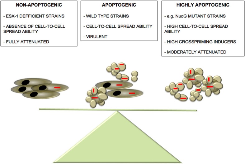
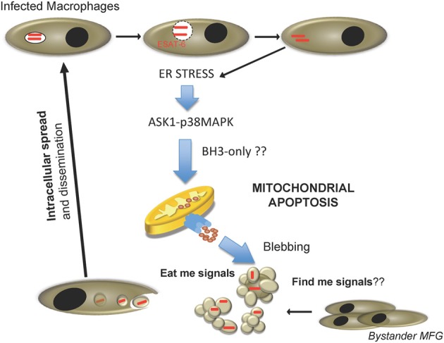
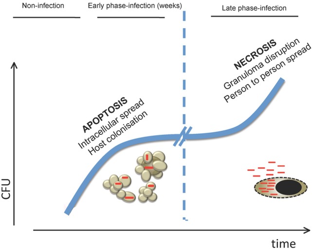
The major Mycobacterium tuberculosis virulence factor ESAT-6 exported by the ESX-1 secretion system has been described as a pro-apoptotic factor by several independent groups in recent years, sustaining a role for apoptosis in M. tuberculosis pathogenesis. This role has been supported by independent studies in which apoptosis has been shown as a hallmark feature in human and mouse lungs infected with virulent strains. Nevertheless, the role of apoptosis during mycobacterial infection is subject to an intense debate. Several works maintain that apoptosis is more evident with attenuated strains, whereas virulent mycobacteria tend to inhibit this process, suggesting that apoptosis induction may be a host mechanism to control infection. In this review, we summarize the evidences that support the involvement of ESX-1-induced apoptosis in virulence, intending to provide a rational treatise for the role of programmed cell death during M. tuberculosis infection.

Keywords: Mycobacterium tuberculosis; apoptosis; attenuated strains; cell death; necrosis; virulence.


Figures

Figure 1
Figure 1
Balance between M. tuberculosis-induced apoptosis and virulence. We propose the existence of a delicate balance between mycobacteria-induced apoptosis and pathogenesis. Absence of apoptosis observed in ESX-1-deficient strains leads to a sound attenuation phenotype accompanied by abrogated cell-to-cell spread capacity (Aguilo et al., 2013). On the other hand, although highly apoptogenic strains show an increased cell-to-cell spread capacity compared to wild-type strains, they might present higher visibility to the host favoring an accelerated establishment of host immune defences, thus, tipping the balance to the favor of the host. In the “golden mean” between the two extremes of apoptosis induction, virulent wild-type strains have active both pro- and anti-apoptotic mechanisms that even though partially limit cell-to-cell infectivity, they impair the rapid establishment of host immune response, thus, favoring M. tuberculosis pathogenesis.
Figure 2
Figure 2
Proposed cell-to-cell spread model for M. tuberculosis during host cell infection. Internalized M. tuberculosis disrupts phagosome in an ESAT-6 dependent manner. After reaching the cytosol, mycobacteria trigger ER stress, and activate pro-apoptotic cellular routes involving the ASK1-p38MAPK signaling axis, and causing host cell apoptosis by activation of the mitochondrial apoptotic pathway. Consequently, fresh macrophages are recruited and M. tuberculosis-containing apoptotic bodies are phagocytosed by new cells, re-starting the infection cycle.
Figure 3
Figure 3
Model of how cell death is involved in the different stages of M. tuberculosis host infection. In the early stages of infection, killing of macrophages by apoptosis would allow M. tuberculosis to spread cell-to-cell in the lung in the absence of inflammatory mediators associated with necrotic release of intracellular content. This would permit bacteria to replicate in the preferred intracellular environment prior to establishment of an effective adaptive immune response. In the latest stages of infection, when bacterial burden is high, M. tuberculosis would induce necrosis on infected cells leading to mycobacterial release to the extracellular medium. As a consequence, granuloma is disrupted allowing aerosol transmission to new hosts.

References

    1. Abarca-Rojano E., Rosas-Medina P., Zamudio-Cortez P., Mondragon-Flores R., Sanchez-Garcia F. J. (2003). Mycobacterium tuberculosis virulence correlates with mitochondrial cytochrome c release in infected macrophages. Scand. J. Immunol. 58, 419–427 10.1046/j.1365-3083.2003.01318.x - DOI - PubMed
    1. Adams J. M., Cory S. (2007). Bcl-2-regulated apoptosis: mechanism and therapeutic potential. Curr. Opin. Immunol. 19, 488–496 10.1016/j.coi.2007.05.004 - DOI - PMC - PubMed
    1. Aguilo J. I., Alonso H., Uranga S., Marinova D., Arbues A., de Martino A., et al. (2013). ESX-1-induced apoptosis is involved in cell-to-cell spread of Mycobacterium tuberculosis. Cell. Microbiol. 15, 1994–2005 10.1111/cmi.12169 - DOI - PubMed
    1. Aleman M., Schierloh P., de la Barrera S. S., Musella R. M., Saab M. A., Baldini M., et al. (2004). Mycobacterium tuberculosis triggers apoptosis in peripheral neutrophils involving toll-like receptor 2 and p38 mitogen protein kinase in tuberculosis patients. Infect. Immun. 72, 5150–5158 10.1128/IAI.72.9.5150-5158.2004 - DOI - PMC - PubMed
    1. Aporta A., Arbues A., Aguilo J. I., Monzon M., Badiola J. J., de Martino A., et al. (2012). Attenuated Mycobacterium tuberculosis SO2 vaccine candidate is unable to induce cell death. PLoS ONE 7:e45213 10.1371/journal.pone.0045213 - DOI - PMC - PubMed

Publication types