Comment
. 2014 Jan;155(1):1-5.
doi: 10.1210/en.2013-2079.

Allosteric modulators hit the TSH receptor

Affiliations
Comment

Allosteric modulators hit the TSH receptor

Terry F Davies et al. Endocrinology. 2014 Jan.
No abstract available


Figures

Figure 1.
Figure 1.
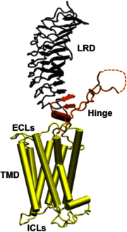
An homology model of the entire TSH holoreceptor This model highlights the tripartite structure of the TSHR. The ectodomain, shown in gray/black, is made up of 10 leucine-rich repeat domains (LRD) characterized as a “scythe-blade”-shaped structure with loops and β-pleated sheets obtained from the published crystal structure (3) (PDB:3G04). The region connecting the LRD and TMD, known as the “hinge” region, has recently been crystallized for the FSH receptor (46) (PDB:4AY9) and is shown as a looped structure (orange) with a helix conformation close to the carboxyl end of the LRD. The hinge in the TSHR has an additional sequence insert and is larger than in the FSH receptor. Therefore, amino acids 305–381 are missing in the illustrated model (47), and this insert is depicted as a closed dotted loop. The TMD (yellow), with its 7 helices, is depicted as cylindrical structures connected to each other by the specific TSHR intra- and ECLs. The TMD is the region that harbors the allosteric binding pockets for the SMLs. ICL, intracellular loops; PDB, Protein Data Base. ECL, extracellular loops.
Figure 2.
Figure 2.
Interaction of SMLs with the TSHR Similar to most GPCRs, the basal state of TSHR activity is hypothesized to be constrained by polar interactions between different TMHs forming an “ionic lock” between an arginine (R) at the base of transmembrane TMH3 and partly conserved residues such as glutamate (E) or aspartic acid (D) at the base of TM6 as shown in red (left panel). When a SML antagonist binds to the allosteric pocket (purple) on the TMD, the ionic lock remains intact even if TSH ligand binds to the ectodomain because there can be no outward movement of TMH6. Hence, the basal conformation of the receptor is locked and stabilized. In contrast, when a SML agonist binds to the allosteric pocket (right panel), there is an outward movement of TMH6 as a result of destabilization of the ionic lock and loss of constrained polar interactions. This movement causes conformational changes in the intracellular loops, which, in turn, allow binding of G proteins and β-arrestin activation leading to signal transduction. ICL, intracellular loops; ECL, extracellular loops.

Comment on

Similar articles

Cited by

References

    1. Adams DD, Purves HD. Abnormal responses in the assay of thyrotropin. Proceedings of the University of Otago Medical School. 1956;34:11–12
    1. Smith BR, Hall R. Thyroid-stimulating immunoglobulins in Graves' disease. Lancet. 1974;2(7878):427–429 - PubMed
    1. Sanders J, Chirgadze DY, Sanders P, et al. Crystal structure of the TSH receptor in complex with a thyroid-stimulating autoantibody. Thyroid. 2007;17(5):395–410 - PubMed
    1. Calebiro D, Nikolaev VO, Persani L, Lohse MJ. Signaling by internalized G-protein-coupled receptors. Trends Pharmacol Sci. 2010;31(5):221–228 - PubMed
    1. Tanaka K, Chazenbalk GD, McLachlan SM, Rapoport B. Subunit structure of thyrotropin receptors expressed on the cell surface. J Biol Chem. 1999;274(48):33979–33984 - PubMed

Publication types

MeSH terms

Substances