. 2013 Dec 23:10:359.
doi: 10.1186/1743-422X-10-359.

In-vitro renal epithelial cell infection reveals a viral kidney tropism as a potential mechanism for acute renal failure during Middle East Respiratory Syndrome (MERS) Coronavirus infection

Affiliations

In-vitro renal epithelial cell infection reveals a viral kidney tropism as a potential mechanism for acute renal failure during Middle East Respiratory Syndrome (MERS) Coronavirus infection

Isabella Eckerle et al. Virol J. .

Abstract

Background: The Middle East Respiratory Syndrome Coronavirus (MERS-CoV) causes symptoms similar to Severe Acute Respiratory Syndrome Coronavirus (SARS-CoV), yet involving an additional component of acute renal failure (ARF) according to several published case reports. Impairment of the kidney is not typically seen in Coronavirus infections. The role of kidney infection in MERS is not understood.

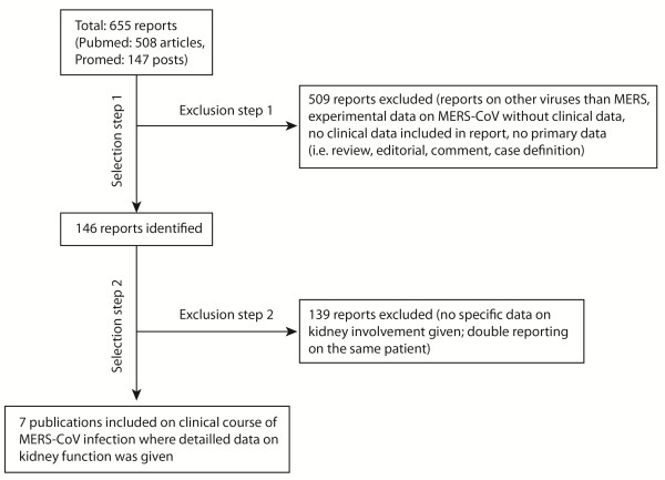
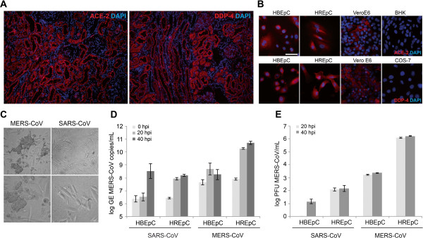
Findings: A systematic review of communicated and peer-reviewed case reports revealed differences in descriptions of kidney involvement in MERS versus SARS patients. In particular, ARF in MERS patients occurred considerably earlier after a median time to onset of 11 days (SD ±2,0 days) as opposed to 20 days for SARS, according to the literature. In-situ histological staining of the respective cellular receptors for MERS- and SARS-Coronavirus showed highly similar staining patterns with a focus of a receptor-specific signal in kidney epithelial cells. Comparative infection experiments with SARS- and MERS-CoV in primary human kidney cells versus primary human bronchial epithelial cells showed cytopathogenic infection only in kidney cells, and only if infected with MERS-CoV. Kidney epithelial cells produced almost 1000-fold more infectious MERS-CoV progeny than bronchial epithelial cells, while only a small difference was seen between cell types when infected with SARS-CoV.

Conclusion: Epidemiological studies should analyze kidney impairment and its characteristics in MERS-CoV. Virus replication in the kidney with potential shedding in urine might constitute a way of transmission, and could explain untraceable transmission chains leading to new cases. Individual patients might benefit from early induction of renoprotective treatment.


Figures

Figure 1
Figure 1
Flowchart of study selection. Systematic search strategies were applied to identify all reports on patients’ kidney function during MERS-CoV infection. Data sources used were Pubmed (http://www.ncbi.nlm.nih.gov/pubmed/) and Promed.Mail (http://www.promedmail.org) by searching the term “coronavirus” with a date range from 15th September 2012 (date of the first MERS-CoV report) to 16th September 2013. All identified publications were reviewed for clinical data on kidney function (irrespective of pointing to normal or impaired function). Data on patient sex and age, country of patient origin and travel route, onset of illness, presence or absence of ARF, date of onset of ARF, day of illness on which ARF occurred, as well as clinical outcome (death or survival) were extracted. In total, 7 publications were finally included that described a total of 12 MERS-CoV patients and their kidney function during the course of disease.
Figure 2
Figure 2
SARS- and MERS-CoV receptor expression and virus infection experiments in human primary cells. (A) SARS- and MERS-CoV receptor expression of human Angiotensin-converting-enzyme-2 (ACE-2) and Dipeptidyl-peptidase-4 (DPP-4), respectively, in cryosections of a healthy human kidney and (B) in primary bronchial (HBEpC) and renal (HREpC) epithelial cells. For positive controls, ACE2- and DPP-4-expressing primate cells (kidney cells from African green monkey [Vero E6]) were stained in parallel. Cell lines known to be negative for ACE-2 (kidney cells from Syrian hamster [BHK]) or DPP-4 (kidney cells from African green monkey [COS-7]) were used as negative controls. The white bar represents 50 μm. (C) Cell morphology and cytopathic effect (CPE) formation of human primary renal epithelial cells (HREpC) infected with MERS-CoV or SARS-CoV with 0.5 plaque-forming units of either virus per cell. A pronounced CPE formation 20 hours post infection (hpi) was seen only after infection with MERS-CoV in HREpC but not in cells infected with SARS-CoV. No CPE formation was seen in HBEpC infected with MERS-CoV or SARS-CoV (data not shown). Upper row: 100-fold magnification, lower row: 400-fold magnification, bright field microscopy (D) Replication of SARS- and MERS-CoV on HBEpC and HREpC determined by real time RT-PCR after 0, 20 and 40 hpi (E) Progeny virus measured by titration of supernatants in duplicates in a plaque assay in Vero cells. MERS-CoV replicates in HREpC with peak titers of 6.2 log plaque forming units (PFU)/mL, showing a 2,9-fold log difference between HREpC and HBEpC, while replication of SARS-CoV showed only a 1-fold log difference between bronchial and renal primary cells. Replication levels for each virus used are given as log of the genome equivalents (GEs) (D) or as plaque-forming units (PFUs) (E). All virus infection experiments were performed in triplicates. Bars represent mean values, error bars represent standard deviation of triplicates.

References

    1. Peiris JS, Guan Y, Yuen KY. Severe acute respiratory syndrome. Nat Med. 2004;10:S88–S97. doi: 10.1038/nm1143. - DOI - PMC - PubMed
    1. Zaki AM, van Boheemen S, Bestebroer TM, Osterhaus AD, Fouchier RA. Isolation of a novel coronavirus from a man with pneumonia in Saudi Arabia. N Engl J Med. 2012;10:1814–1820. doi: 10.1056/NEJMoa1211721. - DOI - PubMed
    1. Memish ZA, Zumla AI, Al-Hakeem RF, Al-Rabeeah AA, Stephens GM. Family cluster of Middle East respiratory syndrome coronavirus infections. N Engl J Med. 2013;10:2487–2494. doi: 10.1056/NEJMoa1303729. - DOI - PubMed
    1. Assiri A, McGeer A, Perl TM, Price CS, Al Rabeeah AA, Cummings DA, Alabdullatif ZN, Assad M, Almulhim A, Makhdoom H. et al. Hospital outbreak of Middle East respiratory syndrome coronavirus. N Engl J Med. 2013;10:407–416. doi: 10.1056/NEJMoa1306742. - DOI - PMC - PubMed
    1. Drosten C, Seilmaier M, Corman VM, Hartmann W, Scheible G, Sack S, Guggemos W, Kallies R, Muth D, Junglen S. et al. Clinical features and virological analysis of a case of Middle East respiratory syndrome coronavirus infection. Lancet Infect Dis. 2013;10:745–751. doi: 10.1016/S1473-3099(13)70154-3. - DOI - PMC - PubMed

Publication types