

A mouse model links asthma susceptibility to prenatal exposure to diesel exhaust
- PMID: 24365139
- PMCID: PMC4065237
- DOI: 10.1016/j.jaci.2013.10.047
A mouse model links asthma susceptibility to prenatal exposure to diesel exhaust
Abstract
Background: Most asthma begins in the first years of life. This early onset cannot be attributed merely to genetic factors because the prevalence of asthma is increasing. Epidemiologic studies have indicated roles for prenatal and early childhood exposures, including exposure to diesel exhaust. However, little is known about the mechanisms. This is largely due to a paucity of animal models.
Objective: We aimed to develop a mouse model of asthma susceptibility through prenatal exposure to diesel exhaust.
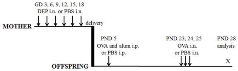
Methods: Pregnant C57BL/6 female mice were given repeated intranasal applications of diesel exhaust particles (DEPs) or PBS. Offspring underwent suboptimal immunization and challenge with ovalbumin (OVA) or received PBS. Pups were examined for features of asthma; lung and liver tissues were analyzed for transcription of DEP-regulated genes.
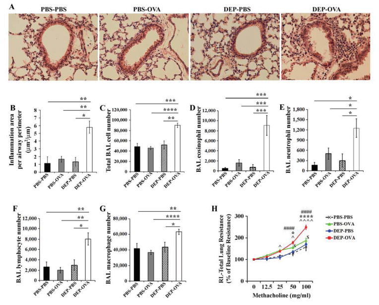
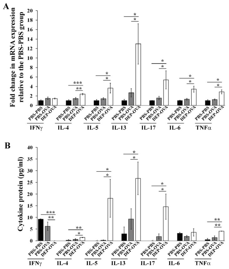
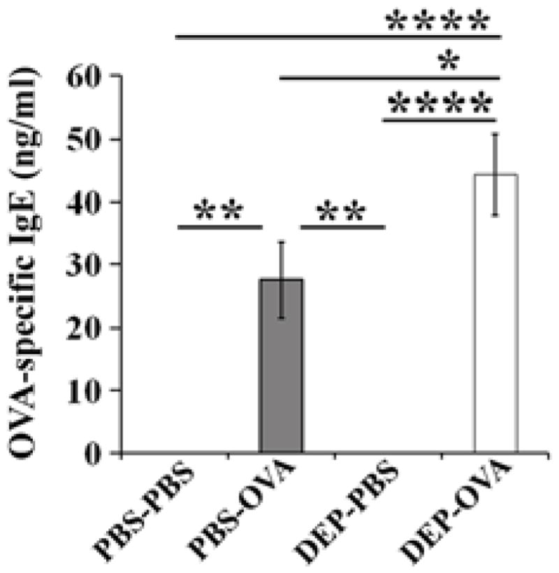
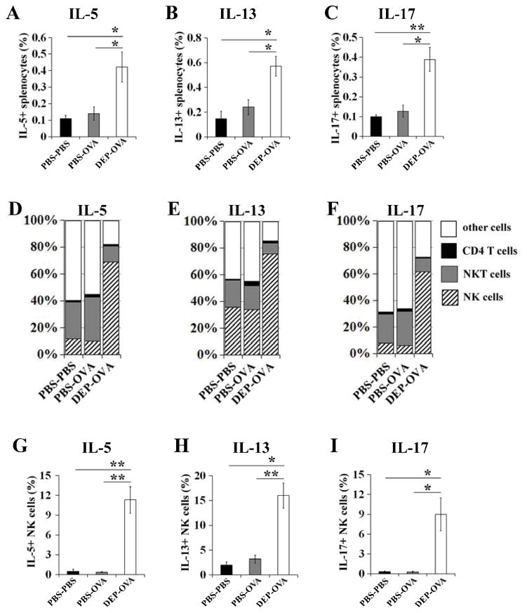
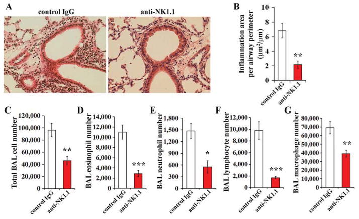
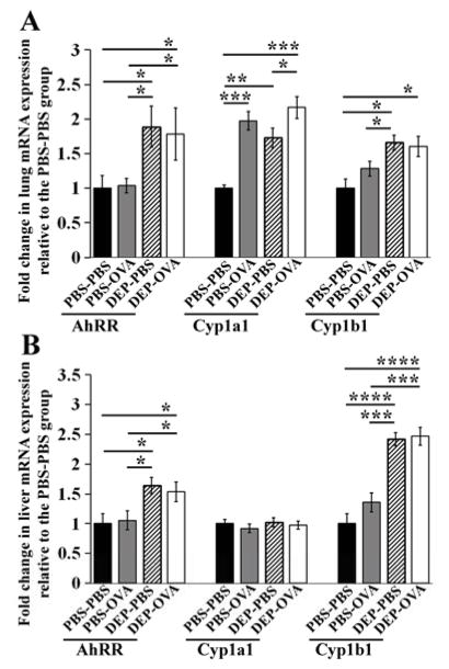
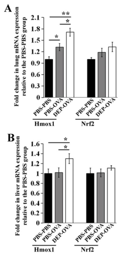
Results: Offspring of mice exposed to DEPs were hypersensitive to OVA, as indicated by airway inflammation and hyperresponsiveness, increased serum OVA-specific IgE levels, and increased pulmonary and systemic TH2 and TH17 cytokine levels. These cytokines were primarily produced by natural killer (NK) cells. Antibody-mediated depletion of NK cells prevented airway inflammation. Asthma susceptibility was associated with increased transcription of genes known to be specifically regulated by the aryl hydrocarbon receptor and oxidative stress. Features of asthma were either marginal or absent in OVA-treated pups of PBS-exposed mice.
Conclusion: We created a mouse model that linked maternal exposure to DEPs with asthma susceptibility in offspring. Development of asthma was dependent on NK cells and associated with increased transcription from aryl hydrocarbon receptor- and oxidative stress-regulated genes.
Keywords: IL-13; IL-17; IL-5; Prenatal exposure; aryl hydrocarbon receptor; asthma; diesel exhaust particles; mouse model; natural killer cells.
Copyright © 2013 American Academy of Allergy, Asthma & Immunology. Published by Mosby, Inc. All rights reserved.
Figures
Comment in
-
Diesel exhaust particle exposure during pregnancy promotes development of asthma and atopy.J Allergy Clin Immunol. 2014 Jul;134(1):73-4. doi: 10.1016/j.jaci.2014.04.002. Epub 2014 May 13. J Allergy Clin Immunol. 2014. PMID: 24835501 No abstract available.
References
-
- Moorman JE, Akinbami LJ, Bailey CM, et al. National Surveillance of Asthma: United States, 2001–2010. [Accessed June 23, 2013];National Center for Health Statistics. Vital Health Stat. 2012 3(35) Available at: http://www.cdc.gov/nchs/data/series/sr_03/sr03_035.pdf. - PubMed
-
- Litonjua AA, Carey VJ, Burge HA, Weiss ST, Gold DR. Parental history and the risk for childhood asthma. Does mother confer more risk than father? Am J Respir Crit Care Med. 1998;158:176–81. - PubMed
-
- Ruiz RG, Kemeny DM, Price JF. Higher risk of infantile atopic dermatitis from maternal atopy than from paternal atopy. Clin Exp Allergy. 1992;22:762–6. - PubMed
Publication types
MeSH terms
Substances
Grants and funding
LinkOut - more resources
Full Text Sources
Other Literature Sources
Medical
