Review
. 2013 Dec 23;369(1635):20120510.
doi: 10.1098/rstb.2012.0510. Print 2014 Feb 5.

Space in the brain: how the hippocampal formation supports spatial cognition

Affiliations
Review

Space in the brain: how the hippocampal formation supports spatial cognition

Tom Hartley et al. Philos Trans R Soc Lond B Biol Sci. .

Abstract

Over the past four decades, research has revealed that cells in the hippocampal formation provide an exquisitely detailed representation of an animal's current location and heading. These findings have provided the foundations for a growing understanding of the mechanisms of spatial cognition in mammals, including humans. We describe the key properties of the major categories of spatial cells: place cells, head direction cells, grid cells and boundary cells, each of which has a characteristic firing pattern that encodes spatial parameters relating to the animal's current position and orientation. These properties also include the theta oscillation, which appears to play a functional role in the representation and processing of spatial information. Reviewing recent work, we identify some themes of current research and introduce approaches to computational modelling that have helped to bridge the different levels of description at which these mechanisms have been investigated. These range from the level of molecular biology and genetics to the behaviour and brain activity of entire organisms. We argue that the neuroscience of spatial cognition is emerging as an exceptionally integrative field which provides an ideal test-bed for theories linking neural coding, learning, memory and cognition.

Keywords: boundary cells; entorhinal cortex; grid cells; head direction cells; hippocampus; place cells.


Figures

Figure 1.
Figure 1.
Schematic overview of major anatomical pathways in the hippocampal formation of the rat. Left-hand side of figure emphasizes gross morphology (rat brain) of cell layers in hippocampus and dentate gyrus and long-established unidirectional projections. Classic trisynaptic pathway consists of projection from entorhinal cortex (LEC: lateral entorhinal cortex; MEC: medial entorhinal cortex) to dentate gyrus (DG), from DG to CA3, and from CA3 to CA1. Entorhinal input also consists of direct monosynaptic LEC and MEC projections to CA3, to CA1, and to subiculum (Sb). CA1 projection to Sb and to LEC/MEC, and Sb projections to LEC/MEC, complete the circuit. Other circuits involve projections from subiculum to presubiculum (PreSb) and to parasubiculum (ParaSb), and projections from PreSb to MEC, and ParaSb to both LEC and MEC. Arrows indicate the direction of projection, and circles indicate cell bodies. For simplicity in this highly schematic figure, omissions include the following: dendrites and dendritic location of axonal termination zones; commissural projections connecting left and right hemispheres; CA2-involving projections. Additional guidance. The term ‘hippocampal formation’ applies to regions contained within dashed box. Entorhinal pathways to DG, CA3, CA1 and Sb known as perforant pathway, DG to CA3 pathway as mossy fibre projection, CA3 to CA1 pathway as Schaffer collaterals. As well as projecting in feed-forward manner to CA1, the CA3 pyramidal cells project to other CA3 pyramidal cells; these recurrent collaterals were proposed by Marr to underlie pattern completion (the ‘collaterals effect’ [14]). Postrhinal cortex is rat analogue of primate parahippocampal cortex (PHC), strongly implicated in visuospatial processing. In rodents, term ‘postsubiculum’ (containing many HD cells) refers to dorsal portion of presubiculum. Two parallel pathways formed by projections from postrhinal cortex and presubiculum to MEC, and perirhinal cortex to LEC, are not fully illustrated. Inspired by [15].
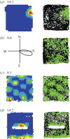
Figure 2.
Figure 2.
Four types of fundamental spatial cell. Figure shows one example of each type of fundamental spatial cell: (a) place cell; (b) HD cell; (c) grid cell; (d) boundary cell. For each cell: left-hand column shows locational firing ratemap (a,c,d) or directional firing polar plot (b), with peak firing rate in hertz shown top left of rate map/polar plot; right-hand column depicts path taken over whole trial (black line), on which are plotted the locations at which spikes were recorded (green squares). In firing rate maps, one of five colours in locational bin indicates spatially smoothed firing rate in that bin (autoscaled to firing rate peak; dark blue, 0–20%; light blue, 20–40%; green, 40–60%; yellow, 60–80%; red, 80–100%). HD, grid and boundary cell recorded in 1 × 1 m (place cell: 62 × 62 cm) square-walled box with 50 cm-high walls. For boundary cell, 50 cm-long barrier inserted into box elicits the second field along north side of barrier (as predicted by the boundary vector cell (BVC) model [53]) in addition to original field along south wall. Cells provided by Sarah Stewart and Colin Lever.
Figure 3.
Figure 3.
Theta phase precession of place cell firing. (a) As a rat runs along a linear track, a place cell in the hippocampus fires as the animal moves through the firing field (b). The firing rate code for location is also a temporal code (c): spikes (ticks) are fired at successively earlier phases of the theta rhythm of the local field potential (blue trace), referred to as ‘theta phase precession’. The theta phase of firing correlates with the distance travelled through the place field (d), even when pooled over runs that might be fast or slow. Adapted from [70].

Similar articles

Cited by

References

    1. Hubel DH, Wiesel TN. 1962. Receptive fields, binocular interaction and functional architecture in the cat's visual cortex. J. Physiol. 160, 106–154. - PMC - PubMed
    1. Kaas JH, Nelson RJ, Sur M, Lin C-S, Merzenich MM. 1979. Multiple representations of the body within the primary somatosensory cortex of primates. Science 204, 521–523. (10.1126/science.107591) - DOI - PubMed
    1. Georgopoulos AP, Schwartz AB, Kettner RE. 1986. Neuronal population coding of movement direction. Science 233, 1416–1419. (10.1126/science.3749885) - DOI - PubMed
    1. Colby CL. 1998. Action-oriented spatial reference frames in cortex. Neuron 20, 15–24. (10.1016/S0896-6273(00)80429-8) - DOI - PubMed
    1. Andersen RA. 1997. Multimodal integration for the representation of space in the posterior parietal cortex. Phil. Trans. R. Soc. Lond. B 352, 1421–1428. (10.1098/rstb.1997.0128) - DOI - PMC - PubMed

Publication types