Review
. 2013 Dec 23;369(1635):20120515.
doi: 10.1098/rstb.2012.0515. Print 2014 Feb 5.

Architecture of spatial circuits in the hippocampal region

Affiliations
Review

Architecture of spatial circuits in the hippocampal region

Menno P Witter et al. Philos Trans R Soc Lond B Biol Sci. .

Abstract

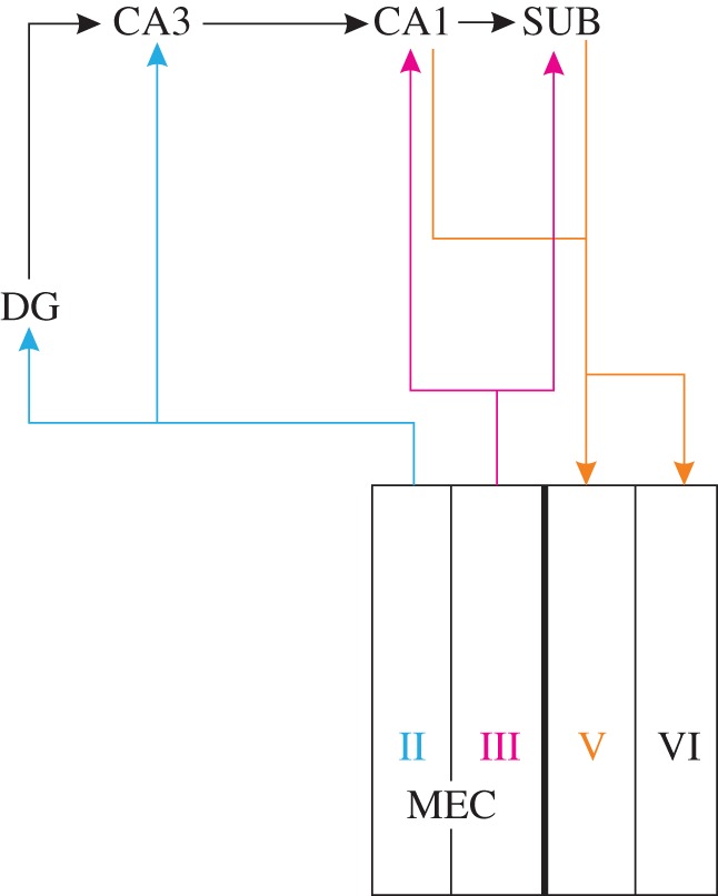
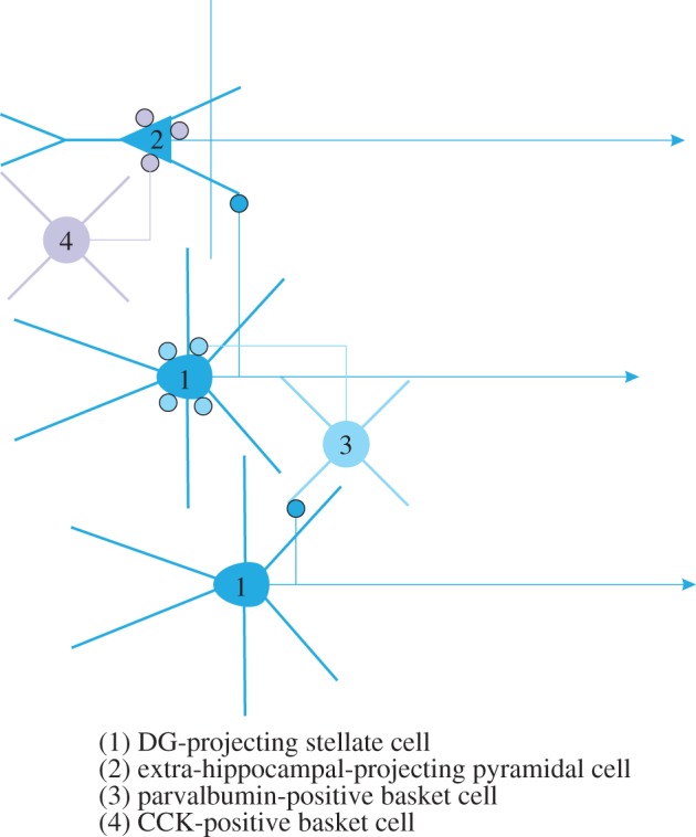
The hippocampal region contains several principal neuron types, some of which show distinct spatial firing patterns. The region is also known for its diversity in neural circuits and many have attempted to causally relate network architecture within and between these unique circuits to functional outcome. Still, much is unknown about the mechanisms or network properties by which the functionally specific spatial firing profiles of neurons are generated, let alone how they are integrated into a coherently functioning meta-network. In this review, we explore the architecture of local networks and address how they may interact within the context of an overarching space circuit, aiming to provide directions for future successful explorations.

Keywords: development; entorhinal cortex; hippocampus; parasubiculum; presubiculum.


Figures

Figure 1.
Figure 1.
The ‘classical’ hippocampal–parahippocampal network. Neurons in MEC layer II provide inputs DG and CA3, feeding into the trisynaptic pathway that includes projections from DG to CA3 to CA1. Layer III neurons project to CA1 and SUB, which both reciprocate with projections to layer V (and weaker to VI). (Online version in colour.)
Figure 2.
Figure 2.
Extended hippocampal–parahippocampal network. The ‘classical’ scheme with added local connectivity of PrS and PaS and the reciprocal layer II–CA2 pathway. (Online version in colour.)
Figure 3.
Figure 3.
Medial entorhinal circuits. Principal cells in all layers receive monosynaptic convergent inputs from PrS and PaS. Axons of layer V pyramidal cells target superficial layers, making synaptic contacts onto principal cells in layers II and III. These layer V cells are likely contacted by local axons of layer II and III cells, and cells in layer II presumably contact layer III cells. Although no neurons are indicated in layer VI, synaptic contacts from PrS and PaS have been reported [34] and are schematically indicated. (Online version in colour.)
Figure 4.
Figure 4.
Proposed local circuits in medial entorhinal layer II. (1) DG projecting stellate cells make synaptic contacts with fast-spiking, (3) parvalbumin-positive interneurons that innervate the somata of stellate cells. Stellate-to-stellate synaptic contacts are non-existent. (2) Non-hippocampal projecting neurons, likely pyramidal cells receive input from stellate cells but do not reciprocate that input. The pyramidal cells are innervated by (4) CCK-positive basket cells. Other interneuron types, such as chandelier, goblet and multipolar cells, are not indicated. CCK, cholecystokinin. (Online version in colour.)

Similar articles

Cited by

References

    1. O'Keefe J, Dostrovsky J. 1971. The hippocampus as a spatial map. Preliminary evidence from unit activity in the freely-moving rat. Brain Res. 34, 171–175. (10.1016/0006-8993(71)90358-1) - DOI - PubMed
    1. Ranck JB. 1985. Head direction cells in the deep cell layer of dorsal presubiculum in freely moving rats. In Electrical activity of the archicortex (eds Buzsaki G, Vanderwolf CH.), pp. 217–220. Budapest, Hungary: Akademiai Kiado.
    1. Taube JS, Muller RU, Ranck JB., Jr 1990. Head-direction cells recorded from the postsubiculum in freely moving rats. I. Description and quantitative analysis. J. Neurosci. 10, 420–435. - PMC - PubMed
    1. Fyhn M, et al. 2004. Spatial representation in the entorhinal cortex. Science 305, 1258–1264. (10.1126/science.1099901) - DOI - PubMed
    1. Hafting T, Fyhn M, Molden S, Moser M-B, Moser EI. 2005. Microstructure of a spatial map in the entorhinal cortex. Nature 436, 801–806. (10.1038/nature03721) - DOI - PubMed

Publication types

LinkOut - more resources