Review
. 2013 Dec 23;369(1635):20130369.
doi: 10.1098/rstb.2013.0369. Print 2014 Feb 5.

Functional correlates of the lateral and medial entorhinal cortex: objects, path integration and local-global reference frames

Affiliations
Review

Functional correlates of the lateral and medial entorhinal cortex: objects, path integration and local-global reference frames

James J Knierim et al. Philos Trans R Soc Lond B Biol Sci. .

Abstract

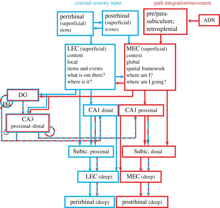
The hippocampus receives its major cortical input from the medial entorhinal cortex (MEC) and the lateral entorhinal cortex (LEC). It is commonly believed that the MEC provides spatial input to the hippocampus, whereas the LEC provides non-spatial input. We review new data which suggest that this simple dichotomy between 'where' versus 'what' needs revision. We propose a refinement of this model, which is more complex than the simple spatial-non-spatial dichotomy. MEC is proposed to be involved in path integration computations based on a global frame of reference, primarily using internally generated, self-motion cues and external input about environmental boundaries and scenes; it provides the hippocampus with a coordinate system that underlies the spatial context of an experience. LEC is proposed to process information about individual items and locations based on a local frame of reference, primarily using external sensory input; it provides the hippocampus with information about the content of an experience.

Keywords: episodic memory; lateral entorhinal cortex; medial entorhinal cortex; memory; path integration.


Figures

Figure 1.
Figure 1.
Parallel processing streams into the hippocampus. This wiring diagram is a simplified version of the real anatomy, leaving out a number of projections. The structure of the diagram emphasizes the dual processing streams that pass through the LEC and MEC. Prior diagrams of these processing streams stressed their origins in the perirhinal-LEC and postrhinal-MEC connections [–8]. Here, we add the critical connectivity between the MEC and limbic regions involved in movement, location and head direction processing (presubiculum, parasubiculum, retrosplenial cortex and anterior dorsal thalamus). (A different subregion of the parasubiculum projects to the LEC, not shown on this diagram [9].) The LEC and MEC connect to distinct regions of CA1 and subiculum, segregated along the transverse axis of the hippocampus (proximal–distal relative to the DG). CA1 and subiculum send return projections to the deep layers of the entorhinal cortex (EC), completing a processing loop. There is crosstalk along these pathways, both prior to their entry into the hippocampus and especially in the convergent projections to the DG and CA3. Although this article does not discuss DG and CA3 properties in detail, these areas are included in the diagram because they are major components of the classic ‘trisynaptic loop’ circuit of the hippocampus and it is important to place this circuit within the larger context of the MEC–LEC parallel streams. In this illustration, the DG and CA3 is represented as a ‘side loop’ of processing, in which the MEC and LEC streams are merged onto the same CA3 pyramidal cells and DG granule cells and the combined representations are then merged in CA1 with the separate input streams from the direct EC–CA1 projections. Specific mnemonic properties of the DG and CA3 regions are thought to be supported by the recurrent feedback loops represented by the dashed circles. In CA3, the recurrent connections are more prominent in the distal than the proximal regions [10]; moreover, the distal CA3 projects more strongly to proximal CA1 (which receives MEC input), and proximal CA3 projects more strongly to distal CA1 (which receives LEC input). The DG receives feedback from CA3 [11], and a disynaptic recurrent loop via mossy cells of the hilus is also present [12] For a more detailed explanation and references to the primary literature on these anatomical connections, the reader is referred to a number of review articles [,–17]. ADN, anterior dorsal nucleus of the thalamus; DG, dentate gyrus; Hil, hilus; Subic., subiculum. (Online version in colour.)

References

    1. O'Keefe J, Nadel L. 1978. The hippocampus as a cognitive map. Oxford, UK: Clarendon Press.
    1. O'Keefe J. 1976. Place units in the hippocampus of the freely moving rat . Exp. Neurol. 51, 78–109. ( 10.1016/0014-4886(76)90055-8) - DOI - PubMed
    1. O'Keefe J. 1999. Do hippocampal pyramidal cells signal non-spatial as well as spatial information? Hippocampus 9, 352–364. ( 10.1002/(SICI)1098-1063(1999)9:4<352::AID-HIPO3>3.0.CO;2-1) - DOI - PubMed
    1. Shapiro ML, Eichenbaum H. 1999. Hippocampus as a memory map: synaptic plasticity and memory encoding by hippocampal neurons . Hippocampus 9, 365–384. ( 10.1002/(SICI)1098-1063(1999)9:4<365::AID-HIPO4>3.0.CO;2-T) - DOI - PubMed
    1. Burwell RD. 2000. The parahippocampal region: corticocortical connectivity . Ann. NY Acad. Sci. 911, 25–42. ( 10.1111/j.1749-6632.2000.tb06717.x) - DOI - PubMed

Publication types

LinkOut - more resources