Review
. 2013 Dec 23;369(1635):20130409.
doi: 10.1098/rstb.2013.0409. Print 2014 Feb 5.

The development of spatial behaviour and the hippocampal neural representation of space

Affiliations
Review

The development of spatial behaviour and the hippocampal neural representation of space

Thomas J Wills et al. Philos Trans R Soc Lond B Biol Sci. .

Abstract

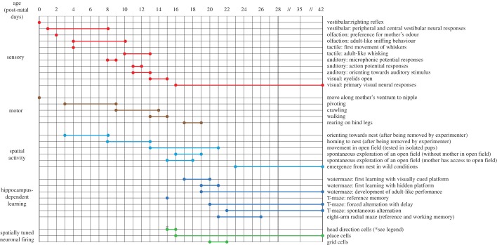
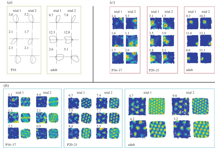
The role of the hippocampal formation in spatial cognition is thought to be supported by distinct classes of neurons whose firing is tuned to an organism's position and orientation in space. In this article, we review recent research focused on how and when this neural representation of space emerges during development: each class of spatially tuned neurons appears at a different age, and matures at a different rate, but all the main spatial responses tested so far are present by three weeks of age in the rat. We also summarize the development of spatial behaviour in the rat, describing how active exploration of space emerges during the third week of life, the first evidence of learning in formal tests of hippocampus-dependent spatial cognition is observed in the fourth week, whereas fully adult-like spatial cognitive abilities require another few weeks to be achieved. We argue that the development of spatially tuned neurons needs to be considered within the context of the development of spatial behaviour in order to achieve an integrated understanding of the emergence of hippocampal function and spatial cognition.

Keywords: development; grid cell; hippocampus; navigation; place cell; spatial.


Figures

Figure 1.
Figure 1.
Summary timeline of sensory, motor and spatial cognition development in the rat. Each horizontal line represents the development of a particular spatial behaviour, motor ability or sensory function. Vertical lines indicate age between postnatal days 0 and 42 (P0–P42); note the compressed horizontal scale between P28 and P42. Bold horizontal lines (ending in circles) represent the beginning and the end of the developmental emergence of each trait. Single circles indicate the existence of a spatial behaviour, motor ability or sensory function at a given age. Different colours indicate different developmental trends: red, sensory; brown, motor; light blue, spontaneous spatial activity; dark blue, hippocampus-dependent spatial learning; green, spatially tuned neuronal firing in the hippocampal formation. Asterisk denotes that the age of the earliest emergence of head direction cells is not yet known. (Online version in colour.)
Figure 2.
Figure 2.
The development of spatially responsive neurons in the hippocampal formation. (a) Three HD cells recorded from medial entorhinal cortex at P16 (left), and from adults (right). Each polar plot represents the firing rate (action potentials per seconds of dwell time) for each directional heading, the peak firing rate is shown at the top-left corner. Each row represents one cell, each column a separate recording trial (separated by 15 min). Note the similarity between P16 and adult HD cells, in terms of directional selectivity and the stability of the preferred direction of firing. (b) Medial entorhinal cortex cells recorded at P16–17, P20–21 and from adult rats. Each row shows a cell, each pair of columns a trial. For each trial, the left column shows a false-colour firing rate map, the right column a spatial auto-correlogram of the firing rate map, which highlights hexagonal grid structure. Peak rate (action potentials per seconds of dwell time) is shown in the top-left corner of the rate map. Note the absence of stable and regularly symmetrical grid firing fields at P16–17. Note also that adult recording arenas are larger than those for immature rats; rate maps are shown to scale. (c) Three place cells recorded from CA1 at P16–17, P20–21 and from adult rats. Each row shows a cell, each column a trial. Peak rate (action potentials per seconds of dwell time) is shown in the top-left corner of the rate map. Note the gradual improvement in the specificity of spatial tuning, and the stability of the place field position. (Online version in colour.)

Similar articles

Cited by

References

    1. O'Keefe J, Nadel L. 1978. The hippocampus as a cognitive map. Oxford, UK: Oxford University Press.
    1. Morris RGM, Garrud P, Rawlins JN, O'Keefe J. 1982. Place navigation impaired in rats with hippocampal lesions. Nature 297, 681–683. (10.1038/297681a0) - DOI - PubMed
    1. Packard MG, McGaugh JL. 1996. Inactivation of hippocampus or caudate nucleus with lidocaine differentially affects expression of place and response learning. Neurobiol. Learn. Mem. 65, 65–72. (10.1006/nlme.1996.0007) - DOI - PubMed
    1. Burgess N, Maguire EA, O'Keefe J. 2002. The human hippocampus and spatial and episodic memory. Neuron 35, 625–641. (10.1016/S0896-6273(02)00830-9) - DOI - PubMed
    1. Rodriguez F, Lopez JC, Vargas JP, Gomez Y, Broglio C, Salas C. 2002. Conservation of spatial memory function in the pallial forebrain of reptiles and ray-finned fishes. J. Neurosci. 22, 2894–2903. - PMC - PubMed

Publication types

LinkOut - more resources