. 2014 Feb 14;114(4):650-9.
doi: 10.1161/CIRCRESAHA.114.302884. Epub 2013 Dec 23.

Preclinical evaluation of the engineered stem cell chemokine stromal cell-derived factor 1α analog in a translational ovine myocardial infarction model

Affiliations

Preclinical evaluation of the engineered stem cell chemokine stromal cell-derived factor 1α analog in a translational ovine myocardial infarction model

John W Macarthur Jr et al. Circ Res. .

Abstract

Rationale: After myocardial infarction, there is an inadequate blood supply to the myocardium, and the surrounding borderzone becomes hypocontractile.

Objective: To develop a clinically translatable therapy, we hypothesized that in a preclinical ovine model of myocardial infarction, the modified endothelial progenitor stem cell chemokine, engineered stromal cell-derived factor 1α analog (ESA), would induce endothelial progenitor stem cell chemotaxis, limit adverse ventricular remodeling, and preserve borderzone contractility.

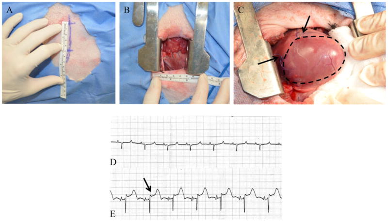
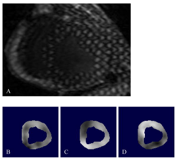
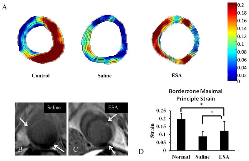
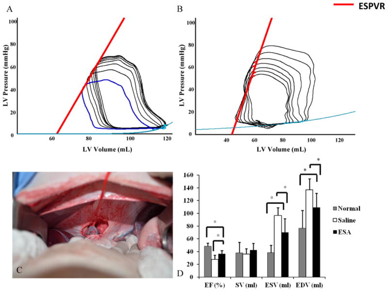
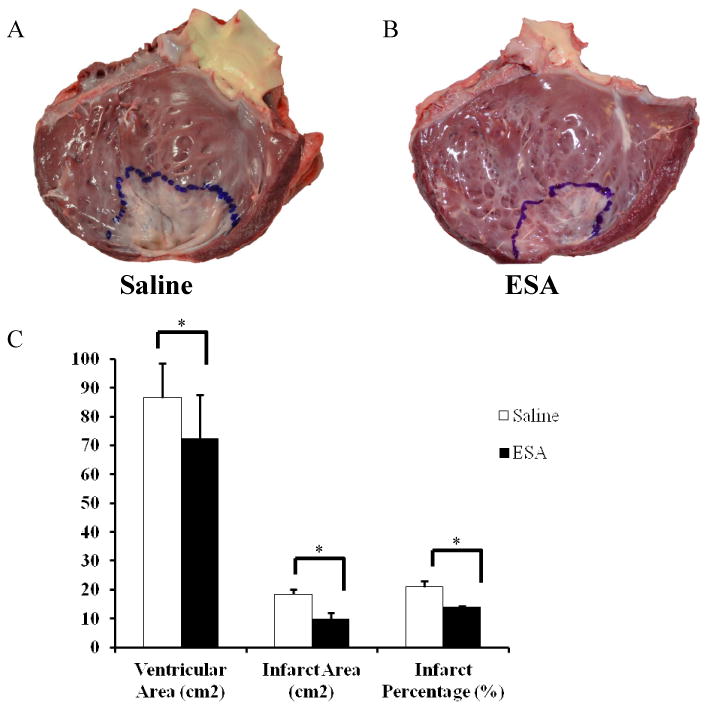
Methods and results: Thirty-six adult male Dorset sheep underwent permanent ligation of the left anterior descending coronary artery, inducing an anteroapical infarction, and were randomized to borderzone injection of saline (n=18) or ESA (n=18). Ventricular function, geometry, and regional strain were assessed using cardiac MRI and pressure-volume catheter transduction. Bone marrow was harvested for in vitro analysis, and myocardial biopsies were taken for mRNA, protein, and immunohistochemical analysis. ESA induced greater chemotaxis of endothelial progenitor stem cells compared with saline (P<0.01) and was equivalent to recombinant stromal cell-derived factor 1α (P=0.27). Analysis of mRNA expression and protein levels in ESA-treated animals revealed reduced matrix metalloproteinase 2 in the borderzone (P<0.05), with elevated levels of tissue inhibitor of matrix metalloproteinase 1 and elastin in the infarct (P<0.05), whereas immunohistochemical analysis of borderzone myocardium showed increased capillary and arteriolar density in the ESA group (P<0.01). Animals in the ESA treatment group also had significant reductions in infarct size (P<0.01), increased maximal principle strain in the borderzone (P<0.01), and a steeper slope of the end-systolic pressure-volume relationship (P=0.01).

Conclusions: The novel, biomolecularly designed peptide ESA induces chemotaxis of endothelial progenitor stem cells, stimulates neovasculogenesis, limits infarct expansion, and preserves contractility in an ovine model of myocardial infarction.

Keywords: bioengineering; magnetic resonance imaging; myocardial infarction; translational research.


Figures

Figure 1
Figure 1. Operative Exposure and Induction of MI
A left anterior, 5 cm minithoracotomy was used to enter the chest and expose the heart (A, B). C, The LAD and D2 branches were ligated with polypropylene suture, creating an anteroapical infarct (black arrows denote site of suture ligation, infarct outlined in black). Pre (D) and post (E) ligation ECGs were recorded and printed. ST segment elevation (black arrow in panel E) confirmed transmural infarction in all animals.
Figure 2
Figure 2. 3D SPAMM Acquisition and the Optical Flow Technique
A, SPAMM creates a periodic modulation of magnetization along a desired direction by applying a series of non-selective excitation pulses, creating tagged lines that move with the myocardium, providing a 3D sample of cardiac motion. This is a representative cross sectional 3D SPAMM tagged image of the LV, where three tag planes are utilized; two through plane tags and one oblique tag plane. Blood appears black and the myocardium is bright. In order to measure systolic regional strain, the LV myocardium was contoured to create an image mask and a custom optical flow plug-in for Image J was used to calculate x, y, and z displacement flow fields (B,C, and D, respectively).
Figure 3
Figure 3. Transwell Migration Assay
A, Representative images of modified transwell Boyden chamber migration assay filters demonstrating EPC migration in saline, SDF, and ESA gradients. (B) The graph summarizes EPC migration between groups. *p<0.05, scale bar = 75μm.
Figure 4
Figure 4. LV mRNA and Protein Expression Profile
A-F, mRNA analysis of MMP, TIMP, elastin and transforming growth factor beta (Tgfb) were performed on myocardial biopsies from infarct, borderzone, and remote areas of the LV. Samples were standardized to mRNA of GAPDH (RFU = ratio of fluorescence units). (B) MMP-2 levels are decreased in the borderzone of ESA treated animals and (D) TIMP-1 and elastin levels are increased in the infarct of ESA treated animals. In order to confirm that protein expression correlated to the observed mRNA expression, immunoblots were performed on myocardial biopsies from infarct, borderzone, and remote areas of the LV. Representative immunoblots show (G) decreased expression of MMP-2 in the borderzone and (H) increased expression of TIMP-1 and elastin in the infarct region of ESA treated animals compared to those treated with saline. I, Values are presented as a ratio of β-actin expression. *p<0.05, BZ = borderzone.
Figure 5
Figure 5. Vascular Density and Myocardial Perfusion
Immunohistochemical light microscope images of borderzone myocardium using anti-vWF antibody (A-B) and anti-αSMA antibody (C-D). Analysis of immunofluorescent expression of vWF and αSMA revealed a significant increase in signal in the ESA group compared to saline (E-F, respectively). Coronary angiograms were reviewed and assessed for collateral filling using Rentrop scores in an effort to evaluate whether or not the increased vascularity seen from the immunohistochemical data resulted in increased myocardial perfusion. G, Coronary angiograms from animals in the ESA group had significantly higher Rentrop scores, and hence better collateral filling, compared to angiograms from animals in the saline group. *p<0.05, scale bar = 75μm.
Figure 6
Figure 6. Maximal Principle Strain Analysis
A, Color map of regional maximum principle strain from cross sections of the LV. Representative color maps of hearts from ESA treated animals show elevated (red on color map) regional strain about the borderzone. Hearts from saline treated animals show decreased (green to blue) regional strain about the borderzone. Representative color map of a heart from a normal animal without MI shows a characteristic strain pattern with elevated strain throughout the free wall of the LV. B, Cross sectional late gadolinium enhanced MRI images corresponding to the strain maps from the saline and ESA groups (white arrows delineate boundary of infarct; scar tissue shows up white from gadolinium contrast). C, Regional strain at the borderzone is significantly elevated in hearts from animals treated with ESA compared to hearts from animals treated with saline, and closer to healthy myocardium. *p<0.05.
Figure 7
Figure 7. Hemodynamic Assessment
Pressure-volume loops were obtained during IVC occlusion for variable load data. (A-B) Representative PV loops from animals in the saline group and ESA group. Note the increased slope of the ESPVR (red line) in the ESA group, a pre-load independent measure of myocardial contractility. C, Before PV loop acquisition, a small upper midline laparotomy was performed and the suprahepatic IVC was dissected free and encircled with a vessel loop. This is a representative image of complete IVC occlusion by gentle retraction on the vessel loop. D, Animals in the ESA group had improved EF, lower ESV, and lower EDV compared to animals in the saline group, representing preserved LV function and limited LV remodeling. *p<0.05. EF = ejection fraction, SV = stroke volume, ESV = end systolic volume, EDV = end diastolic volume.
Figure 8
Figure 8. LV Infarct Area
Hearts were explanted and opened longitudinally. The infarct was outlined and photographed for quantification. Representative images of a heart from an animal in the saline group (A) and a heart from an animal in the ESA treated group (B). C, Animals in the ESA group had significantly reduced surface area of the LV and smaller infarct sizes when compared to animals in the saline group. *p<0.05.

Similar articles

Cited by

References

    1. Go AS, Mozaffarian D, Roger VL, Benjamin EJ, Berry JD, Borden WB, Bravata DM, Dai S, Ford ES, Fox CS, Franco S, Fullerton HJ, Gillespie C, Hailpern SM, Heit JA, Howard VJ, Huffman MD, Kissela BM, Kittner SJ, Lackland DT, Lichtman JH, Lisabeth LD, Magid D, Marcus GM, Marelli A, Matchar DB, McGuire DK, Mohler ER, Moy CS, Mussolino ME, Nichol G, Paynter NP, Schreiner PJ, Sorlie PD, Stein J, Turan TN, Virani SS, Wong ND, Woo D, Turner MB American Heart Association Statistics C; Stroke Statistics S. Heart disease and stroke statistics--2013 update: A report from the american heart association. Circulation. 2013;127:e6–e245. - PMC - PubMed
    1. Heidenreich PA, Trogdon JG, Khavjou OA, Butler J, Dracup K, Ezekowitz MD, Finkelstein EA, Hong Y, Johnston SC, Khera A, Lloyd-Jones DM, Nelson SA, Nichol G, Orenstein D, Wilson PW, Woo YJ American Heart Association Advocacy Coordinating C, Stroke C, Council on Cardiovascular R Intervention, Council on Clinical C, Council on E Prevention, Council on A, Thrombosis Vascular B, Council on C, Critical C, Perioperative, Resuscitation, Council on Cardiovascular N, Council on the Kidney in Cardiovascular D, Council on Cardiovascular S, Anesthesia, Interdisciplinary Council on Quality of C, Outcomes R. Forecasting the future of cardiovascular disease in the united states: A policy statement from the american heart association. Circulation. 2011;123:933–944. - PubMed
    1. Yankey GK, Li T, Kilic A, Cheng G, Satpute A, Savai K, Li S, Moainie SL, Prastein D, DeFillipi C, Wu ZJ, Griffith BP. Regional remodeling strain and its association with myocardial apoptosis after myocardial infarction in an ovine model. The Journal of thoracic and cardiovascular surgery. 2008;135:991–998. 998 e991-992. - PubMed
    1. Jackson BM, Gorman JH, Moainie SL, Guy TS, Narula N, Narula J, John-Sutton MG, Edmunds LH, Jr, Gorman RC. Extension of borderzone myocardium in postinfarction dilated cardiomyopathy. Journal of the American College of Cardiology. 2002;40:1160–1167. discussion 1168-1171. - PubMed
    1. Jayasankar V, Woo YJ, Bish LT, Pirolli TJ, Berry MF, Burdick J, Bhalla RC, Sharma RV, Gardner TJ, Sweeney HL. Inhibition of matrix metalloproteinase activity by timp-1 gene transfer effectively treats ischemic cardiomyopathy. Circulation. 2004;110:II180–186. - PubMed

Publication types

MeSH terms

Substances