. 2014 Mar;82(3):1112-22.
doi: 10.1128/IAI.01459-13. Epub 2013 Dec 23.

The Nod1, Nod2, and Rip2 axis contributes to host immune defense against intracellular Acinetobacter baumannii infection

Affiliations

The Nod1, Nod2, and Rip2 axis contributes to host immune defense against intracellular Acinetobacter baumannii infection

Pradeep Bist et al. Infect Immun. 2014 Mar.

Abstract

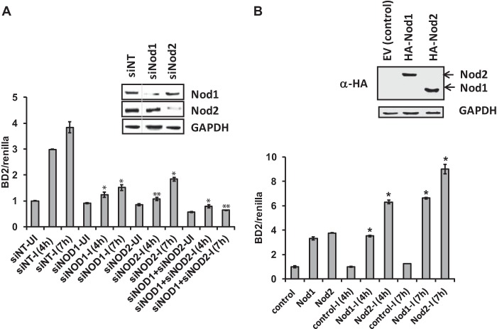
Acinetobacter baumannii is a major extensively drug-resistant lethal human nosocomial bacterium. However, the host innate immune mechanisms controlling A. baumannii are not well understood. Although viewed as an extracellular pathogen, A. baumannii can also invade and survive intracellularly. However, whether host innate immune pathways sensing intracellular bacteria contribute to immunity against A. baumannii is not known. Here, we provide evidence for the first time that intracellular antibacterial innate immune receptors Nod1 and Nod2, and their adaptor Rip2, play critical roles in the sensing and clearance of A. baumannii by human airway epithelial cells in vitro. A. baumannii infection upregulated Rip2 expression. Silencing of Nod1, Nod2, and Rip2 expression profoundly increased intracellular invasion and prolonged the multiplication and survival of A. baumannii in lung epithelial cells. Notably, the Nod1/2-Rip2 axis did not contribute to the control of A. baumannii infection of human macrophages, indicating that they play cell type-specific roles. The Nod1/2-Rip2 axis was needed for A. baumannii infection-induced activation of NF-κB but not mitogen-activated protein kinases. Moreover, the Nod1/2-Rip2 axis was critical to induce optimal cytokine and chemokine responses to A. baumannii infection. Mechanistic studies showed that the Nod1/2 pathway contributed to the innate control of A. baumannii infection through the production of β-defensin 2 by airway epithelial cells. This study revealed new insights into the immune control of A. baumannii and may contribute to the development of effective immune therapeutics and vaccines against A. baumannii.


Figures

FIG 1
FIG 1
Rip2 is important for the innate immune control of A. baumannii infection. (A) A. baumannii infection leads to the transcriptional upregulation of Rip2. Human lung epithelial cell line A549 was infected with A. baumannii for 4 h, and the expression levels of Rip2 were measured using qRT-PCR. The results are expressed as the number of copies of Rip2 mRNA normalized to β-actin (*, P < 0.05 versus I at 0 h). I, infected; UI, uninfected. (B) Infection kinetics of A. baumannii in A549 cells. A549 cells were infected with A. baumannii (MOI, 100) for 2 h and gentamicin treated for 2 h to eliminate the extracellular bacteria. The infection was then allowed to proceed for various times (4 h, 24 h, 48 h, and 72 h). Intracellular bacterial levels were calculated at each time point using a CFU assay as described in Materials and Methods. (C, D, and E) Silencing of Rip2 leads to increased bacterial load in host cells. Rip2 expression was silenced in A549 and NHBE cells using siRNA and standard protocols. Forty-eight h following transfection, the cells were infected with A. baumannii (MOI, 100) for either 30 min (to measure entry) or 2 h and gentamicin treated, and infection was allowed to proceed for various times (4 h, 24 h, and 48 h). Intracellular bacterial levels were calculated at each time point using a CFU assay. (C) Gene silencing was confirmed by measuring Rip2 transcript levels by qRT-PCR. The result is expressed as a percentage of the nontargeting siRNA control (NT) following normalization with β-actin mRNA transcripts. (D and E) Intracellular bacterial load represented as CFU/ml in A549 (P < 0.05 [*] and P < 0.01 [**] versus NT values at the corresponding time points) and NHBE (*, P < 0.05 versus NT) cells, respectively. Untreated represents the sample treated without siRNA. (F) Overexpression of Rip2 made A549 cells resistant to A. baumannii infection. A549 cells were transfected with Rip2-expressing plasmid or vector control plasmid for 24 h, infected with A. baumannii for 2 h, and gentamicin treated. The number of intracellular bacteria was calculated (CFU/ml) at 24 h postinfection (*, P < 0.05 versus the vector control). All of the results are expressed as means ± SD from three independent experiments, and statistical significance was calculated using Student's t test.
FIG 2
FIG 2
Rip2-mediated innate immune response to A. baumannii infection is induced through Nod1 and Nod2. (A, top) Rip2 is required for NF-κB but not MAPK activation during A baumannii infection in A549 cells. Rip2-silenced A549 cells were infected with A. baumannii for various times, and activation of NF-κB and MAPK (p38 and Erk1/2) was determined by Western blotting using phosphospecific antibodies. NT represents the nontargeting siRNA control. GAPDH was used as an internal control. IB, immunoblot. (Bottom) Quantitation of the Western blots using the software ImageJ. Relative densitometric data (respective protein/GAPDH) are shown. Values shown represent means ± standard deviations from results of three experiments (*, P < 0.05 versus siNT values at the corresponding time points). (B and C) Rip2 is required for cytokine and chemokine responses during A. baumannii infection in lung cells. Rip2-silenced A549 cells were infected with A. baumannii for 24 h, and the culture supernatant was collected. IL-8 (B) and TNF-α (C) levels were measured using ELISA (*, P < 0.05 versus NT-I). (D to G) Both Nod1 and Nod2 are involved in immunity to A. baumannii infection. Nod1- and Nod2-silenced A549 cells were infected with A. baumannii, and the intracellular bacterial levels were assessed at various time points postinfection (30 min and 4, 24, and 48 h) using the CFU assay as described in Materials and Methods. (D) Gene silencing was confirmed by measuring Nod1 and Nod2 transcript levels by qRT-PCR. The result is expressed as a percentage of the NT control following normalization with β-actin mRNA transcripts. (E) Intracellular bacterial load represented as CFU/ml (P < 0.05 [*] and P < 0.01 [**] versus NT values at the corresponding time points). The results shown are expressed as means ± SD from a representative experiment performed in triplicate, and statistical significance was calculated using Student's t test. (F) Ectopic expression of Nod1 and Nod2 leads to the enhancement of A. baumannii-induced NF-κB activation in HEK293T cells. Cells were cotransfected with the NF-κB-dependent luciferase reporter, Renilla luciferase (for normalization), and Nod1- or Nod2-expressing plasmids for 24 h. The cells were then stimulated with A. baumannii (MOI, 25 and 100), and the luciferase assay was performed. The results are expressed after normalization with a constitutively expressing Renilla luciferase expression plasmid. The results shown are expressed as means ± SD from a representative experiment performed in triplicate, and statistical significance was calculated using Student's t test (*, P < 0.05). (G, top) Nod1 and Nod2 are required for NF-κB but not MAPK activation during A. baumannii infection in A549 cells. Gene-silenced A549 cells were infected with A. baumannii for various time points, and activation of NF-κB and MAPK (p38 and Erk1/2) was determined by Western blotting using phosphospecific antibodies. GAPDH was used as an internal control. The Western blots shown are representative of three independent experiments. (Bottom) Quantitation of the Western blots using the software ImageJ. Relative densitometric data (respective protein/GAPDH) are shown. Values shown represent means ± standard deviations from results of three experiments (*, P < 0.05 versus siNT values at the corresponding time points). NT-UI, nontargeting siRNA-uninfected sample; NT-I, nontargeting siRNA-infected sample.
FIG 3
FIG 3
A. baumannii infection promotes Nod1/2-Rip2 signalosome assembly. A549 cells were infected with A. baumannii, MDP (positive control for Nod2 pathway stimulation), or iE-DAP (positive control for Nod1 pathway stimulation) for various times. Rip2 was immunoprecipitated using RIP2 antibody (as described in Materials and Methods) and immunoblotted for Nod1, Nod2, and Nemo (IKKγ). (A) Results of immunoprecipitation (top) and quantitation of the Western blots using the software ImageJ (bottom). Relative densitometric data (Rip2 binding to respective proteins) are shown. Values shown represent means ± standard deviations from results of three experiments. (*, P < 0.05 versus siNT values at the corresponding time points). (B) Total cell lysates probed for various proteins. GAPDH was used as a control. The images are representative of three independent experiments.
FIG 4
FIG 4
Cell type-specific role of Nod1/2-Rip2 in the immune control of A. baumannii infection. (A) Nod-Rip2 pathway was dispensable for immune control of A. baumannii in macrophages. Nod1, Nod2, and Rip2 were silenced in human monocytic cell line THP-1 as described in Materials and Methods. The cells were differentiated into macrophages followed by infection with A. baumannii for various times. The number of intracellular bacteria was calculated and is represented as CFU/ml. (B and C) Rip2 pathway was essential for the cytokine/chemokine response by the macrophages following A. baumannii infection. THP-1 cells silenced for Rip2 were infected with A. baumannii for 24 h, and the culture supernatant was collected to measure the levels of IL-8 (B) and TNF-α (C) (*, P < 0.05 versus NT-I). (D) Bone marrow-derived macrophages were isolated from Nod2-deficient mice (Nod2−/−) and age- and sex-matched control mice (WT) using standard protocols. The cells were infected with A. baumannii for various times, and the number of intracellular bacteria was calculated and is represented as CFU/ml. The culture supernatant was collected to measure the levels of MIP-2 (E) and TNF-α (F) (*, P < 0.05 versus WT-I). The results shown are expressed as means ± SD from a representative experiment performed in triplicate. The statistical significance was calculated using Student's t test.
FIG 5
FIG 5
Prestimulation of Nod1/2 pathway enhances the host cellular resistance to A. baumannii infection. A549 cells were pretreated with medium alone (no-MDP sample) or with different concentrations of the Nod2 ligand MDP (10 μg/ml and 50 μg/ml) (A) or Nod1 ligand iE-DAP (10 μg/ml and 50 μg/ml) (B) for 4 h, followed by infection with A. baumannii for 2 h and 2 h of gentamicin treatment. Cells were lysed, and the number of intracellular bacteria was calculated and is represented as CFU/ml. The results shown are expressed as means ± SD from a representative experiment performed in triplicate. The statistical significance was calculated using Student's t test. *, P < 0.05; ***, P < 0.001.
FIG 6
FIG 6
Nod1/2 pathway contributes to beta defensin 2 production during A. baumannii infection. (A) Nod1 and Nod2 genes were silenced in A549 cells. Thirty-six h following knockdown, the cells were cotransfected with BD2 luciferase reporter, Renilla luciferase (for normalization), and Nod1- or Nod2-expressing plasmids for a further 24 h. The cells were then stimulated with A. baumannii (MOI, 100) for 4 h or 7 h, and the luciferase assay was performed. (B) Ectopic expression of Nod1 and Nod2 leads to the enhancement of A. baumannii-induced BD2 activation in HEK293T cells. Cells were cotransfected with the BD2-dependent luciferase reporter Renilla luciferase (for normalization) and with Nod1-, Nod2-, or control vector-expressing plasmids for 24 h. The cells were then stimulated with A. baumannii (MOI, 100) and the luciferase assay was performed. The results are expressed after normalization with a constitutively expressing Renilla luciferase expression plasmid. The results shown are expressed as means ± SD from a representative experiment performed in triplicate, and statistical significance was calculated using Student's t test (P < 0.05 [*] and P < 0.01 [**] versus siNT/control vector values at the corresponding time points).

Similar articles

Cited by

References

    1. McConnell MJ, Actis L, Pachon J. 2012. Acinetobacter baumannii: human infections, factors contributing to pathogenesis and animal models. FEMS Microbiol. Rev. 37:130–155. 10.1111/j.1574-6976.2012.00344.x - DOI - PubMed
    1. Cerqueira GM, Peleg AY. 2011. Insights into Acinetobacter baumannii pathogenicity. IUBMB Life 63:1055–1060. 10.1002/iub.533 - DOI - PubMed
    1. McConnell MJ, Dominguez-Herrera J, Smani Y, Lopez-Rojas R, Docobo-Perez F, Pachon J. 2011. Vaccination with outer membrane complexes elicits rapid protective immunity to multidrug-resistant Acinetobacter baumannii. Infect. Immun. 79:518–526. 10.1128/IAI.00741-10 - DOI - PMC - PubMed
    1. Talbot GH, Bradley J, Edwards JE, Jr, Gilbert D, Scheld M, Bartlett JG. 2006. Bad bugs need drugs: an update on the development pipeline from the Antimicrobial Availability Task Force of the Infectious Diseases Society of America. Clin. Infect. Dis. 42:657–668. 10.1086/499819 - DOI - PubMed
    1. van Faassen H, KuoLee R, Harris G, Zhao X, Conlan JW, Chen W. 2007. Neutrophils play an important role in host resistance to respiratory infection with Acinetobacter baumannii in mice. Infect. Immun. 75:5597–5608. 10.1128/IAI.00762-07 - DOI - PMC - PubMed

Publication types

MeSH terms

LinkOut - more resources