. 2014 Mar 15;592(6):1199-211.
doi: 10.1113/jphysiol.2013.268540. Epub 2013 Dec 23.

Inhibition of cardiac pacemaker channel hHCN2 depends on intercalation of lipopolysaccharide into channel-containing membrane microdomains

Affiliations

Inhibition of cardiac pacemaker channel hHCN2 depends on intercalation of lipopolysaccharide into channel-containing membrane microdomains

Udo Klöckner et al. J Physiol. .

Abstract

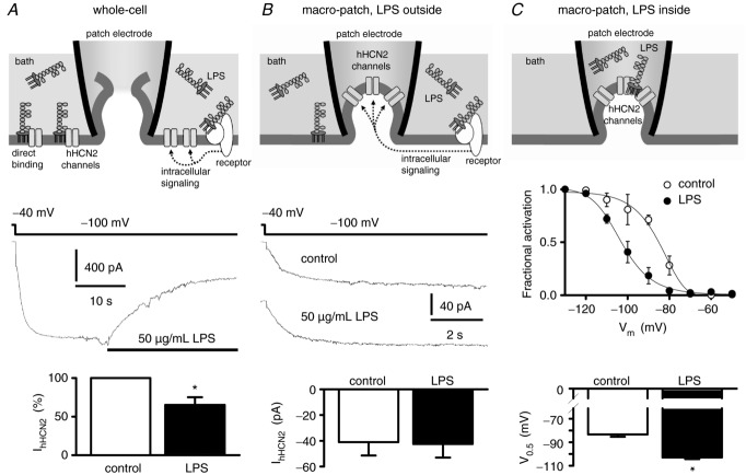
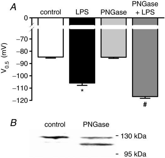
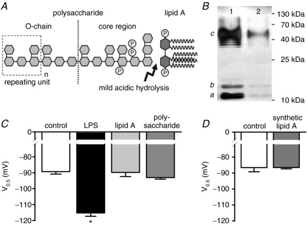
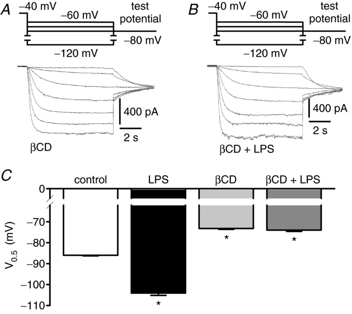
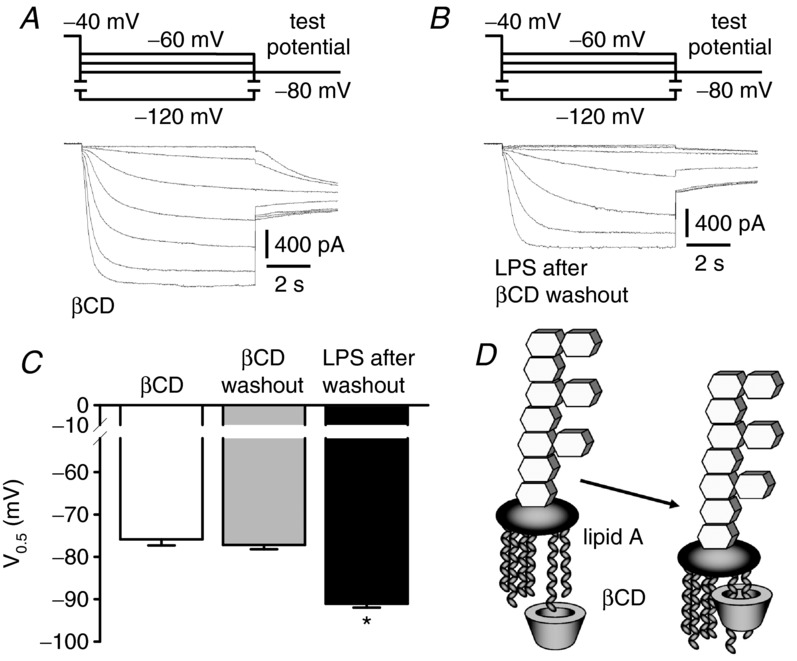
Depressed heart rate variability in severe inflammatory diseases can be partially explained by the lipopolysaccharide (LPS)-dependent modulation of cardiac pacemaker channels. Recently, we showed that LPS inhibits pacemaker current in sinoatrial node cells and in HEK293 cells expressing cloned pacemaker channels, respectively. The present study was designed to verify whether this inhibition involves LPS-dependent intracellular signalling and to identify structures of LPS responsible for pacemaker current modulation. We examined the effect of LPS on the activity of human hyperpolarization-activated cyclic nucleotide-gated channel 2 (hHCN2) stably expressed in HEK293 cells. In whole-cell recordings, bath application of LPS decreased pacemaker current (IhHCN2) amplitude. The same protocol had no effect on channel activity in cell-attached patch recordings, in which channels are protected from the LPS-containing bath solution. This demonstrates that LPS must interact directly with or close to the channel protein. After cleavage of LPS into lipid A and the polysaccharide chain, neither of them alone impaired IhHCN2, which suggests that modulation of channel activity critically depends on the integrity of the entire LPS molecule. We furthermore showed that β-cyclodextrin interfered with LPS-dependent channel modulation predominantly via scavenging of lipid A, thereby abrogating the capability of LPS to intercalate into target cell membranes. We conclude that LPS impairs IhHCN2 by a local mechanism that is restricted to the vicinity of the channels. Furthermore, intercalation of lipid A into target cell membranes is a prerequisite for the inhibition that is suggested to depend on the direct interaction of the LPS polysaccharide chain with cardiac pacemaker channels.


Figures

Figure 1
Figure 1
A, whole-cell configuration. Application of 50 μg ml−1 LPS decreased mean whole-cell current IhHCN2 at a test potential of −100 mV to about 65% (n = 4 each). B, cell-attached recordings (macro-patch) with LPS applied via the bath solution. The patch electrode protects the channels under investigation from direct interaction with LPS. However, LPS remained able to induce receptor-mediated intracellular signalling, thereby modulating channel activity. Representative IhHCN2 traces were recorded in the absence of LPS (control) and after application of 50 μg ml−1 LPS. Mean current amplitude of IhHCN2 did not differ between control and LPS treatments (n = 8 each). C, cell-attached recordings with LPS applied via the pipette solution. Action of LPS is spatially restricted to the vicinity of the channels under investigation. This experimental configuration does not allow for paired experiments. Therefore, activation curves were recorded with or without 50 μg ml−1 LPS included in the pipette solution (n = 3 each). Potential of half-maximal activation (V0.5) is shifted by LPS to more negative potentials. *P < 0.05 versus control.
Figure 2
Figure 2
A, effect of channel deglycosylation on LPS-dependent modulation of IhHCN2. Cells were left untreated (control) or were incubated with peptide N-glycosidase F (PNGase F, 1000 U ml−1, 37°C, 1 h). Mean V0.5 recorded under control conditions (open bar, n = 7), in the presence of 50 μg ml−1 LPS (black bar, n = 7), after incubation of cells with PNGase F (light grey bar, n = 7) and in the presence of 50 μg ml−1 LPS after incubation with PNGase F (dark grey bar, n = 7). LPS-dependent modulation of IhHCN2 is not attenuated by channel deglycosylation. Rather, it sensitizes channels to LPS-dependent modulation because the shift in V0.5 is augmented after PNGase F treatment of cells. B, a representative Western blot of total lysates from control and PNGase F-treated hHCN2-expressing HEK293 cells. The intensity of the upper band is reduced, whereas a lower band appears by PNGase F treatment, indicating channel deglycosylation. *P < 0.05 versus control; #P < 0.05 versus PNGase F; §P < 0.05 versus LPS.
Figure 3
Figure 3
A, schematic chemical structure of lipopolysaccharide (LPS) (according to Rietschel et al. 1994). The arrow indicates the chemical bond, which is cleaved by mild acetic acid hydrolysis, resulting in a separation of the polysaccharide moiety containing the O-chain from the hydrophobic lipid A. B, 7.5 μg of untreated (lane 1) or hydrolysed (lane 2) LPS were subjected to 12% SDS-PAGE and silver-stained. The typical staining pattern of S-type LPS is characterized by intermediates of the full-length LPS representing lipid A with the core oligosaccharide only (a), lipid A and core with one repeating unit (b), and lipid A and core with different numbers of the repeating unit (c) (according to Palva & Makela, 1980). Because only intact LPS can be separated and visualized by this method, the reduction in staining intensity indicates 90% hydrolysis of LPS. C, mean V0.5 recorded under control conditions (open bar, n = 11), in the presence of 50 μg ml−1 LPS (black bar, n = 11) and in the presence of degradation products derived from 50 μg ml−1 LPS exposed to mild acidic hydrolysis (lipid A: light grey bar, n = 10; polysaccharide: dark grey bar, n = 10). Only the intact LPS molecule affects V0.5 of IhHCN2. D, compared to control (open bar, n = 6), V0.5 of IhHCN2 is not modulated by 10 μg ml−1 synthetic lipid A (grey bar, n = 10). *P < 0.05 versus control.
Figure 4
Figure 4
A, current traces recorded after incubation of cells with 10 mm βCD (90 min). B, current traces obtained after co-incubation of cells with 10 mm βCD and 50 μg ml−1 LPS (90 min). C, mean V0.5 recorded under control conditions (open bar, n = 15), in the presence of 50 μg ml−1 LPS (black bar, n = 8), after incubation of cells with 10 mm βCD (60–120 min, light grey bar, n = 10) and after co-incubation of cells with 10 mm βCD and 50 μg ml−1 LPS (60–120 min, dark grey bar, n = 13). βCD shifts V0.5 of IhHCN2 to less negative potentials. However, in the presence of βCD, LPS does not modulate IhHCN2. *P < 0.05 versus control.
Figure 5
Figure 5
A, current traces recorded in the presence of 10 mm βCD. B, current traces recorded in the presence of 50 μg ml−1 LPS, 60 min after washout of βCD. C, mean V0.5 obtained in the presence of 10 mm βCD (white bar, n = 9), 60 min after washout of βCD (grey bar, n = 7) and in the presence of 50 μg ml−1 LPS, 60 min after washout of βCD (black bar, n = 9). The effect of βCD on V0.5 of IhHCN2 is still present 60 min after βCD washout, indicating that cell membranes are still cholesterol-depleted. The LPS-dependent shift of V0.5 to more negative potentials is reduced but still detectable after βCD is washed out. D, schematic diagram of the interaction of the acyl chains of lipid A with the hydrophobic cavity of βCD (according to Sakata et al. 2011). *P < 0.05 versus βCD washout.

Comment in

  • The 'funny' side of sepsis.
    Barbuti A, DiFrancesco D. Barbuti A, et al. J Physiol. 2014 Mar 15;592(6):1171. doi: 10.1113/jphysiol.2014.271171. J Physiol. 2014. PMID: 24634015 Free PMC article. No abstract available.

Similar articles

Cited by

References

    1. Alam JM, Yamazaki M. Spontaneous insertion of lipopolysaccharide into lipid membranes from aqueous solution. Chem Phys Lipids. 2011;164:166–174. - PubMed
    1. Allen JA, Halverson-Tamboli RA, Rasenick MM. Lipid raft microdomains and neurotransmitter signalling. Nat Rev Neurosci. 2007;8:128–140. - PubMed
    1. Barbuti A, Gravante B, Riolfo M, Milanesi R, Terragni B, DiFrancesco D. Localization of pacemaker channels in lipid rafts regulates channel kinetics. Circ Res. 2004;94:1325–1331. - PubMed
    1. Barbuti A, Scavone A, Mazzocchi N, Terragni B, Baruscotti M, DiFrancesco D. A caveolin-binding domain in the HCN4 channels mediates functional interaction with caveolin proteins. J Mol Cell Cardiol. 2012;53:187–195. - PubMed
    1. Baruscotti M, Bucchi A, Viscomi C, Mandelli G, Consalez G, Gnecchi-Rusconi T, Montano N, Casali KR, Micheloni S, Barbuti A, DiFrancesco D. Deep bradycardia and heart block caused by inducible cardiac-specific knock-out of the pacemaker channel gene Hcn4. Proc Natl Acad Sci U S A. 2011;108:1705–1710. - PMC - PubMed

Publication types

MeSH terms

LinkOut - more resources