. 2014 Apr 1;592(7):1505-18.
doi: 10.1113/jphysiol.2013.268342. Epub 2013 Dec 23.

Huntingtin-associated protein 1 regulates exocytosis, vesicle docking, readily releasable pool size and fusion pore stability in mouse chromaffin cells

Affiliations

Huntingtin-associated protein 1 regulates exocytosis, vesicle docking, readily releasable pool size and fusion pore stability in mouse chromaffin cells

Kimberly D Mackenzie et al. J Physiol. .

Abstract

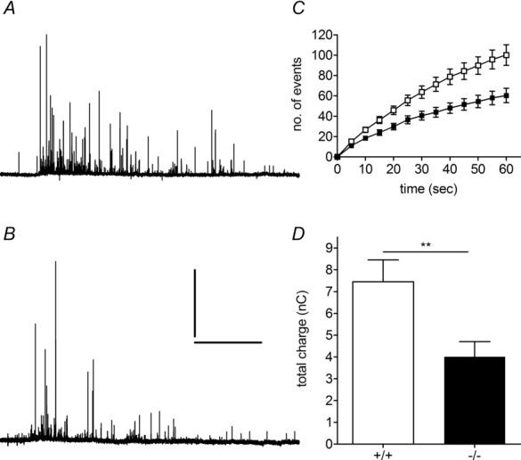
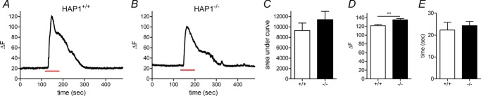
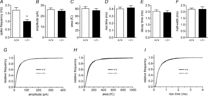
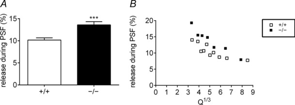
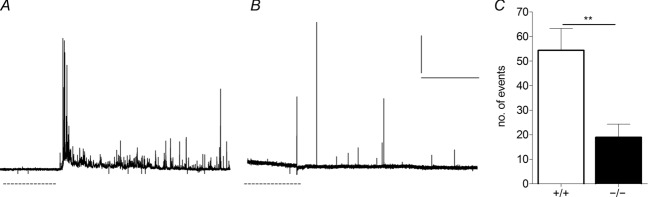
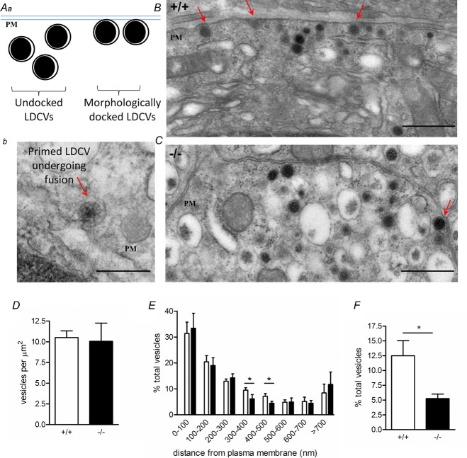
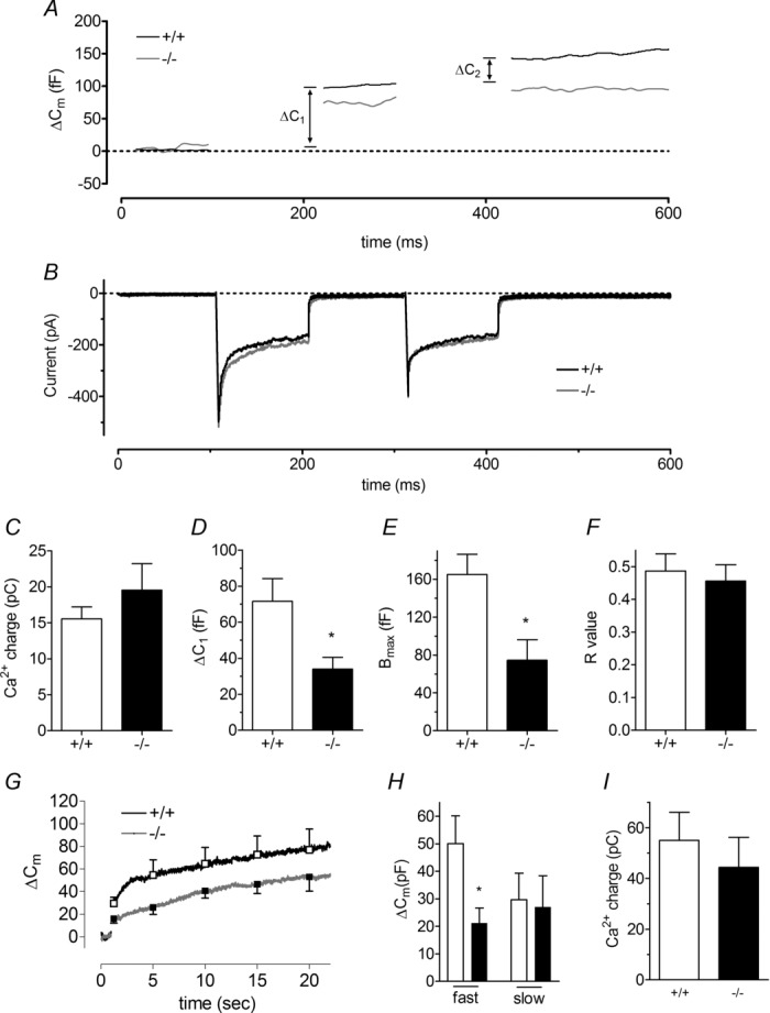
Huntingtin-associated protein 1 (HAP1) was initially established as a neuronal binding partner of huntingtin, mutations in which underlie Huntington's disease. Subcellular localization and protein interaction data indicate that HAP1 may be important in vesicle trafficking and cell signalling. In this study, we establish that HAP1 is important in several steps of exocytosis in adrenal chromaffin cells. Using carbon-fibre amperometry, we measured single vesicle exocytosis in chromaffin cells obtained from HAP1(-/-) and HAP1(+/+) littermate mice. Numbers of Ca(2+)-dependent and Ca(2+)-independent full fusion events in HAP1(-/-) cells are significantly decreased compared with those in HAP1(+/+) cells. We observed no change in the frequency of 'kiss-and-run' fusion events or in Ca(2+) entry. Whereas release per full fusion event is unchanged in HAP1(-/-) cells, early fusion pore duration is prolonged, as indicated by the increased duration of pre-spike foot signals. Kiss-and-run events have a shorter duration, indicating opposing roles for HAP1 in the stabilization of the fusion pore during full fusion and transient fusion, respectively. We use electron microscopy to demonstrate a reduction in the number of vesicles docked at the plasma membrane of HAP1(-/-) cells, where membrane capacitance measurements reveal the readily releasable pool of vesicles to be reduced in size. Our study therefore illustrates that HAP1 regulates exocytosis by influencing the morphological docking of vesicles at the plasma membrane, the ability of vesicles to be released rapidly upon stimulation, and the early stages of fusion pore formation.


Figures

Figure 1
Figure 1
A, B, example amperometric traces from HAP1+/+ (A) and HAP1−/− (B) chromaffin cells. Scale bar in (B) = 20 s/100 pA for (A) and (B). C, rate of HAP1+/+ (white squares, n = 29 cells) and HAP1−/− (black squares, n = 35 cells) exocytosis in chromaffin cells over 1 min. D, average total charge released per cell after 1 min of stimulation in each group (**< 0.01).
Figure 2
Figure 2
A, B, cells were stimulated with 70 mm K+ solution for 1 min (red lines below traces) causing a reversible increase in Fluo-4 fluorescence in single HAP1+/+ (A) and HAP1−/− (B) cells. CE, the average area under the curve (C), peak fluorescence change (D) and time to peak fluorescence (E) were calculated for each genotype (n = 13 cells in each genotype; **P < 0.01).
Figure 3
Figure 3
AF, the number of amperometric spikes per recording is reduced in HAP1−/− cells (A), but no changes are observed in mean spike amplitude (B), area (C), rise time (D), decay time (E) or half-width (F). GI, between-groups comparisons for these parameters show no difference in amplitude (G), area (H) or rise time (I). (**< 0.01.)
Figure 4
Figure 4
A, a full fusion event preceded by a pre-spike foot (PSF) signal (arrow, dotted line indicates PSF duration) representing catecholamine release during fusion pore formation and stabilization. B, when a fusion pore does not stabilize into a full fusion pore but instead reverses and closes, only a stand-alone foot (SAF) signal is observed. C, D, there is no change in the average number of SAF events (C), and SAF events are more frequent in relation to full fusion events in HAP1−/− cells (D). E, F, in HAP1−/− cells, PSF amplitude is unchanged (E), but SAF amplitude is increased (F). G, H, PSF duration is longer in HAP1−/− cells (G), but SAF duration is shorter (H). I, J, in HAP1−/− cells, PSF area is larger (I), but SAF charge is unchanged (J). (*< 0.05, ***< 0.001; n = 523 PSF and n = 186 SAF in HAP1+/+ cells; n = 326 PSF and n = 192 SAF in HAP1−/− cells.)
Figure 5
Figure 5
A, B, the fractional release from the PSF is larger in HAP1−/− cells, as illustrated by the average of the cell mean values (A) and plots of specific kinetic parameters at different values of Q (B).
Figure 6
Figure 6
A, B, HAP1+/+ (A) and HAP1−/− (B) cells were exposed for 10 s to a hyperosmotic solution (dashed line) and rapidly returned to isotonic solution to measure the size of the readily releasable pool (RRP). C, the mean number of vesicles in the RRP is lower in HAP1−/− cells than in HAP1+/+ cells. (**< 0.01; n = 7 HAP1+/+ cells, n = 9 HAP1−/− cells.)
Figure 7
Figure 7
Aa, undocked and morphologically docked large dense core vesicles (LDCVs) at the plasma membrane (PM). Ab, an electron micrograph shows a primed vesicle undergoing fusion. B, C, electron micrographs from HAP1+/+ (B) and HAP1−/− (C) chromaffin cells clearly identify LDCVs; red arrows indicate morphologically docked LDCVs. D, E, there are no changes in the total number of vesicles per unit area (D) or the distance of vesicles from the plasma membrane (in 100 nm bins) except in those 300–500 nm from the PM (E). F, the number of vesicles morphologically docked at the plasma membrane is significantly lower in HAP1−/− cells. (*< 0.05; n = 9 HAP1+/+ cells, n = 7 HAP1−/− cells. Scale bars: Ab, 100 nm; B, C, 500 nm. Data represent the mean ± s.e.m.)
Figure 8
Figure 8
AC, in the absence of HAP1, exocytosis is decreased during a dual-pulse stimulation (A), whereas the size of the integrated Ca2+ current is unchanged (B, C). DF, there is a significant difference in both ΔC1 (D), and the maximal size of the readily releasable pool (RRP) (Bmax) (E), whereas the ratio of ΔC1 to ΔC2 (R) is unchanged (F) (n = 17 HAP1+/+ cells, n = 4 HAP1−/− cells). G, fast (<3 s) and slow (>3 s) phases of exocytosis are triggered by a single 200 ms voltage pulse to 0 mV. H, the fast but not the slow component of exocytosis is significantly reduced in HAP1−/− cells. I, these changes do not reflect reduced Ca2+ current size in HAP1−/− cells. (*< 0.05; n = 6 HAP1+/+ cells, n = 9 HAP1−/− cells.)

Similar articles

Cited by

References

    1. Aminoff MJ, Trenchard A, Turner P, Wood WG, Hills M. Plasma uptake of dopamine and 5-hydroxytryptamine and plasma-catecholamine levels in patients with Huntington's chorea. Lancet. 1974;2:1115–1116. - PubMed
    1. Berberian K, Torres AJ, Fang Q, Kisler K, Lindau M. F-actin and myosin II accelerate catecholamine release from chromaffin granules. J Neurosci. 2009;29:863–870. - PMC - PubMed
    1. Bertaux F, Sharp AH, Ross CA, Lehrach H, Bates GP, Wanker E. HAP1–huntingtin interactions do not contribute to the molecular pathology in Huntington's disease transgenic mice. FEBS Letters. 1998;426:229–232. - PubMed
    1. Bjorkqvist M, Fex M, Renstrom E, Wierup N, Petersen A, Gil J, Bacos K, Popovic N, Li JY, Sundler F, Brundin P, Mulder H. The R6/2 transgenic mouse model of Huntington's disease develops diabetes due to deficient β-cell mass and exocytosis. Hum Mol Genet. 2005;14:565–574. - PubMed
    1. Block-Galarza J, Chase KO, Sapp E, Vaughn KT, Vallee RB, DiFiglia M, Aronin N. Fast transport and retrograde movement of huntingtin and HAP 1 in axons. Neuroreport. 1997;8:2247–2251. - PubMed

Publication types