. 2014 Apr;24(4):400-16.
doi: 10.1038/cr.2013.170. Epub 2013 Dec 24.

USP3 inhibits type I interferon signaling by deubiquitinating RIG-I-like receptors

Affiliations

USP3 inhibits type I interferon signaling by deubiquitinating RIG-I-like receptors

Jun Cui et al. Cell Res. 2014 Apr.

Abstract

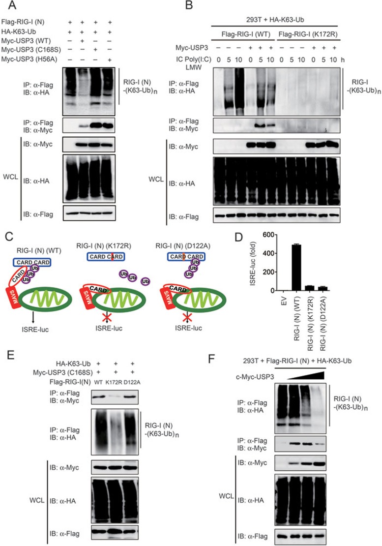
Lysine 63 (K63)-linked ubiquitination of RIG-I plays a critical role in the activation of type I interferon pathway, yet the molecular mechanism responsible for its deubiquitination is still poorly understood. Here we report that the deubiquitination enzyme ubiquitin-specific protease 3 (USP3) negatively regulates the activation of type I interferon signaling by targeting RIG-I. Knockdown of USP3 specifically enhanced K63-linked ubiquitination of RIG-I, upregulated the phosphorylation of IRF3 and augmented the production of type I interferon cytokines and antiviral immunity. We further show that there is no interaction between USP3 and RIG-I-like receptors (RLRs) in unstimulated or uninfected cells, but upon viral infection or ligand stimulation, USP3 binds to the caspase activation recruitment domain of RLRs and then cleaves polyubiquitin chains through cooperation of its zinc-finger Ub-binding domain and USP catalytic domains. Mutation analysis reveals that binding of USP3 to polyubiquitin chains on RIG-I is a prerequisite step for its cleavage of polyubiquitin chains. Our findings identify a previously unrecognized role of USP3 in RIG-I activation and provide insights into the mechanisms by which USP3 inhibits RIG-I signaling and antiviral immunity.


Figures

Figure 1
Figure 1
USP3 negatively regulates the type I IFN signaling pathway. (A, B) Luciferase activity in 293T cells transfected with plasmid encoding a luciferase reporter for IFN-β (IFN-β-luc) (A) or ISRE (ISRE-luc; 100 ng each) (B), together with empty vector (no wedge) or an expression vector for USP3 (0, 200 and 400 ng; wedge), followed by no treatment or treatment with intracellular (IC) low molecular weight (LMW) poly(I:C) (1 μg/ml), IC high molecular weight (HMW) poly(I:C) (1 μg/ml), poly(dA:dT) (1 μg/ml) or VSV-eGFP (MOI = 0.1). Data are presented relative to Renilla luciferase activity. (C) Immunoblot analysis of total and phosphorylated (p-) IRF3 in 293T cells transfected with empty vector or Myc-tagged USP3, followed by the same treatment set as in A. (D) Phase-contrast (PH) and fluorescence microscopy analyses of 293T cells transfected with empty vector (EV) or an expression vector of USP3, and then infected with VSV-eGFP at an MOI of 0.01 at the indicated time. Original magnification, ×10. Data in A, B are presented as the means ± SD of three independent experiments. *P < 0.05, **P < 0.01 and ***P < 0.001, versus cells with the same treatment without USP3 expression (Student's t-test).
Figure 2
Figure 2
Knockdown of Usp3 enhances IFN-β expression as well as antiviral responses. (A) Immunoblot analysis of the knockdown of exogenous USP3 in 293T cells expressing Myc-USP3 (top) or endogenous (endo) USP3 in 293T cells (bottom) treated with Usp3-specific shRNAs or control (Ctrl) shRNA. β-actin serves as a loading control throughout. (B) Immunoblot analysis of total and phosphorylated (p-) IRF3 in 293T cells transfected with control or Usp3-specific shRNA, followed by treatment with intracellular poly(I:C) (LMW or HMW) or infection with VSV-eGFP at different time points. (C) Luciferase activity in 293T cells transfected with Usp3-specific or ctrl shRNA, together with an ISRE luciferase reporter, then left untreated (UT) or treated with intracellular poly(I:C) (LMW), poly(I:C) (HMW), poly(dA:dT) or VSV-eGFP. (D) Real-time PCR analysis of IFNB, IFIT2 and CCL5 mRNA in 293T cells treated with Usp3-specific or control shRNA, followed by the same treatment set as in C. (E) Enzyme-linked immunosorbent assay of IFN-β protein in 293T cells, THP-1 and PBMCs treated with Usp3-specific or control siRNA or shRNA, followed by the VSV-eGFP infection. (F, G) Phase-contrast (PH) and fluorescence microscopy (F) and flow cytometry (G) assessing the infection of 293T cells left untreated or treated with Usp3-specific or control siRNA, and then infected with VSV-eGFP at an MOI of 0.05. Original magnification (F), ×10. Numbers above bracketed lines (G) indicate the percentage of cells expressing eGFP (infected cells). Data in panels C-E are presented as the means ± SD of three independent experiments. *P < 0.05, **P < 0.01 and ***P < 0.001, versus cells transfected with control siRNA (Student's t-test).
Figure 3
Figure 3
USP3 regulates type I IFN signaling through RIG-I and MDA5. (A) Luciferase activity in 293T cells transfected with plasmid encoding a luciferase reporter for ISRE together with empty vector (no wedge) or an expression vector for USP3 (0, 200 and 400 ng; wedge) and RIG-I or MDA5, followed by treatment with intracellular low molecular weight (LMW) poly(I:C) (1 μg/ml) or high molecular weight (HMW) poly(I:C) (1 μg/ml). Results are presented relative to Renilla luciferase activity. (B, C) USP3 inhibits RIG-I-, MDA5-mediated ISRE activation but not MAVS-, TBK1- and IRF3-mediated ISRE activation. 293T cells were transfected with an ISRE luciferase reporter, together with vector for RIG-I (N), MDA5 (N) (B), MAVS, TBK1 or IRF3 (C), along with empty vector (no wedge) or increasing amounts (wedge) of expression vector for USP3. (D) USP3 inhibits RIG-I- and MDA5-mediated activation but not MAVS-mediated activation of the IFN-β promoter. The experiments were similarly performed as in B. (E) Knockdown of USP3 enhances RIG-I- and MDA5-mediated ISRE activation but not MAVS-, TBK1- and IRF3-mediated ISRE activation. 293T cells were transfected with an ISRE luciferase reporter, together with vector for RIG-I (N), MDA5 (N), MAVS, TBK1 or IRF3, along with control (ctrl) shRNA or Usp3-specific shRNA. (F, G) Immunoblot analysis of total and phosphorylated (p-) IRF3 in 293T cells transfected with various combinations (above lanes) of plasmid for Flag-tagged RIG-I (N) or MDA5 (N) plus vector for Myc-tagged USP3 (F) or USP3 shRNA and control shRNA (G). Data in panels A-E are presented as the means ± SD of three independent experiments. *P < 0.05, **P < 0.01 and ***P < 0.001, versus overexpression of RIG-I, MDA5, MAVS, TBK1 or IRF3 alone (Student's t-test).
Figure 4
Figure 4
USP3 interacts with RIG-I and MDA5 after dsRNA stimulation and viral infection. (A) 293T cells were transfected with vectors for Myc-USP3 and Flag-RIG-I, followed by the treatment with intracellular Poly(I:C) and immunoprecipitation (IP) with anti-Flag beads and immunoblot analysis with anti-Myc. WCL, immunoblot analysis of whole-cell lysates without immunoprecipitation (throughout). (B, C) Extracts of THP-1 cells (B) or PBMCs (C) infected with VSV-eGFP for various times (above lanes) or treated with intracellular Poly(I:C) were subjected to immunoprecipitation with anti-USP3 and immunoblot analysis (antibodies, shown on the left). (D, E) Coimmunoprecipitation and immunoblot analysis of 293T cells transfected with deletion mutants of RIG-I (D) or MDA5 (E) along with vector for Myc-USP3.
Figure 5
Figure 5
USP3 inhibits IRF3 activation by removing K63-linked ubiquitin chain on RIG-I. (A) Lysates of 293T cells transfected with plasmid for Flag-RIG-I (N) and HA-tagged ubiquitin (HA-Ub), HA-tagged K63-linked ubiquitin (HA-K63-Ub) or HA-tagged K48-linked ubiquitin (HA-K48-Ub), together with the empty vector or expression vector of Myc-USP3 were immunoprecipitated with anti-Flag and immunoblotted with anti-HA. (B) Lysates of 293T cells transfected with plasmid for HA-K63-Ub and Flag-RIG-I, together with the empty vector or expression vector of Myc-USP3, and treated with intracellular poly(I:C) LMW were subjected to immunoprecipitation with anti-Flag and immunoblot analysis with anti-HA. (C) Lysates of 293T cells transfected with plasmid for HA-K63-Ub, Flag-RIG-I, together with the control shRNA or USP3-specific shRNA, and treated with intracellular poly(I:C) LMW were immunoprecipitated with anti-Flag and immunoblotted with anti-HA. (D) Extracts of THP-1 cells transfected with control or USP3-specific siRNA and infected with VSV-eGFP were subjected to immunoprecipitation with anti-RIG-I and immunoblot analysis with antibody against K63-linked ubiquitin. (E) Ubiquitinated RIG-I was incubated with immunopurified Flag-USP3 in vitro in deubiquitinating buffer. The immunoblot was probed with anti-HA.
Figure 6
Figure 6
Both ZnF and UCH domains are required for USP3-mediated inhibition of type I interferon signaling. (A) Constructs of full-length USP3 (USP3 (FL)) or USP3 containing only ZnF (USP3 (ZnF)) or UCH domain (USP3 (UCH)). (B) Coimmunoprecipitation and immunoblot analysis of 293T cells transfected with various combinations (above lanes) of plasmid for Flag-RIG-I (N), Flag-MDA5 (N) and the Myc-tagged USP3 constructs shown in A. (C) Luciferase activity of 293T cells transfected with expression vector for RIG-I (N) and an ISRE luciferase reporter, together with empty vector (EV) or vectors for the USP3 constructs. (D) Ubiquitinated RIG-I was incubated with immunopurified Flag-USP3, Flag-USP3(C168S) or Flag-USP3 (H56A) in vitro in deubiquitinating buffer. The immunoblot was probed with anti-K63-ubiquitin. (E, F) Luciferase activity of 293T cells treated with intracellular poly(I:C) (E) or transfected with expression vector for RIG-I (N) or MDA5 (N) (F) and an ISRE luciferase reporter, together with empty vector (EV) or vectors for the USP3 mutants. Data in panels C, E and F are reported as the means ± SD of three independent experiments. *P < 0.05, **P < 0.01, ***P< 0.001, versus control cells expressing RIG-I (N) or treated with intracellular poly(I:C) alone.
Figure 7
Figure 7
USP3 binds to the ubiquitinated RIG-I and cleaves the K63-linked ubiquitin chains in a “cut and run” manner. (A) Extracts of 293T cells transfected with various combinations of plasmid for Flag-RIG-I (N), Myc-USP3 or it mutants, and HA-tagged K63 ubiquitin (HA-K63 Ub) were subjected to immunoprecipitation with anti-Flag beads and immunoblot analysis with anti-HA or anti-Myc. (B) Lysates of 293T cells transfected with plasmid for HA-K63-Ub and Flag-RIG-I or Flag-RIG-I (K172R), together with the empty vector or expression vector of Myc-USP3, and treated with intracellular poly(I:C) LMW were immunoprecipitated with anti-Flag and immunoblotted with anti-HA or anti-Myc. (C) Proposed model illustrating how RIG-I CARD and its mutants regulate type I IFN signaling pathways. (D) Luciferase activity of 293T cells transfected with expression vectors for RIG-I (N), RIG-I (N) (K172R) or RIG-I (N) (D122A) and an ISRE luciferase reporter. (E) Extracts of 293T cells transfected with various combinations of plasmid for Myc-USP(C168S), Flag-RIG-I (N) or its mutants, and HA-K63-Ub were subjected to immunoprecipitation with anti-Flag beads and immunoblot analysis with anti-HA or anti-Myc. (F) Extracts of 293T cells transfected with expression plasmid for Flag-RIG-I (N), HA-K63-Ub and increasing amount of Myc-USP3 (wedge) were subjected to immunoprecipitation with anti-Flag beads and immunoblot analysis with anti-HA or anti-Myc.

Similar articles

Cited by

References

    1. Takeuchi O, Akira S. Pattern recognition receptors and inflammation. Cell. 2010;140:805–820. - PubMed
    1. Loo YM, Gale M., Jr Immune signaling by RIG-I-like receptors. Immunity. 2011;34:680–692. - PMC - PubMed
    1. Barbalat R, Ewald SE, Mouchess ML, Barton GM. Nucleic acid recognition by the innate immune system. Annu Rev Immunol. 2011;29:185–214. - PubMed
    1. Chiu YH, Macmillan JB, Chen ZJ. RNA polymerase III detects cytosolic DNA and induces type I interferons through the RIG-I pathway. Cell. 2009;138:576–591. - PMC - PubMed
    1. Ablasser A, Bauernfeind F, Hartmann G, Latz E, Fitzgerald KA, Hornung V. RIG-I-dependent sensing of poly(dA:dT) through the induction of an RNA polymerase III-transcribed RNA intermediate. Nat Immunol. 2009;10:1065–1072. - PMC - PubMed

Publication types