. 2014 Jan;24(1):105-21.
doi: 10.1038/cr.2013.171. Epub 2013 Dec 24.

Translocation of mixed lineage kinase domain-like protein to plasma membrane leads to necrotic cell death

Affiliations

Translocation of mixed lineage kinase domain-like protein to plasma membrane leads to necrotic cell death

Xin Chen et al. Cell Res. 2014 Jan.

Abstract

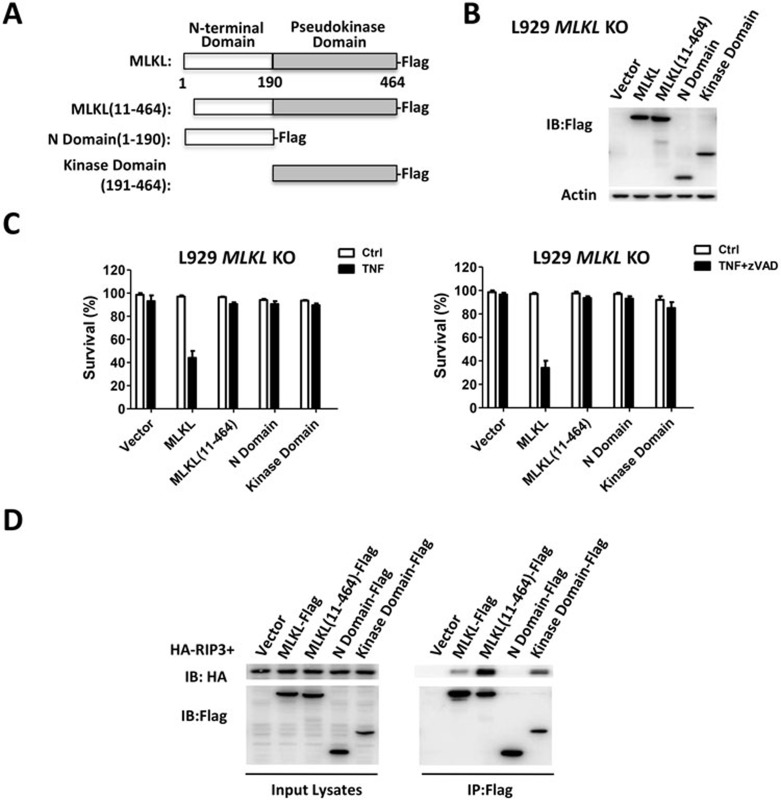
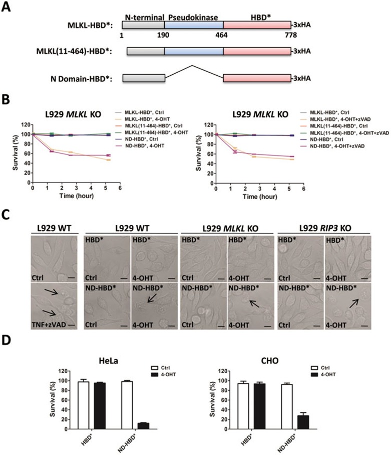
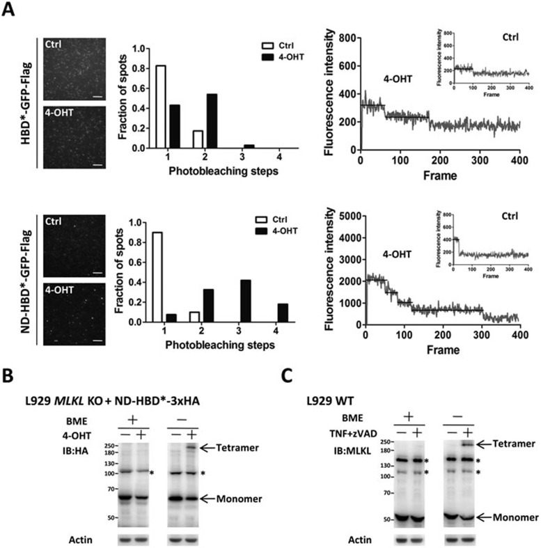
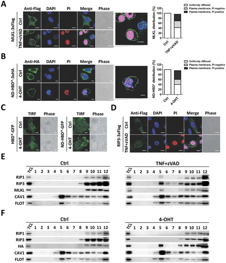
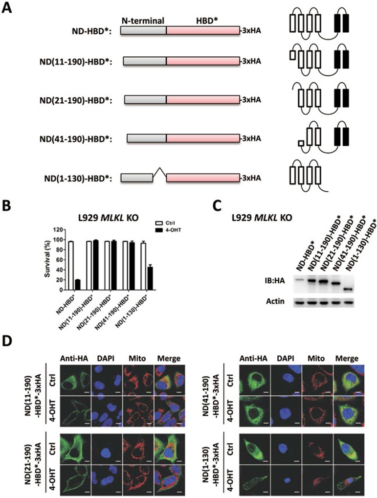
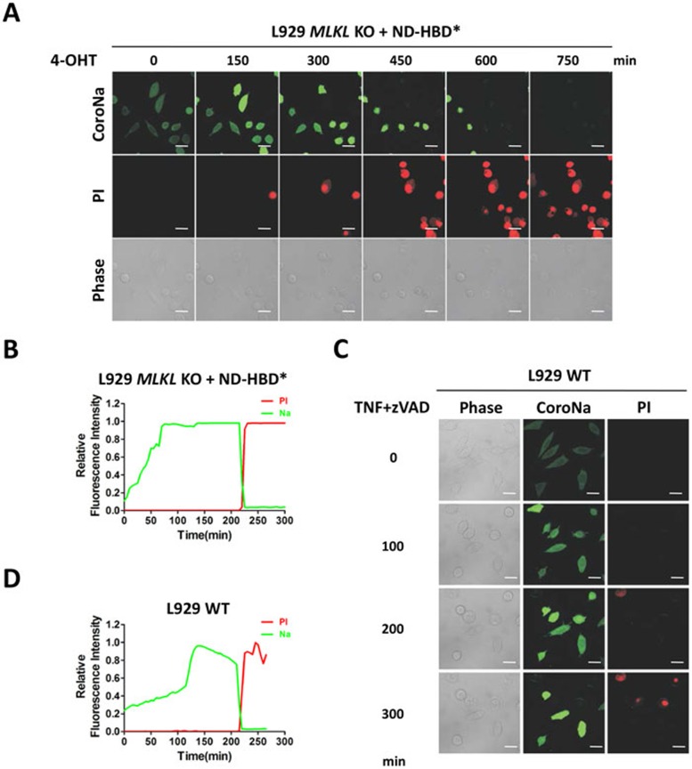
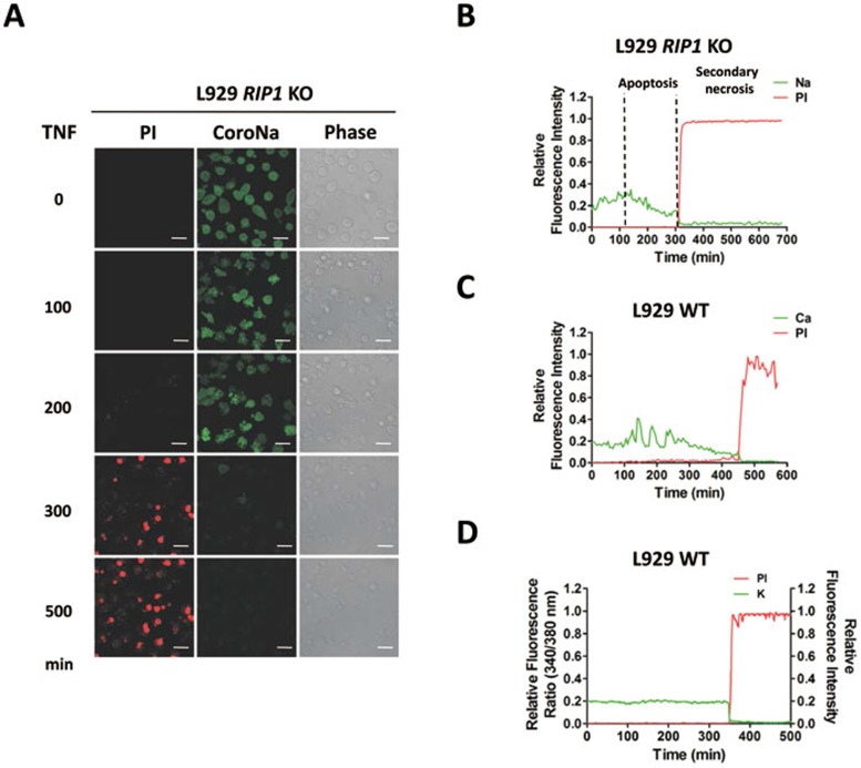
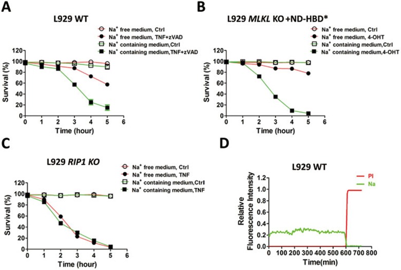
Mixed lineage kinase domain-like protein (MLKL) was identified to function downstream of receptor interacting protein 3 (RIP3) in tumor necrosis factor-α (TNF)-induced necrosis (also called necroptosis). However, how MLKL functions to mediate necroptosis is unknown. By reconstitution of MLKL function in MLKL-knockout cells, we showed that the N-terminus of MLKL is required for its function in necroptosis. The oligomerization of MLKL in TNF-treated cells is essential for necroptosis, as artificially forcing MLKL together by using the hormone-binding domain (HBD*) triggers necroptosis. Notably, forcing together the N-terminal domain (ND) but not the C-terminal kinase domain of MLKL causes necroptosis. Further deletion analysis showed that the four-α-helix bundle of MLKL (1-130 amino acids) is sufficient to trigger necroptosis. Both the HBD*-mediated and TNF-induced complexes of MLKL(ND) or MLKL are tetramers, and translocation of these complexes to lipid rafts of the plasma membrane precedes cell death. The homo-oligomerization is required for MLKL translocation and the signal sequence for plasma membrane location is located in the junction of the first and second α-helices of MLKL. The plasma membrane translocation of MLKL or MLKL(ND) leads to sodium influx, and depletion of sodium from the cell culture medium inhibits necroptosis. All of the above phenomena were not seen in apoptosis. Thus, the MLKL oligomerization leads to translocation of MLKL to lipid rafts of plasma membrane, and the plasma membrane MLKL complex acts either by itself or via other proteins to increase the sodium influx, which increases osmotic pressure, eventually leading to membrane rupture.


Figures

Figure 1
Figure 1
The N-terminus of MLKL is required for its function in necroptosis, and the N-terminal domain (ND) of MLKL is responsible for triggering necroptosis. (A) Schematic representation of full-length and truncated murine MLKL. (B) Lentiviral vector was used to express MLKL and its mutants in MLKL KO L929 cells. The expression of full-length and truncated MLKL proteins was analyzed by immunoblotting with the anti-Flag antibody 48 h after infection. (C) The cells described in B were treated with TNF (10 ng/ml) or TNF + zVAD (20 μM) for 12 h and 4 h, respectively. Viabilities of the cells were measured by PI exclusion. The data represented the mean ± SD of triplicates, and was representative of three independent experiments. (D) Flag-tagged full-length and truncated MLKL were cotransfected with HA-tagged RIP3 in HEK293T cells. The cell lysates were prepared 24 h after transfection and the immunoprecipitations were carried out by using anti-Flag antibody beads. The immunoprecipitates and cell lysates were subjected to immunoblotting with antibodies against HA and Flag.
Figure 2
Figure 2
The HBD*-mediated interaction of MLKL or ND of MLKL can bypass RIP3 and directly induce necroptosis. (A) Schematic representation of full-length and truncated murine MLKL fused with HBD*. (B) Viabilities of MLKL KO L929 cells expressing MLKL-HBD*, MLKL(11-464)-HBD* or ND-HBD* after treatment with or without 4-OHT (1 μM) or 4-OHT + zVAD (20 μM) for different periods of time as indicated. (C) WT, MLKL KO and RIP3 KO L929 cells expressing HBD* or ND-HBD* were treated with or without 4-OHT (1 μM) for 1 h and then imaged using a confocal microscope. WT L929 cells stimulated with or without TNF + zVAD (10 ng/ml + 20 μM) for 4 h were included as a control. The black arrows indicate necrotic cells and the scale bars represent 10 μm. (D) Viabilities of HeLa and CHO cells expressing HBD* or ND-HBD* after treatment with or without 4-OHT (1 μM) for 2 h. Data represented the mean ± SD of triplicates (B, D), and were representative of three independent experiments.
Figure 3
Figure 3
Both 4-OHT-induced oligomerization of ND-HBD* and TNF-induced oligomerization of MLKL produce tetramers. (A) HeLa cells were infected with a lentiviral vector encoding HBD*-GFP-Flag or ND-HBD*-GFP-Flag for 24 h, and then treated with 4-OHT (1 μM) or vehicle (Ctrl) for 1 h. Cells were lysed and subjected to the SiMPull assay as described in Materials and Methods. Left panels are TIRF microscopy images showing HBD*-GFP-Flag and ND-HBD*-GFP-Flag fluorescence spots on the coverslips. Each scale bar represents 5 μm. Examples of observed bleaching steps from HBD*-GFP-Flag- or ND-HBD*-GFP-Flag-expressing cells treated with or without 4-OHT (1 μM) were shown in right panels. The distribution of observed bleaching step numbers from HBD*-GFP-Flag- or ND-HBD*-GFP-Flag-expressing cells treated with or without 4-OHT (1 μM) was summarized in the middle panels. (B) MLKL KO L929 cells expressing ND-HBD*-3×HA were treated with or without 4-OHT (1 μM) for 1 h, and the lysates were subjected to SDS-PAGE with or without β-mercaptoethanol (BME), and analyzed by immunoblotting using the anti-HA antibody. (C) To collect intact cells for analysis, L929 cells were stimulated with or without TNF + zVAD (10 ng/ml + 20 μM) for 3 h, lysed and subjected to reducing or non-reducing SDS-PAGE as in B, and analyzed by immunoblotting using the anti-MLKL antibody. Arrows indicate the monomer or tetramer of ND-HBD*-3×HA or MLKL. The asterisk denotes non-specific bands.
Figure 4
Figure 4
Both MLKL and ND-HBD* translocated to the plasma membrane and were enriched in lipid raft microdomain during necroptosis. (A) MLKL KO L929 cells reconstituted with C-terminal 3×Flag-tagged MLKL were treated with or without TNF + zVAD (10 ng/ml + 20 μM) for 3.5 h, and then immunostained for Flag and counterstained with DAPI. PI was added to culture medium before cell fixation. The merge image of MLKL-3×Flag located in the plasma membrane was zoomed in in the middle panel. The distributions of MLKL in cells are defined as three types: uniformly diffused, plasma membrane location with PI negative and plasma membrane location with PI positive. (B) HeLa cells expressing ND-HBD*-3×HA were treated with or without 4-OHT (1 μM) for 1 h and analyzed as described in A. (C) HeLa cells expressing HBD*-GFP or ND-HBD*-GFP were stimulated with or without 4-OHT (1 μM) for 1 h and then imaged by TIRF microscopy. (D) RIP3 KO L929 cells reconstituted with 3×Flag-tagged RIP3 were treated with or without TNF + zVAD (10 ng/ml + 20 μM) for 3.5 h and analyzed as described in A. (E) L929 cells were treated with or without TNF + zVAD (10 ng/ml + 20 μM) for 2 h, and then the total cell lysates (TCL) were fractionated to isolate lipid rafts as described in Materials and Methods. TCL and the fractions collected were immunoblotted with antibodies against RIP1, RIP3 and MLKL. Lipid raft markers CAV1 and FLOT were immunoblotted to identify lipid raft fractions. (F) MLKL KO L929 cells expressing ND-HBD*-3×HA were treated with or without 4-OHT (1 μM) for 1 h. TCL were then fractionated and analyzed as described in E. Each scale bar indicates 10 μm.
Figure 5
Figure 5
Translocation of the N-terminal four-α-helix bundle of MLKL to plasma membrane is necessary and sufficient for triggering necroptosis, and targeting to plasma membrane is determined by the certain sequence within the α-helix bundle. (A) Schematic representation of full-length and truncated ND-HBD*-3×HA. The corresponding positions of the different deletions in α-helices are shown in the right panels. (B) Viabilities of MLKL KO L929 cells expressing ND-HBD* or truncated ND-HBD* (11-190, 21-190, 41-190, 1-130) as described in A after stimulation with or without 4-OHT (1 μM) for 2 h. Data represented the mean ± SD of triplicates, and were representative of three independent experiments. (C) Expression levels of ND-HBD* and its truncated forms in cells described in B were determined by immunoblotting with the anti-HA antibody. (D) MLKL KO L929 cells reconstituted with full-length or truncated ND-HBD*-3×HA were treated with or without 4-OHT (1 μM) for 2 h and then immunostained with anti-HA antibody and counterstained with DAPI and Mitotracker Red. Each scale bar represents 10 μm.
Figure 6
Figure 6
Sodium influx occurs before cell membrane breakage in necroptosis. (A) Representative time-lapse images of ND-HBD*-expressing MLKL KO L929 cells undergoing necroptosis upon treatment of 4-OHT (1 μM). The intracellular sodium concentration was measured by fluorescence sodium indicator CoroNa Green, and cell membrane integrity was monitored by PI uptake. Elapsed time is indicated above each frame and each scale bar represents 15 μm. (B) Relative changes in CoroNa Green and PI fluorescence over time in one representative cell shown in A were graphed. (C) Representative time-lapse images of L929 cells undergoing necroptosis induced by TNF + zVAD (10 ng/ml + 20 μM). Intracellular sodium concentration and PI uptake were monitored as described in A. Elapsed time is indicated on the left side of each frame and each scale bar represents 15 μm. (D) Relative changes in CoroNa Green and PI fluorescence over time in one representative cell shown in C were graphed.
Figure 7
Figure 7
Sodium influx does not occur in the course of apoptosis, and potassium and calcium influx are not detected during necroptosis of L929 cells. (A) Representative time-lapse images of RIP1 KO L929 cells undergoing apoptosis induced by TNF (10 ng/ml). The intracellular sodium concentration was measured by fluorescence sodium indicator CoroNa Green and cell membrane integrity was monitored by PI uptake. Elapsed time is indicated on the left side of each frame and each scale bar represents 15 μm. (B) Relative changes in CoroNa Green and PI fluorescence over time in one representative cell shown in A were graphed. The stages of apoptosis and secondary necrosis of the selected single cell from A were indicated above dotted lines. (C) L929 cells loaded with calcium indicator Fluo-4, as described in Materials and Methods, were treated with TNF + zVAD (10 ng/ml + 20 μM). Relative changes in calcium indicator Fluo-4 and PI fluorescence over time in one representative cell were graphed. (D) Relative changes in potassium indicator PBFI and PI fluorescence over time in one representative L929 cell were graphed.
Figure 8
Figure 8
Sodium depletion inhibits necroptosis, but not apoptosis. (A) Viabilities of L929 cells treated with or without TNF + zVAD (10 ng/ml + 20 μM) in sodium-free or sodium-containing medium. (B) Viabilities of MLKL KO L929 cells expressing ND-HBD* treated with or without 4-OHT (1 μM) in sodium-free or sodium-containing medium. (C) Viabilities of RIP1 KO L929 cells treated with or without TNF (10 ng/ml) in sodium-free or sodium-containing medium. (D) L929 cells were treated with TNF + zVAD (10 ng/ml + 20 μM) in sodium-free medium, and relative changes in CoroNa Green and PI fluorescence over time in one representative cell were graphed. Data shown represented the mean ± SD of triplicates (A-C), and were representative of three independent experiments.

Similar articles

Cited by

References

    1. Laster SM, Wood JG, Gooding LR. Tumor necrosis factor can induce both apoptic and necrotic forms of cell lysis. J Immunol. 1988;141:2629–2634. - PubMed
    1. Vandenabeele P, Galluzzi L, Vanden Berghe T, Kroemer G. Molecular mechanisms of necroptosis: an ordered cellular explosion. Nat Rev Mol Cell Biol. 2010;11:700–714. - PubMed
    1. Christofferson DE, Yuan J. Necroptosis as an alternative form of programmed cell death. Curr Opin Cell Biol. 2010;22:263–268. - PMC - PubMed
    1. Degterev A, Huang Z, Boyce M, et al. Chemical inhibitor of nonapoptotic cell death with therapeutic potential for ischemic brain injury. Nat Chem Biol. 2005;1:112–119. - PubMed
    1. Haas TL, Emmerich CH, Gerlach B, et al. Recruitment of the linear ubiquitin chain assembly complex stabilizes the TNF-R1 signaling complex and is required for TNF-mediated gene induction. Mol Cell. 2009;36:831–844. - PubMed

Publication types

MeSH terms