Review
. 2014 May 1;123(18):2768-76.
doi: 10.1182/blood-2013-10-463646. Epub 2013 Dec 23.

Thrombosis: tangled up in NETs

Affiliations
Review

Thrombosis: tangled up in NETs

Kimberly Martinod et al. Blood. .

Abstract

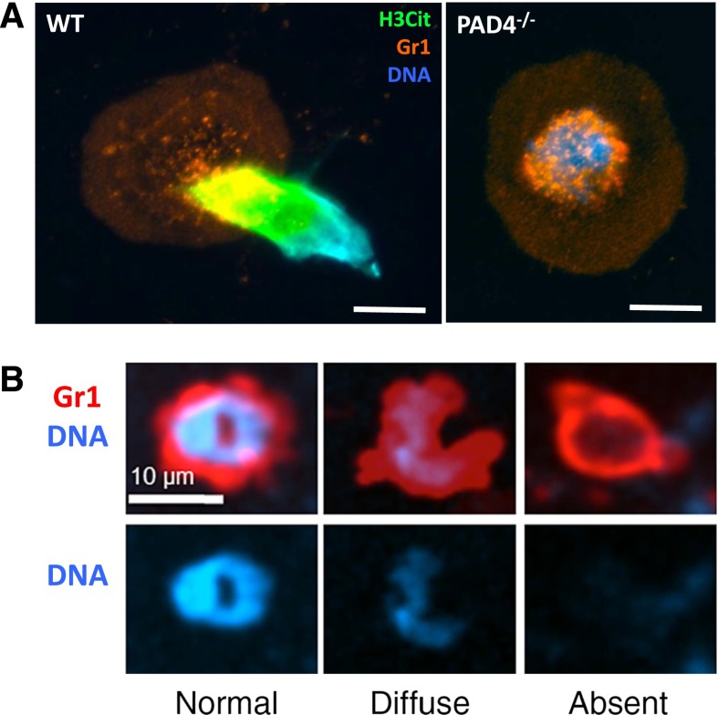
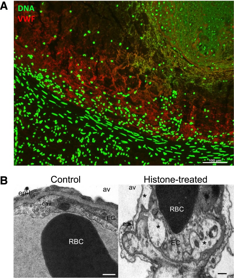
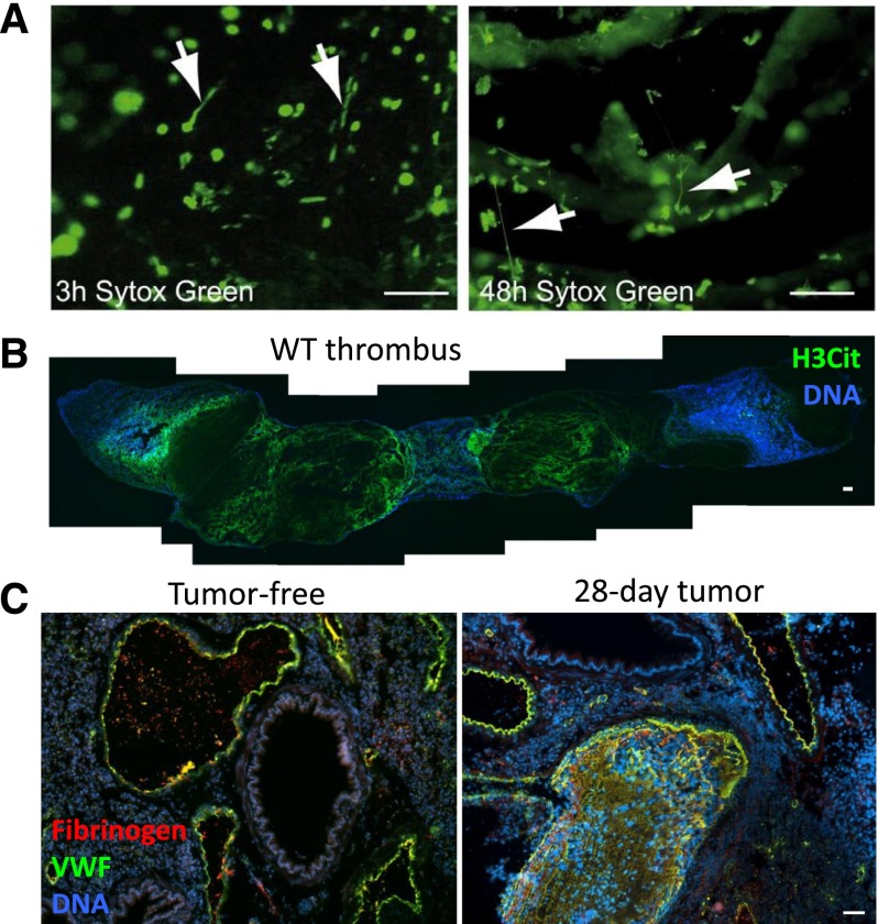
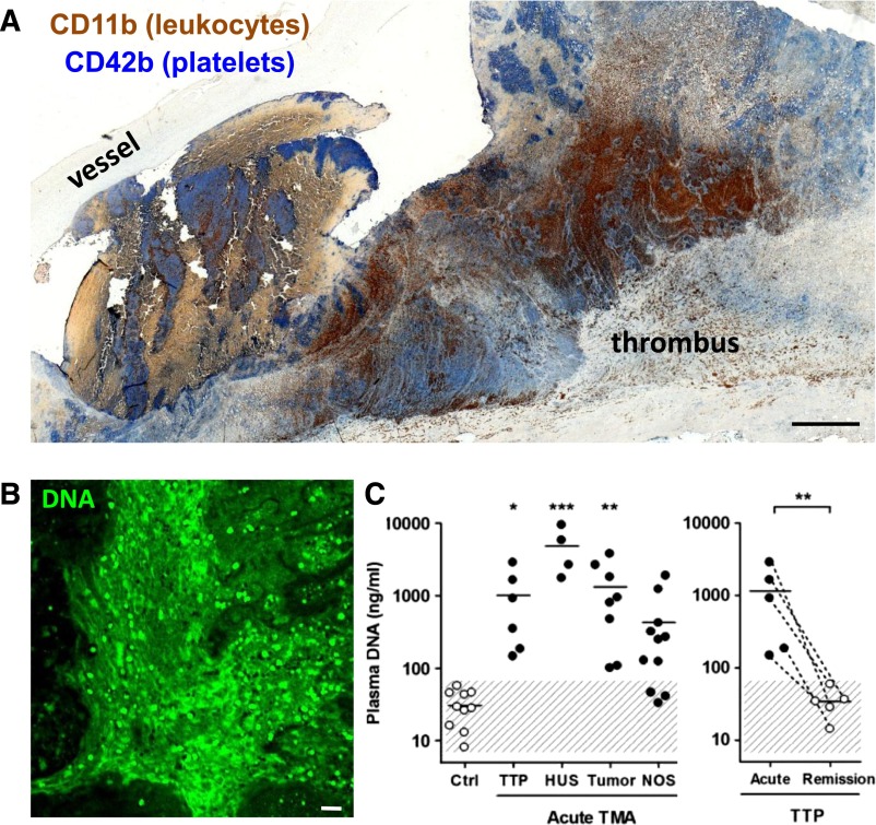
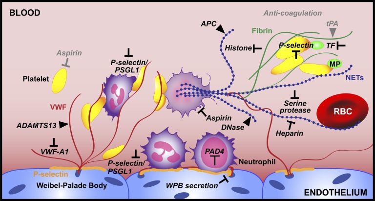
The contributions by blood cells to pathological venous thrombosis were only recently appreciated. Both platelets and neutrophils are now recognized as crucial for thrombus initiation and progression. Here we review the most recent findings regarding the role of neutrophil extracellular traps (NETs) in thrombosis. We describe the biological process of NET formation (NETosis) and how the extracellular release of DNA and protein components of NETs, such as histones and serine proteases, contributes to coagulation and platelet aggregation. Animal models have unveiled conditions in which NETs form and their relation to thrombogenesis. Genetically engineered mice enable further elucidation of the pathways contributing to NETosis at the molecular level. Peptidylarginine deiminase 4, an enzyme that mediates chromatin decondensation, was identified to regulate both NETosis and pathological thrombosis. A growing body of evidence reveals that NETs also form in human thrombosis and that NET biomarkers in plasma reflect disease activity. The cell biology of NETosis is still being actively characterized and may provide novel insights for the design of specific inhibitory therapeutics. After a review of the relevant literature, we propose new ways to approach thrombolysis and suggest potential prophylactic and therapeutic agents for thrombosis.


Figures

Figure 1
Figure 1
NETosis is a regulated process. (A) Representative image of a WT or PAD4−/− neutrophil stimulated with calcium ionophore. WT neutrophils undergo histone hypercitrullination (H3Cit, green) and throw NETs, whereas PAD4−/− neutrophils fail to citrullinate histones, decondense chromatin, or release NETs. Reproduced from Martinod et al. Scale bars, 10 μm. (B) In response to S aureus skin infection, neutrophils can secrete their nuclear contents (right) while retaining the ability to crawl and phagocytose, thus multitasking. Reproduced from Yipp et al with permission.
Figure 2
Figure 2
NETs are part of deep vein thrombi and histones produce toxicity in vivo. (A) A deep vein thrombus was formed in an otherwise healthy baboon by balloon catheterization. The thrombus was excised and analyzed for the presence of extracellular DNA (green) and von Willebrand factor (red), which were found to codistribute. Scale bar, 100 μm. Reproduced from Fuchs et al. (B) Intravenous histone infusion was detrimental to both endothelium and epithelium, as shown by vacuolization (stars) of these cells in and around the lung capillaries (right). Av, alveolae. Cav, caveolae. Ep-I, type I epithelial cell. EC, endothelial cell. Scale bars, 500 nm. Reproduced from Xu et al with permission.
Figure 3
Figure 3
NETs form in mouse models of thrombosis and cancer. (A) Intravital microscopy of developing thrombi shows the release of NETs early (3 hours) and more prominently in occlusive thrombi (48 hours). Arrows indicate NETs. Sytox Green, DNA. Scale bars, 50 μm. Reproduced from von Bruhl et al. (B) Composite image of a thrombus formed in a WT mouse 48 hours after IVC stenosis. Mosaic generated using MosaicJ plug-in for ImageJ software. Citrullinated histone H3 (H3Cit) staining (green) shows evidence of a NET meshwork throughout the red portion of the thrombus. Scale bar, 100 μm. (C) Mice bearing a mammary carcinoma develop spontaneous thrombi in the lung (right) after 28 days, a time point when NETs are spontaneously generated in these mice. This does not occur in tumor-free mice (left). VWF, green. Fibrinogen, red. DNA, blue. Scale bar, 50 μm. Reproduced from Demers et al.
Figure 4
Figure 4
Evidence of NETs in human pathological thrombosis. (A) Composite image of a human pulmonary embolism specimen obtained surgically and stained by immunohistochemistry for platelets (blue) and leukocytes (brown) showing that areas of the thrombus are rich in both cell types. Scale bar, 100 μm. Mosaic generated using MosaicJ plug-in for ImageJ. Immunohistochemical analysis by Alexander Savchenko. (B) Representative image of diffuse extracellular DNA staining (green) present in a surgically harvested pulmonary embolism patient specimen. Green, DNA. Scale bar, 20 μm. Anonymous specimens in A and B kindly provided by Richard Mitchell. Most recently, specimens from 11 additional patients were evaluated and biomarkers of NETs were predominantly found in the organizing stage of human venous thromboembolism. (C) Cell-free DNA, a plasma biomarker of NETs, is elevated in patients with thrombotic microangiopathies (left): thrombotic thrombocytopenic purpura (TTP), hemolytic uremic syndrome (HUS), malignancies (tumor), and nonspecified cases (NOS). Patients suffering from acute TTP present significantly elevated plasma DNA compared with when in remission (right). This research was originally published in Blood. Fuchs TA et al. Circulating DNA and myeloperoxidase indicate disease activity in patients with thrombotic microangiopathies. Blood. 2012;120:1157-1164. © American Society of Hematology. Human thrombus samples were obtained by the Wagner Laboratory from Dr Richard N. Mitchell (Brigham and Women's Hospital, Boston, MA) as anonymous tissue specimens. Dr Mitchell's work was approved by Brigham and Women's Hospital Institutional Review Board 2013P000231.
Figure 5
Figure 5
Emerging targets for thrombus prevention and thrombolysis. Here we summarize advances in the field of thrombosis with respect to neutrophil recruitment and NETosis and pinpoint targets that should be investigated as potential therapeutics (black). Existing treatments are in gray. We propose PAD4 inhibition as a way to prevent NET release. For thrombi that have already formed, neutralizing the toxic components of NETs is key. Thrombolytic strategies should involve the targeting of both DNA (blue) and the protein elements (red and green) of the thrombus scaffold.

Similar articles

Cited by

References

    1. Brinkmann V, Reichard U, Goosmann C, et al. Neutrophil extracellular traps kill bacteria. Science. 2004;303(5663):1532–1535. - PubMed
    1. Urban CF, Ermert D, Schmid M, et al. Neutrophil extracellular traps contain calprotectin, a cytosolic protein complex involved in host defense against Candida albicans. PLoS Pathog. 2009;5(10):e1000639. - PMC - PubMed
    1. Urban CF, Reichard U, Brinkmann V, Zychlinsky A. Neutrophil extracellular traps capture and kill Candida albicans yeast and hyphal forms. Cell Microbiol. 2006;8(4):668–676. - PubMed
    1. Narasaraju T, Yang E, Samy RP, et al. Excessive neutrophils and neutrophil extracellular traps contribute to acute lung injury of influenza pneumonitis. Am J Pathol. 2011;179(1):199–210. - PMC - PubMed
    1. Saitoh T, Komano J, Saitoh Y, et al. Neutrophil extracellular traps mediate a host defense response to human immunodeficiency virus-1. Cell Host Microbe. 2012;12(1):109–116. - PubMed

Publication types