Review
. 2014 Jan;127(1):29-51.
doi: 10.1007/s00401-013-1230-6. Epub 2013 Dec 24.

The neuropathology of sport

Affiliations
Review

The neuropathology of sport

Ann C McKee et al. Acta Neuropathol. 2014 Jan.

Abstract

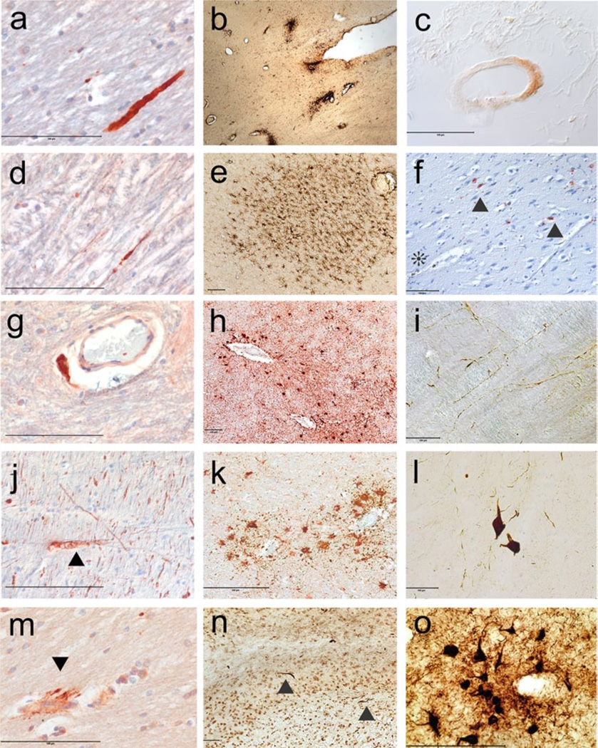
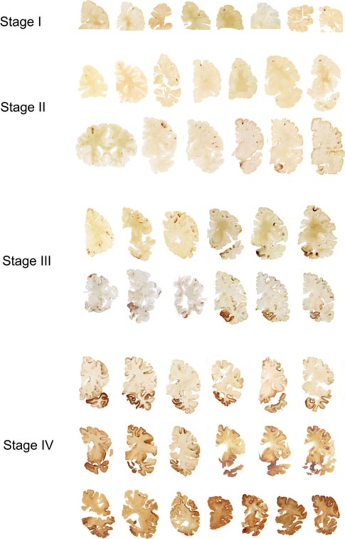
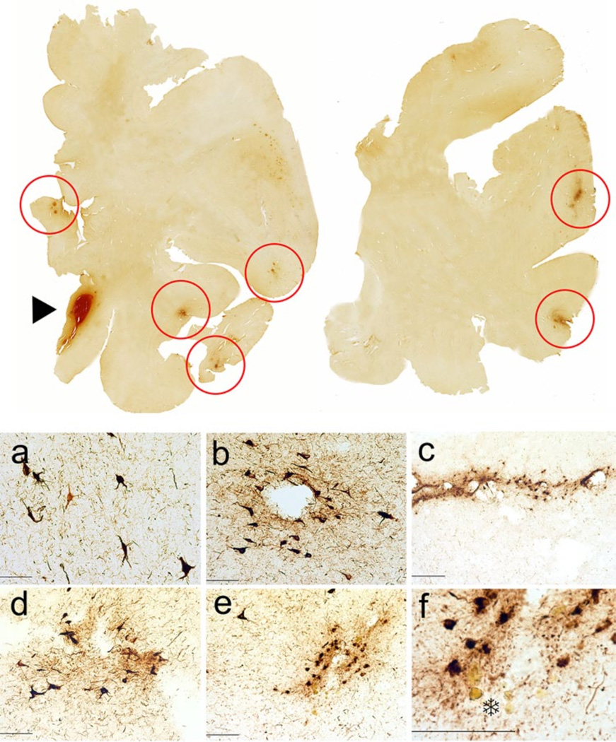
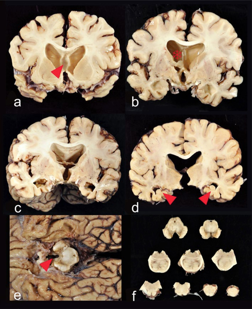
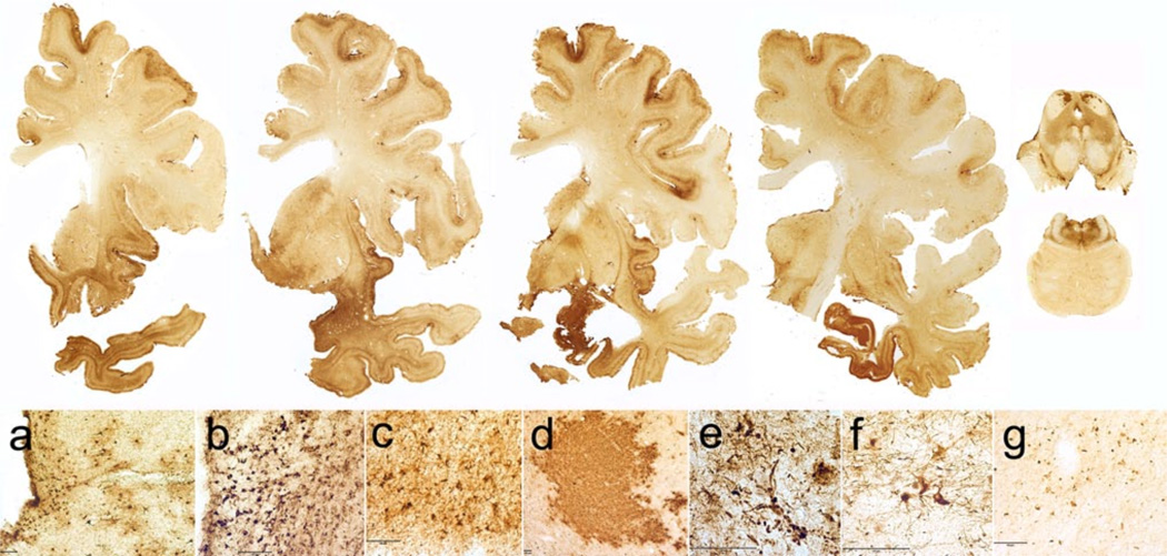
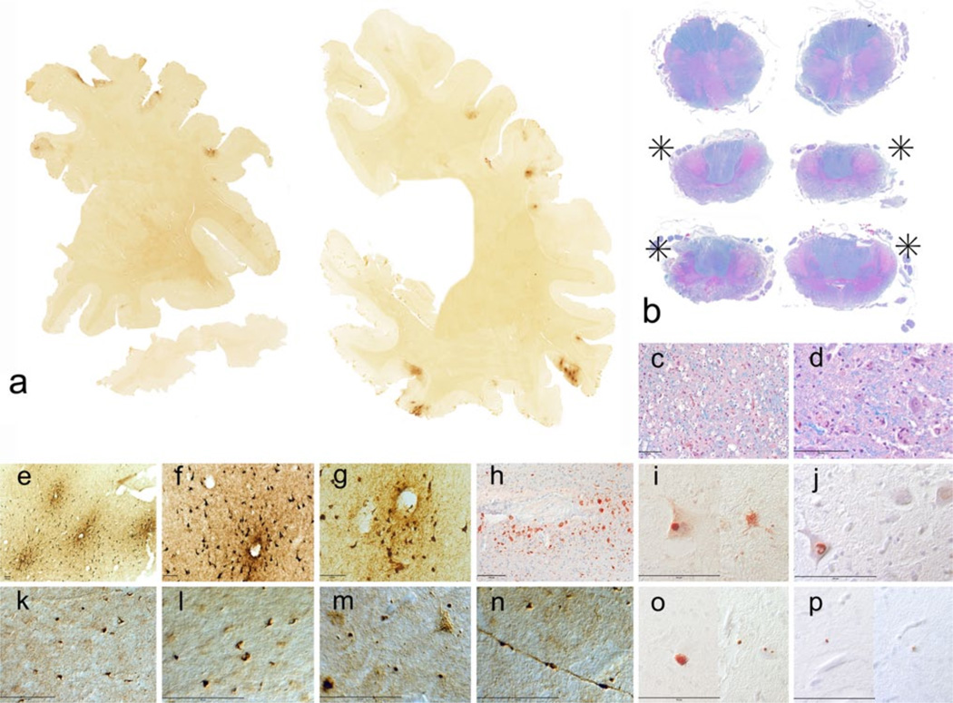
The benefits of regular exercise, physical fitness and sports participation on cardiovascular and brain health are undeniable. Physical activity reduces the risk for cardiovascular disease, type 2 diabetes, hypertension, obesity, and stroke, and produces beneficial effects on cholesterol levels, antioxidant systems, inflammation, and vascular function. Exercise also enhances psychological health, reduces age-related loss of brain volume, improves cognition, reduces the risk of developing dementia, and impedes neurodegeneration. Nonetheless, the play of sports is associated with risks, including a risk for mild TBI (mTBI) and, rarely, catastrophic traumatic injury and death. There is also growing awareness that repetitive mTBIs, such as concussion and subconcussion, can occasionally produce persistent cognitive, behavioral, and psychiatric problems as well as lead to the development of a neurodegeneration, chronic traumatic encephalopathy (CTE). In this review, we summarize the beneficial aspects of sports participation on psychological, emotional, physical and cognitive health, and specifically analyze some of the less common adverse neuropathological outcomes, including concussion, second-impact syndrome, juvenile head trauma syndrome, catastrophic sudden death, and CTE. CTE is a latent neurodegeneration clinically associated with behavioral changes, executive dysfunction and cognitive impairments, and pathologically characterized by frontal and temporal lobe atrophy, neuronal and axonal loss, and abnormal deposits of paired helical filament (PHF)-tau and 43 kDa TAR deoxyribonucleic acid (DNA)-binding protein (TDP-43). CTE often occurs as a sole diagnosis, but may be associated with other neurodegenerative disorders, including motor neuron disease (CTE-MND). Although the incidence and prevalence of CTE are not known, CTE has been reported most frequently in American football players and boxers. Other sports associated with CTE include ice hockey, professional wrestling, soccer, rugby, and baseball.


Figures

Fig. 1
Fig. 1
The neuropathology of acute concussive injury. a Axonal swelling in the corpus callosum (Table 1, case 2), APP immunostaining, 10 m-paraffin section. b Perivascular clusters of activated microglia in the corpus callosum and subcortical white matter (Table 1, case 1), LN3 immunostain, 50 mm free-floating section. c Vascular amyloid in leptomeningeal vessel (Table 1, case 6), Ab 42 immunostain, 10 m paraffin section. d Persistent axonal swelling in the cerebellar white matter (Table 1, case 6), APP immunostain, 10 m paraffin section. e Perivascular microglia around deep white matter vessel (Table 1, case 3), 50 mm free-floating section. f Hemosiderin-laden macrophages around a small blood vessel indicating a remote microbleed (asterisk) in close proximity to focal PHF-tau NFTs and neurites in the uncus (arrowhead) (Table 1, case 5). AT8 immunostain with hematoxylin counterstain, 10 m paraffin section. g Perivascular axonal spheroid in the cerebellar white matter (Table 1, case 6). APP immunostain, 10 m paraffin section. h Perivascular reactive astrocytes in white matter (Table 1, case 3), GFAP immunostain, 10 m paraffin section. i PHF-tau immunoreactive neurites in the medulla (Table 1, case 3), AT8 immunostain, 50 mm free-floating section. j Axonal swelling with digestion chamber in the midbrain tegmentum (Table 1, case 5), SMI 34 immunostain, 10 m-paraffin section. k Perivascular reactive astrocytes in white matter (Table 1, case 2), GFAP immunostaining, 10 m-paraffin section. l PHF-tau immunoreactive NFTs in the reticular formation of the medulla (Table 1, case 3), AT8 immunostain, 50 mm free-floating section. m Perivascular axonal swellings (arrowhead) (Table 1, case 2), APP immunostaining, 10 m-paraffin section. n TDP-43 immunoreactive neurites in the fornix (arrowheads) (Table 1, case 1), TDP-43 immunostain, 50 mm free-floating section. o PHF-tau immunoreactive perivascular NFTs and neurites in the frontal cortex (Table 1, case 3), AT8 immunostain, 50 mm free-floating section. All magnification bars 100 mm
Fig. 2
Fig. 2
The four stages of chronic traumatic encephalopathy. In Stage I CTE, PHF-tau pathology is restricted to discrete foci in the cerebral cortex, most commonly in the superior, dorsolateral or lateral frontal cortices, and typically around small vessels at the depths of sulci. In Stage II CTE, there are multiple epicenters at the depths of the cerebral sulci and spread of neurofibrillary pathology to the superficial layers of adjacent cortex. The medial temporal lobe is spared neurofibrillary PHF-tau pathology in Stage II CTE, although it becomes progressively more involved as disease severity increases. In Stage III, PHF-tau pathology is widespread; the frontal, insular, temporal, and parietal cortices, amygdala, hippocampus, and entorhinal cortex show widespread neurofibrillary pathology. In Stage IV CTE, there is widespread severe PHF-tau pathology affecting most regions of the cerebral cortex and the medial temporal lobe, sparing calcarine cortex in all, but the most severe cases. All images, CP-13 immunostained 50 m tissue sections
Fig. 3
Fig. 3
CTE Stage II in a 36-year-old former Major League baseball player. To p row whole mount sections of occipital and frontal cortex; CP-13 immunostained 50 m tissue sections. Red circles indicate clusters of neurofibrillary pathology, which are primarily perivascular and concentrated at the sulcal depths. The arrowhead shows an area of acute hemorrhage secondary to the fatal gunshot. a–e Neurofibrillary pathology in the frontal and occipital cortices, showing a tendency for the PHF-tau pathology to be perivascular, subpial, and more concentrated at the depths of the cerebral sulci. CP-13 immunostain, 50 mm free-floating sections. f A focus of PHF-tau neurofibrillary pathology surrounding a small blood vessel. There are hematoidin-laden macrophages around the vessel (asterisk) and focal neuronal loss, changes indicative of a remote micro-bleed. CP-13 immunostain, 50 mm free-floating section. All magnification bars 100 mm
Fig. 4
Fig. 4
Gross neuropathological findings in a 77-year-old former Australian Rules rugby player who died with severe dementia and Stage IV CTE. Cognitive problems, memory loss, attention difficulties, and executive dysfunction were first noted in his mid-50s, followed by depression and anxiety, worsening explosivity and impulsivity. By his mid-60s, he was physically and verbally abusive, paranoid, and severely demented. He began playing rugby at age 13, and played for 19 years in U21 and senior leagues. a At autopsy, the brain weighed 1,030 g and showed severe atrophy and ventricular enlargement with a prominent cavum septum pellucidum (arrowhead). bc The mid-portion of the septum pellucidum (asterisk) is reduced to a thin filament with severe atrophy of the fornix, thalamus, hypothalamus, mammillary bodies, amygdala, anterior hippocampus, and entorhinal cortex. d There is bilateral hippocampal atrophy (arrowheads). e The floor of the hypo-thalamus is severely thinned and the mammillary bodies are severely atrophic (arrowhead). f Brainstem sections show pallor of the pars compacta of the substantia nigra and locus coeruleus, with discoloration of the frontal tracts of the cerebral peduncle
Fig. 5
Fig. 5
Microscopic neuropathological findings in a 77-year-old former Australian Rules rugby player with Stage IV CTE. To p row whole mount coronal sections of the brain and brainstem; CP-13 immunostained 50 m tissue sections. There is widespread PHF-tau immunoreactive neurofibrillary pathology. af Microscopic sections show extreme neuronal loss and PHF-tau pathology; CP-13 immunostaining, 50 mm free-floating sections. a. Frontal cortex. b Periventricular caudate. c Temporal cortex. d Superior colliculus. e Substantia nigra pars compacta. f Locus coeruleus. g P-TDP-43 immunoreactive inclusions and neurites in the frontal cortex; pTDP-43 immunostaining, 50 mm free-floating sections. All magni cation bars 100 mm
Fig. 6
Fig. 6
CTE Stage II with motor neuron disease in a 29-year-old soccer player who developed fatigue and weakness of his lower extremities and hands at age 27 and 3 months later was diagnosed with ALS. 21 months after the onset of his symptoms, he died of respiratory insufficiency at age 29. He had played soccer since the age of 3, and started heading the ball at age 5. He played soccer for 12 years in public school, 4 years in college, and 2.5 years as a semi-professional. a Whole mount sections showing clusters of perivascular PHF-tau immunoreactivity preferentially at the sulcal depths of the frontal, temporal, and parietal cortices; CP-13 immunostain. b Sections of lower medulla, cervical thoracic, and lumbar spinal cord show extreme loss of lateral (asterisks) and ventral corticospinal tracts; luxol fast blue, and hematoxylin and eosin stain. c Extreme loss of axons, myelin with macrophage infiltration in lateral corticospinal tracts; luxol fast blue, and hematoxylin and eosin stain. d Severe loss of anterior horn cells in the ventral horn of spinal cord; luxol fast blue, and hematoxylin and eosin stain. e–g Perivascular clusters of PHF-tau immunoreactive NFTs and neurites in cortex; CP-13 immunostain, 50 mm free-floating sections. h Axonal swellings and distorted axons and hemosiderin-laden macrophages around small blood vessel in cortical white matter; SMI-34 immunostain, 10 m paraffin section. i FUS immunoreactive intracytoplasmic inclusions, both rounded and fibrillar; FUS immunostain, 10 m paraffin section. j Ubiquilin immunoreactive nucleus; ubiquilin immunostain, 10 m-paraffin section. k PTDP-43 neurites in frontal cortex; phospho-rylated-TDP-43 immunostain, 50-mm free-floating section. l PTDP-43 neurites in Rolandic cortex; phosphorylated-TDP-43 immunostain, 50-mm free-floating section. m PTDP43 intraneuronal fibrillar inclusions and neurites in lumbar spinal cord; phosphorylated-TDP-43 immunostain, 50 mm free-floating section. n PTDP-43 immuno-reactivity in axon in lumbar spinal cord; phosphorylated-TDP-43 immunostain, 50-mm free-floating section. o PTDP immunoreactive intracytoplasmic inclusions and neurites; phosphorylated-TDP-43 immunostain, 10 m-paraffin section. p PHF-tau immunoreactive intra-cytoplasmic granular inclusions; AT8 immunostain, 10 m-paraffin section

References

    1. Ahlskog JE. Does vigorous exercise have a neuroprotective effect in Parkinson disease? Neurology. 2011;77(3):288–294. - PMC - PubMed
    1. Ang ET, Dawe GS, Wong PT, Moochhala S, Ng YK. Alterations in spatial learning and memory after forced exercise. Brain Res. 2006;1113(1):186–193. - PubMed
    1. Ang ET, Gomez-Pinilla F. Potential therapeutic effects of exercise to the brain. Curr Med Chem. 2007;14(24):2564–2571. - PubMed
    1. Anthony IC, Norrby KE, Dingwall T, Carnie FW, Millar T, Arango JC, Robertson R, Bell JE. Predisposition to accelerated Alzheimer-related changes in the brains of human immunodeficiency virus negative opiate abusers. Brain. 2010;133(12):3685–3698. - PubMed
    1. Aoki Y, Inokuchi R, Gunshin M, Yahagi N, Suwa H. Diffusion tensor imaging studies of mild traumatic brain injury: a meta-analysis. J Neurol Neurosurg Psychiatry. 2012;83(9):870–876. - PMC - PubMed

Publication types