. 2014 Mar;34(5):832-46.
doi: 10.1128/MCB.01191-13. Epub 2013 Dec 23.

Kinetic, thermodynamic, and structural characterizations of the association between Nrf2-DLGex degron and Keap1

Affiliations

Kinetic, thermodynamic, and structural characterizations of the association between Nrf2-DLGex degron and Keap1

Toshiaki Fukutomi et al. Mol Cell Biol. 2014 Mar.

Abstract

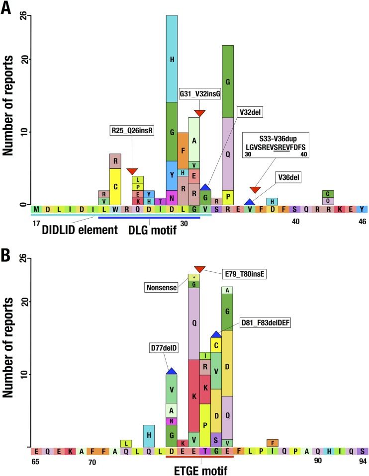
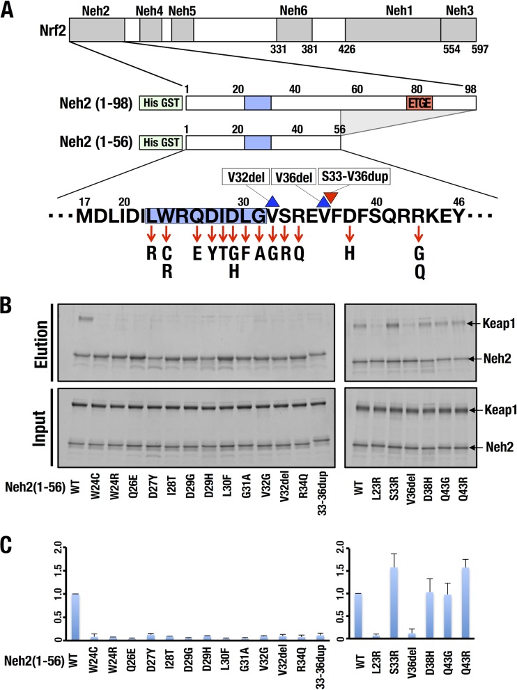
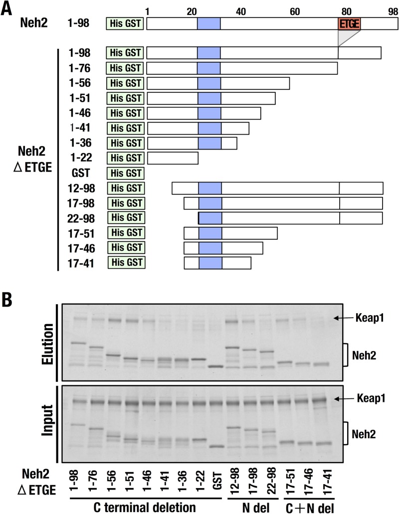
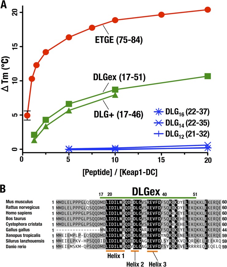
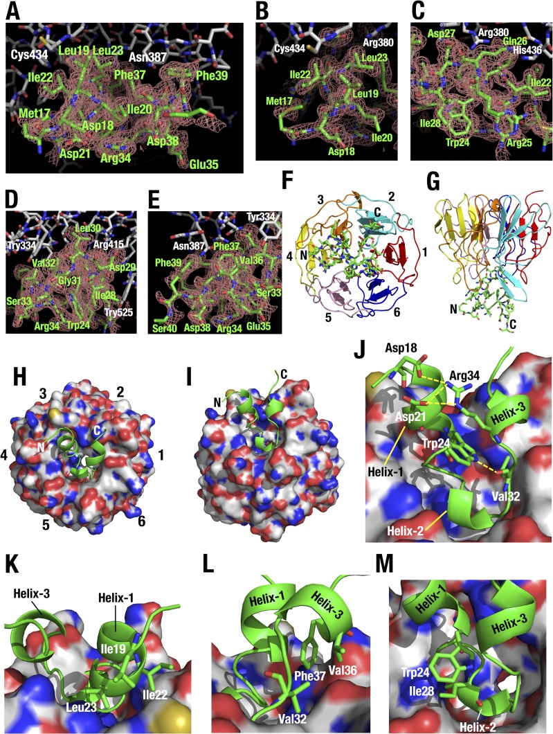
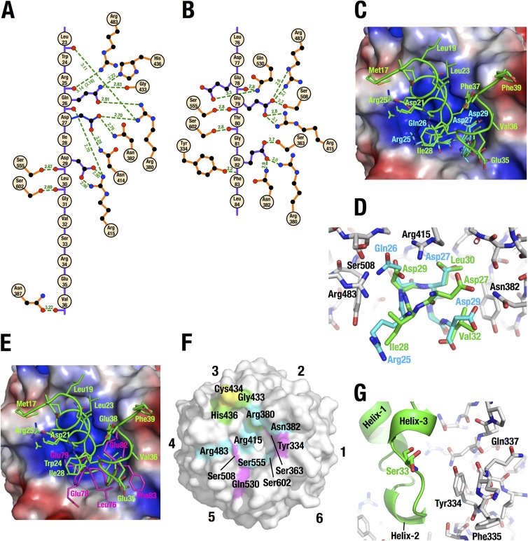
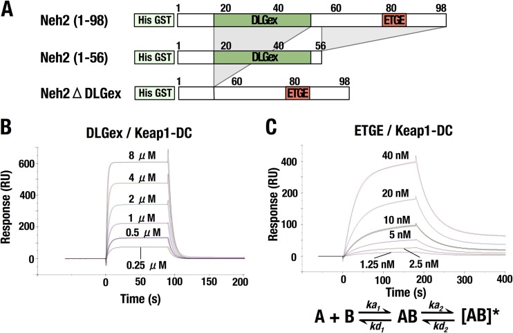
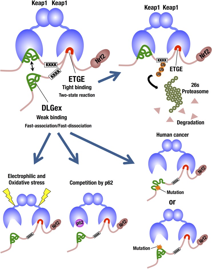
Transcription factor Nrf2 (NF-E2-related factor 2) coordinately regulates cytoprotective gene expression, but under unstressed conditions, Nrf2 is degraded rapidly through Keap1 (Kelch-like ECH-associated protein 1)-mediated ubiquitination. Nrf2 harbors two Keap1-binding motifs, DLG and ETGE. Interactions between these two motifs and Keap1 constitute a key regulatory nexus for cellular Nrf2 activity through the formation of a two-site binding hinge-and-latch mechanism. In this study, we determined the minimum Keap1-binding sequence of the DLG motif, the low-affinity latch site, and defined a new DLGex motif that covers a sequence much longer than that previously defined. We have successfully clarified the crystal structure of the Keap1-DC-DLGex complex at 1.6 Å. DLGex possesses a complicated helix structure, which interprets well the human-cancer-derived loss-of-function mutations in DLGex. In thermodynamic analyses, Keap1-DLGex binding is characterized as enthalpy and entropy driven, while Keap1-ETGE binding is characterized as purely enthalpy driven. In kinetic analyses, Keap1-DLGex binding follows a fast-association and fast-dissociation model, while Keap1-ETGE binding contains a slow-reaction step that leads to a stable conformation. These results demonstrate that the mode of DLGex binding to Keap1 is distinct from that of ETGE structurally, thermodynamically, and kinetically and support our contention that the DLGex motif serves as a converter transmitting environmental stress to Nrf2 induction as the latch site.


Figures

FIG 1
FIG 1
Distribution of somatic mutations within the NRF2 gene encompassing the Nrf2-DLG motif or the DIDLID element (A) and the Nrf2-ETGE motif (B). The horizontal and longitudinal axes represent the amino acid sequence and number of reports, respectively. The boxes, blue triangle, and red triangle indicate substitution mutations, a deletion mutation, and an insertion mutation, respectively. Sky blue, dark blue, and red lines indicate the DIDLID element, the DLG motif, and the ETGE motif, respectively.
FIG 2
FIG 2
Somatic mutations encompassing Nrf2-DLG in human cancers impair the binding of Nrf2-DLG and Keap1. (A) Diagrams of cancer-derived mutation constructs of Nrf2-Neh2(1-56) used for a pulldown assay. The blue boxes and red box indicate the classical DLG motif (amino acids Leu23 to Gly31) and the ETGE motif (amino acids Asp77 to Glu82), respectively. The red arrows, blue triangle, and red triangle indicate substitution mutations, a deletion mutation, and an insertion mutation, respectively. (B) A GST pulldown assay with 6×His-GST-tagged WT Neh2(1-56) or mutant forms thereof with purified recombinant Keap1-His. WT Neh2(1-56) or mutant forms thereof and Keap1-His were mixed and incubated with glutathione magnetic beads. The pulled-down complex was eluted with reduced glutathione, subjected to SDS-PAGE, and visualized by fluorescent staining. (C) Quantification of band intensities in panel B by quantitative densitometry of fluorescent staining. Values relative to the intensity of WT Neh2(1-56) are shown. The average results of three independent experiments are shown with standard deviations.
FIG 3
FIG 3
Minimum Keap1-interacting region of the Nrf2-DLG motif. (A) Diagrams of the deletion-mutation constructs of Nrf2-Neh2ΔETGE used for the pulldown assay. Blue boxes and red box indicate the DLG motif (amino acids Leu23 to Gly31) and the ETGE motif (amino acids Asp77 to Glu82), respectively. (B) A GST pulldown assay with 6×His-GST-tagged Neh2ΔETGE and deletion mutant forms thereof with purified recombinant Keap1-His. Neh2ΔETGE or deletion mutant forms thereof and Keap1-His were mixed and incubated with glutathione magnetic beads. The pulled-down complex was eluted with reduced glutathione, subjected to SDS-PAGE, and visualized by fluorescent staining.
FIG 4
FIG 4
Characterization of the Nrf2-DLGex interaction with Keap1. (A) DSF analysis to determine the Tm of Keap1-DC with various DLG peptides and the ETGE peptide. The horizontal and longitudinal axes indicate peptide concentrations and ΔTms, respectively. ΔTm was generated by subtracting the Tm value without peptide (reference) from the Tm value with each peptide. (B) Alignment of the N-terminal regions of the Nrf2 proteins of various species. Amino acid residues that are conserved completely and partially are shaded black and gray, respectively. Nrf2-DLGex is indicated by the green bar.
FIG 5
FIG 5
Thermodynamic examination of Nrf2-DLGex and Nrf2-ETGE binding to Keap1-DC. (A to C) Representative ITC titration profiles of Keap1-DC with Nrf2-DLGex (A), Neh2ΔETGE (B), and the ETGE peptide (C). (D) Thermodynamic characterization of the modes of Nrf2-DLGex, Neh2ΔETGE, and ETGE peptide binding to Keap1-DC. Bars represent the free-energy change (ΔG), enthalpic component (ΔH), and entropic component (−TΔS). The interrelationships among these parameters are described as ΔG = ΔHTΔS.
FIG 6
FIG 6
Crystal structure of Keap1-DC with the long DLGex peptide. (A to E) Overall (A) and closeup (B to E) views of the interface between Keap1 (white) and DLGex (green). The Fo-Fc omit map of the DLGex peptide is contoured at 2.0 σ. DLGex was omitted from the calculation. (F to I) Overall structure of Keap1-DC (shown as a ribbon model [F and G] and a surface model [H and I]) in complex with the Nrf2 DLGex peptide (Met17 to Gln51) (shown as a stick model [F and G] and a ribbon model [H and I]). The six blades in the β-propeller structure are numbered 1 to 6 (F and H) and are distinguished by separate colors (F and G). (J to M) Closeup view of the DLGex peptide bound to Keap1-DC. (J) Intramolecular electrostatic interactions in DLGex are shown as yellow broken lines. (K to M) Intramolecular hydrophobic interactions in DLGex among the side chains of Ile19, Ile22, and Leu23 (K), among the side chains of Val32, Val36, and Phe37 (L) and between the side chains of Trp24 and Ile28 (M).
FIG 7
FIG 7
Structural characteristics of the Keap1-DC and DLGex peptide interaction. (A and B) Intermolecular hydrogen bonds between Keap1-DC and Nrf2-DLGex (A) and between Keap1-DC and Nrf2-ETGE (PDB accession number 1X2R) (B). Hydrogen bonds (green broken lines) and their distance (in angstroms) are displayed. As Arg25 of DLGex consists of multiple conformers, the distance between the lower-population conformer of Arg25 and Arg483 of Keap1 is displayed in parentheses. (C and E) Superimposition of the Keap1-DLGex peptide complex with the Keap1-short DLG peptide (Ile22 to Val36; DLG15) complex (accession number 2DYH) (C) or with the Keap1-ETGE complex (PDB accession number 1X2R) (E). The backbone and side chains of bound peptides are shown as tubes and sticks, respectively. The DLGex, DLG15, and ETGE peptides are green, cyan, and magenta, respectively. The electrostatic surface potential of the bottom surface of Keap1 was calculated by PYMOL. Red and blue coloring indicates negative and positive charges, respectively. (D) Closeup view of the binding interfaces in panel C. Amino acid residues of Keap1-DC, DLGex, and DLG15 are shown as white, green, and cyan sticks, respectively. (F) Amino acid residues involved in the intermolecular interactions with Nrf2-DLGex, Nrf2-ETGE, and both are green, magenta, and cyan, respectively. Cysteine residues are yellow. (G) Closeup view around Nrf2-Ser33. DLGex is shown as a ribbon model. Amino acid residues of Keap1 and Nrf2-Ser33 are shown as white and green sticks, respectively.
FIG 8
FIG 8
Kinetic analyses of DLGex and Nrf2-ETGE binding to Keap1-DC. (A) Diagrams of Neh2(1-56) and Neh2ΔDLGex used for kinetic analyses. Neh2(1-56) and Neh2ΔDLGex contain the DLGex motif and the ETGE motif, respectively. (B and C) SPR sensorgrams showing the association and dissociation of the DLGex motif and the Keap1-DC domain (B) and of the ETGE motif and the Keap1-DC domain (C). RU, resonance units.
FIG 9
FIG 9
Schematic representation of the distinctive modes of Nrf2 binding to Keap1. We have determined that the DLGex motif corresponds to Met17 to Gln51 (green lines) and covers a much larger sequence (35 aa) than the minimum Keap1-binding sequence of ETGE (9 aa; red lines). Importantly, the binding modes of DLGex and ETGE are quite distinct from each other. Our present Keap1-DLGex complex crystal structure reveals that DLGex possesses a complicated helix structure, which interprets well the human-cancer-derived loss-of-function and gain-of-function mutations in DLGex. Of note, most of the mutations disrupt intramolecular interactions, which gives rise to the abrogation of their Keap1-binding activity. Keap1-DLGex binding is characterized as fast-on and fast-off binding that allows disruptor proteins, such as p62, to intercalate easy. In contrast, the binding of the ETGE motif to the DC domain most properly fits a two-state reaction model, and this two-step binding process of ETGE ensures Keap1-ETGE binding a more stable conformation and contributes to the efficient ubiquitination and degradation of Nrf2. Thus, the DLGex motif consists of efficient sensor machinery acting as a latch that easily dissociates from Keap1-DC upon detecting electrophilic and oxidative stress.

Similar articles

Cited by

References

    1. Motohashi H, O'Connor T, Katsuoka F, Engel JD, Yamamoto M. 2002. Integration and diversity of the regulatory network composed of Maf and CNC families of transcription factors. Gene 294:1–12. 10.1016/S0378-1119(02)00788-6 - DOI - PubMed
    1. Kobayashi M, Yamamoto M. 2006. Nrf2-Keap1 regulation of cellular defense mechanisms against electrophiles and reactive oxygen species. Adv. Enzyme Regul. 46:113–140. 10.1016/j.advenzreg.2006.01.007 - DOI - PubMed
    1. Taguchi K, Motohashi H, Yamamoto M. 2011. Molecular mechanisms of the Keap1-Nrf2 pathway in stress response and cancer evolution. Genes Cells 16:123–140. 10.1111/j.1365-2443.2010.01473.x - DOI - PubMed
    1. Itoh K, Wakabayashi N, Katoh Y, Ishii T, Igarashi K, Engel JD, Yamamoto M. 1999. Keap1 represses nuclear activation of antioxidant responsive elements by Nrf2 through binding to the amino-terminal Neh2 domain. Genes Dev. 13:76–86 - PMC - PubMed
    1. Tong KI, Kobayashi A, Katsuoka F, Yamamoto M. 2006. Two-site substrate recognition model for the Keap1-Nrf2 system: a hinge and latch mechanism. Biol. Chem. 387:1311–1320 - PubMed

Publication types

MeSH terms

Substances