. 2014 Sep;47(1):117-28.
doi: 10.1007/s12020-013-0133-6. Epub 2013 Dec 24.

A critical role for CK2 in cytokine-induced activation of NFκB in pancreatic β cell death

Affiliations

A critical role for CK2 in cytokine-induced activation of NFκB in pancreatic β cell death

Caroline Jaksch et al. Endocrine. 2014 Sep.

Abstract

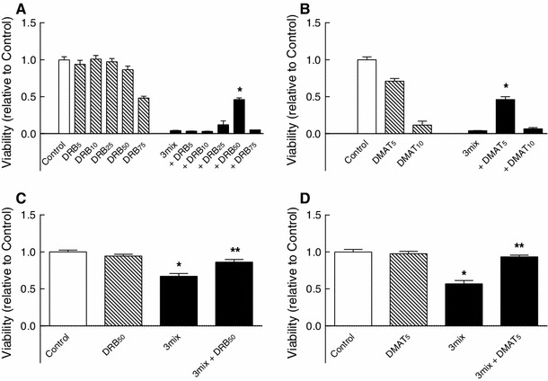
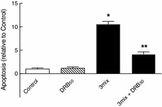
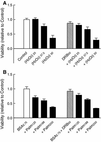
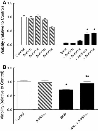
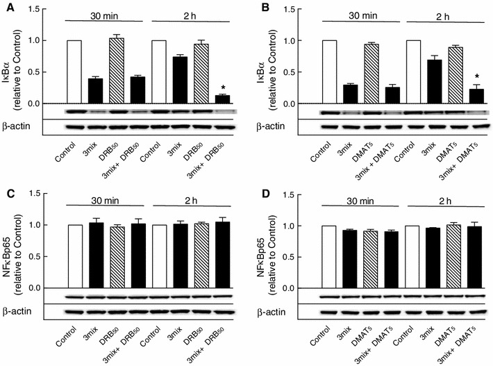
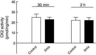
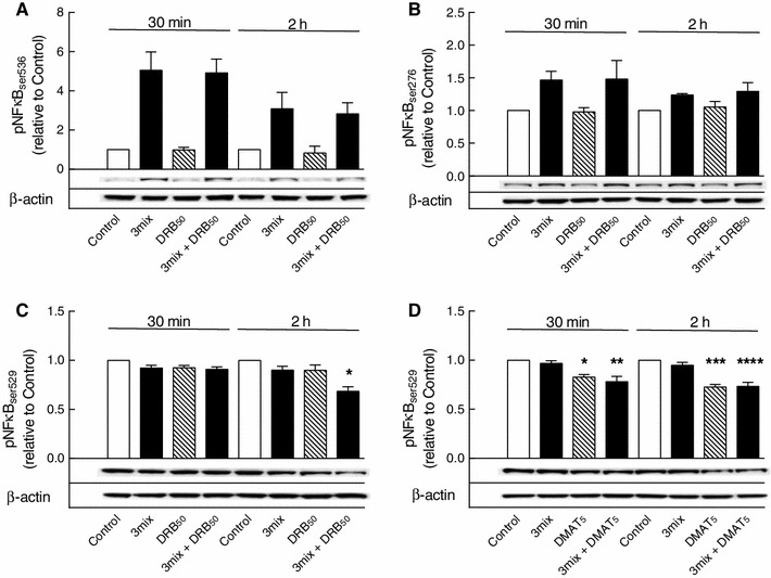
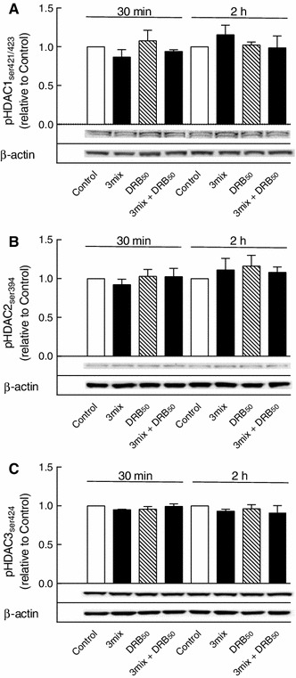
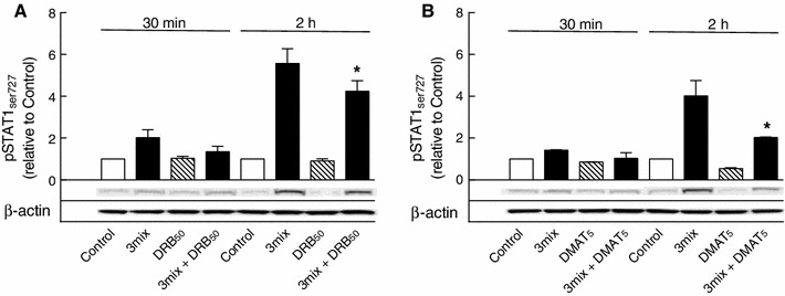
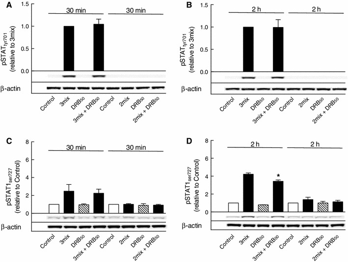
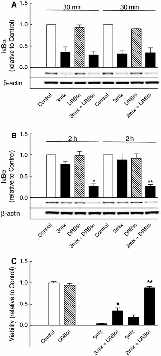
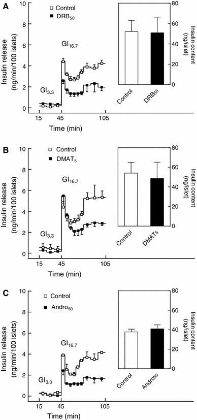
This study aimed to assess the role of constitutive protein kinase CK2 in cytokine-induced activation of NFκB in pancreatic β cell death. The CK2 inhibitors DRB (5,6-dichloro-1-β-D-ribofuranosylbenzimidazole) (50 μM) and DMAT (2-dimethylamino-4,5,6,7-tetrabromo-1H-benzimidazole) (5 μM), which decreased CK2 activity by approx. 65 %, rescued INS-1E β cells and mouse islets from cytokine (IL-1β, TNF-α plus IFN-γ)-induced β cell death without affecting H2O2- or palmitate-induced β cell death. Western blot analysis revealed that while DRB or DMAT did not influence cytokine-induced IκBα degradation, they inhibited NFκB-dependent IκBα resynthesis, demonstrating that cytokine-induced NFκB activity is dependent on CK2. Both DRB and DMAT inhibited the constitutive phosphorylation of NFκB p65 at serine 529, while leaving cytokine-induced phosphorylations of NFκB p65 at serines 276 and 536 unaltered. In comparison, putative phosphorylation sites for CK2 on HDACs 1, 2, and 3 at serines 421/423, 394, and 424, respectively, which may stimulate NFκB transcriptional activity, were unchanged by cytokines and CK2 inhibitors. Whereas IL-1β and TNF-α stimulate IκBα degradation and NFκB activation, IFN-γ potentiates cytokine-induced β cell death through activation of STAT1. DRB and DMAT inhibited IFN-γ-stimulated phosphorylation of STAT1 at serine 727, while leaving IFN-γ-induced phosphorylation of STAT1 at tyrosine 701 unaffected. Inhibition of cytokine-induced β cell death by CK2 inhibitors was, however, not dependent on IFN-γ, and IFN-γ did not affect CK2-dependent IκBα turnover. In conclusion, it is suggested that cytokine-induced activation of NFκB in β cells is dependent on CK2 activity, which phosphorylates NFκB p65 at serine 529.


Figures

Fig. 1
Fig. 1
Effects of DRB and DMAT on β cell survival. a, b INS-1E cells and c, d mouse islets were cultured for 48 h in the absence or presence of cytokines (IL-1β, TNF-α, and IFN-γ) (3mix) and with DRB (5–75 μM) (DRB5–75) or DMAT (5–10 μM) (DMAT5–10) as indicated before determination of cell viability. a, b Results are mean ± SEM (n = 4–12); a *P < 0.001 versus 3mix; b *P < 0.001 versus 3mix. c, d Results are mean ± SEM (n = 4); c *P < 0.001 versus control, **P < 0.001 versus 3mix; d *P < 0.001 versus control, **P < 0.001 versus 3mix
Fig. 2
Fig. 2
Role of CK2 in cytokine-induced β cell apoptosis. Mouse islets were cultured for 48 h in the absence or presence of cytokines (IL-1β, TNF-α, and IFN-γ) (3mix) and with DRB (50 μM) (DRB50) as indicated before determination of apoptosis. Results are mean ± SEM (n = 4), *P < 0.001 versus control, **P < 0.001 versus 3mix
Fig. 3
Fig. 3
Role of CK2 in H2O2- and palmitate-induced β cell death. INS-1E cell were cultured for 24 h with a H2O2 (25–50 μM) [(H2O2)25–50] or b palmitate (125–250 μM) (Palm125–250) plus bovine serum albumin (2.75 mg/ml) (BSA2.75) and with DRB (50 μM) (DRB50) as indicated before determination of cell viability. Results are mean ± SEM (n = 4–6)
Fig. 4
Fig. 4
Role of NFκB in cytokine-induced β cell death. a INS-1E cells and b mouse islets were cultured for 48 h in the absence or presence of cytokines (IL-1β, TNF-α, and IFN-γ) (3mix) and with andrographolide (5–50 μM) (Andro5–50) as indicated before determination of cell viability. a Results are mean ± SEM (n = 8), *P < 0.001 versus 3mix, b Results are mean ± SEM (n = 4), *P < 0.05 versus control, **P < 0.05 versus 3mix
Fig. 5
Fig. 5
Western blots of IκBα and NFκB p65 proteins. INS-1E cells were cultured for 30 min or 2 h in the absence or presence of cytokines (IL-1β, TNF-α, and IFN-γ) (3mix) and DRB (50 μM) (DRB50) or DMAT (5 μM) (DMAT5) as indicated before determination of a, b IκBα and c, d NFκB p65 protein levels. Results are mean ± SEM (n = 3–6) with a representative experiment shown below bars. a *P < 0.001 versus 3mix; b *P < 0.001 versus 3mix
Fig. 6
Fig. 6
Effects of cytokines on CK2 activity in INS-1E cells. INS-1E cells were cultured for 30 min or 2 h in the absence or presence of cytokines (IL-1β, TNF-α, and IFN-γ) (3mix). Results are mean ± SEM (n = 6)
Fig. 7
Fig. 7
Effects of DRB and DMAT on NFκB p65 phosphorylations. INS-1E cells were cultured for 30 min or 2 h in the absence or presence of cytokines (IL-1β, TNF-α, and IFN-γ (3mix) and DRB (50 μM) (DRB50) or DMAT (5 μM) (DMAT5) as indicated. a NFκB p65 phosphoserine 536 (pNFκBser536), b NFκB p65 phosphoserine 276 (pNFκBser276), c, d NFκB p65 phosphoserine 529 (pNFκBser529). Results are mean ± SEM (n = 3–7) with a representative experiment shown below bars. c *P < 0.001 versus control; d *P < 0.01 versus control, **P < 0.01 versus 3mix, ***P < 0.001 versus control, ****P < 0.001 versus 3mix
Fig. 8
Fig. 8
Effects of DRB on HDACs 1–3 phosphorylations. INS-1E cells were cultured for 30 min or 2 h in the absence or presence of cytokines (IL-1β, TNF-α, and IFN-γ) (3mix) and DRB (50 μM) (DRB50) as indicated. a HDAC1 phosphoserine 421/423 (pHDAC1ser421/423), b HDAC2 phosphoserine 394 (pHDAC2ser394), c HDAC3 phosphoserine 424 (pHDAC3ser424). Results are mean ± SEM (n = 3–5) with a representative experiment shown below bars
Fig. 9
Fig. 9
Effects of DRB and DMAT on STAT1 serine 727 phosphorylation. INS-1E cells were cultured for 30 min or 2 h in the absence or presence of cytokines (IL-1β, TNF-α, and IFN-γ) (3mix) and DRB (50 μM) (DRB50) or DMAT (5 μM) (DMAT5) as indicated before determination of STAT1 phosphoserine 727 (pSTAT1ser727). Results are mean ± SEM (n = 3–5) with a representative experiment shown below bars. a *P < 0.01 versus 3mix; b *P < 0.001 versus 3mix
Fig. 10
Fig. 10
Role of IFN-γ in STAT1 phosphorylations. INS-1E cells were cultured for 30 min or 2 h in the absence or presence of IL-1β and TNF-α with IFN-γ (3mix) or without IFN-γ (2mix) and with DRB (50 μM) (DRB50) as indicated before determination of a, b STAT1 phosphotyrosine 701 (pSTATtyr701) and c, d STAT1 phosphoserine 727 (pSTAT1ser727). Results are mean ± SEM (n = 3) with a representative experiment shown below bars. d *P < 0.01 versus 3 mix
Fig. 11
Fig. 11
Role of STAT1 in CK2-dependent NFκB activation and β cell death. a, b INS-1E cells were cultured for 30 min or 2 h in the absence or presence of IL-1β and TNF-α with IFN-γ (3mix) or without IFN-γ (2mix) and with DRB (50 μM) (DRB50) as indicated before determination of IκBα. Results are mean ± SEM (n = 3) with a representative experiment shown below bars. b *P < 0.001 versus 3mix, **P < 0.001 versus 2 mix. c INS-1E cells were cultured for 48 h in the absence or presence of IL-1β, TNF-α with IFN-γ (3mix) or without IFN-γ (2mix) and with DRB (50 μM) (DRB50) as indicated before determination of cell viability. Results are mean ± SEM (n = 7). *P < 0.001 versus 3mix, **P < 0.001 versus 2mix
Fig. 12
Fig. 12
Role of CK2 and NFκB in glucose-induced insulin secretion. Mouse islets were cultured for 24 h in the absence or presence of a DRB (50 μM) (DRB50), b DMAT (5 μM) (DMAT5) or c andrographolide (50 μM) (Andro50) before determination of islet insulin secretion and insulin content as indicated. Islets were perifused for 45 min with 3.3 mM glucose (Gl3.3) and then stimulated for 60 min with 16.7 mM glucose (Gl16.7). Results are mean ± SEM (n = 3–4)

Similar articles

Cited by

References

    1. Cerf ME. Beta cell dynamics: beta cell replenishment, beta cell compensation and diabetes. Endocrine. 2013;44:303–311. doi: 10.1007/s12020-013-9917-y. - DOI - PubMed
    1. Melloul D. Role of NF-κB in β-cell death. Biochem. Soc. Trans. 2008;36:334–339. doi: 10.1042/BST0360334. - DOI - PubMed
    1. Cnop M, Welsh N, Jonas J-C, Jörns A, Lenzen S, Eizirik DL. Mechanisms of pancreatic β-cell death in type 1 and type 2 diabetes. Many differences, few similarities. Diabetes. 2005;54(Suppl. 2):S97–S107. doi: 10.2337/diabetes.54.suppl_2.S97. - DOI - PubMed
    1. Donath MY, Böni-Schnetzler M, Ellingsgaard H, Halban PA, Ehses JA. Cytokine production by islets in health and diabetes: cellular origin, regulation and function. Trends Endocrinol. Metab. 2010;21:261–267. doi: 10.1016/j.tem.2009.12.010. - DOI - PubMed
    1. Singh NN, Ramji DP. Protein kinase CK2, an important regulator of the inflammatory response? J. Mol. Med. 2008;86:887–897. doi: 10.1007/s00109-008-0352-0. - DOI - PubMed

Publication types

MeSH terms

LinkOut - more resources