. 2014 Feb 7;289(6):3126-37.
doi: 10.1074/jbc.M113.508044. Epub 2013 Dec 23.

A novel CXCR3-B chemokine receptor-induced growth-inhibitory signal in cancer cells is mediated through the regulation of Bach-1 protein and Nrf2 protein nuclear translocation

Affiliations

A novel CXCR3-B chemokine receptor-induced growth-inhibitory signal in cancer cells is mediated through the regulation of Bach-1 protein and Nrf2 protein nuclear translocation

Murugabaskar Balan et al. J Biol Chem. .

Erratum in

Abstract

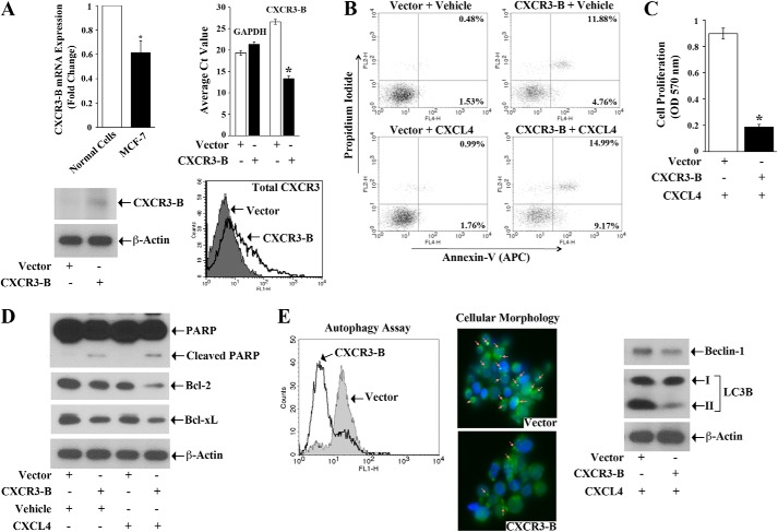
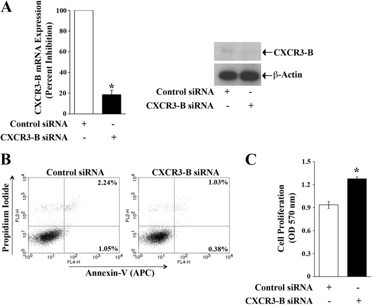
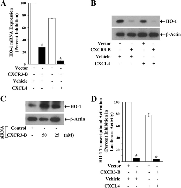
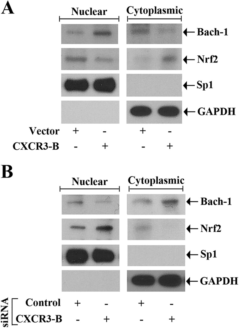
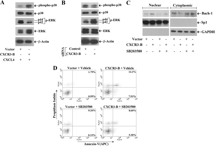
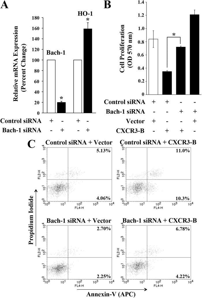
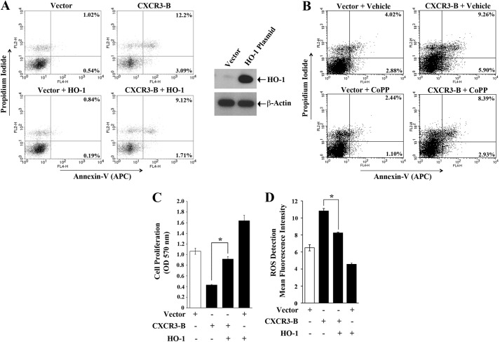
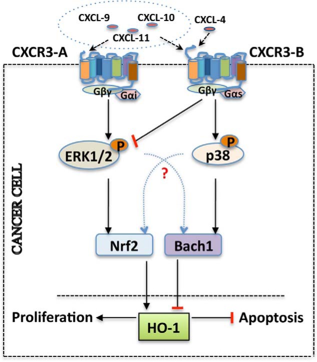
Chemokines and their receptors play diverse roles in regulating cancer growth and progression. The receptor CXCR3 can have two splice variants with opposite functions. CXCR3-A promotes cell growth, whereas CXCR3-B mediates growth-inhibitory signals. However, the negative signals through CXCR3-B in cancer cells are not well characterized. In this study, we found that CXCR3-B-mediated signaling in MCF-7 and T47D breast cancer cells induced apoptotic cell death. Signals through CXCR3-B decreased the levels of the antiapoptotic proteins Bcl-2 and Bcl-xL and increased the expression of apoptotic cleaved poly(ADP-ribose) polymerase. Along with up-regulation in apoptosis, CXCR3-B signals were associated with a decrease in cellular autophagy with reduced levels of the autophagic markers Beclin-1 and LC3B. Notably, CXCR3-B down-regulated the expression of the cytoprotective and antiapoptotic molecule heme oxygenase-1 (HO-1) at the transcriptional level. There was an increased nuclear localization of Bach-1 and nuclear export of Nrf2, which are important negative and positive transcription factors, respectively, for HO-1 expression. We also observed that CXCR3-B promoted the activation of p38 MAPK and the inhibition of ERK-1/2. CXCR3-B could not induce cancer cell apoptosis at the optimal level when we either inhibited p38 activity or knocked down Bach-1. Further, CXCR3-B-induced apoptosis was down-regulated when we overexpressed HO-1. Together, our data suggest that CXCR3-B mediates a growth-inhibitory signal in breast cancer cells through the modulations of nuclear translocation of Bach-1 and Nrf2 and down-regulation of HO-1. We suggest that the induction of CXCR3-B-mediated signaling can serve as a novel therapeutic approach where the goal is to promote tumor cell apoptosis.

Keywords: Apoptosis; Chemokines; Heme Oxygenase; Nrf2; Nuclear Transport; Signal Transduction; Signaling.


Figures

FIGURE 1.
FIGURE 1.
CXCR3-B induces apoptosis and inhibits autophagy of breast cancer cells. A, the mRNA expression of CXCR3-B in normal breast epithelial cells and MCF-7 cells was analyzed by real-time PCR (top left panel). MCF-7 cells were transfected with either the CXCR3-B overexpression plasmid (0.5 μg) or the empty vector. Cells were harvested after 48 h, and the expression of CXCR3-B mRNA, CXCR3-B, and total CXCR3 (A and B) protein were analyzed by real-time PCR (top right panel), Western blot analysis (bottom left panel), and FACS analysis (bottom right panel), respectively. The lower Ct value reflects overexpression of the gene. B, MCF-7 cells were transfected with either the CXCR3-B overexpression plasmid (0.5 μg) or the empty vector and then treated with either CXCL4 (250 ng/ml) or vehicle alone. After 48 h of plasmid transfection, the apoptotic index of cells was determined by annexin V (APC) and propidium iodide staining as described under “Experimental Procedures.” C, MCF-7 cells were transfected with either the CXCR3-B overexpression plasmid (0.5 μg) or the empty vector and then treated with CXCL4 (250 ng/ml). After 72 h, cell proliferation was measured by MTT assay. D, cell lysate from B was used to measure the expression levels of poly(ADP-ribose) polymerase (PARP), Bcl-2, Bcl-xL, and β-actin (internal control) by Western blot analysis. E, MCF-7 cells were transfected with either the CXCR3-B overexpression plasmid (0.5 μg) or the empty vector and then treated with CXCL4 (250 ng/ml). After 48 h, cellular autophagy was measured by flow cytometry by staining the cells with Cyto-ID green autophagy detection reagent (left panel). The morphologies of these Cyto-ID-stained cells were analyzed under a fluorescence microscope (center panel). Cell lysates were used to check the expression of the autophagic markers Beclin-1, LC3B (I and II), and β-actin by Western blot analysis (right panel). A and C, the columns represent the mean ± S.D. of triplicate readings of two samples. *, p < 0.05. B, D, and E, data are representative of three independent experiments.
FIGURE 2.
FIGURE 2.
Knockdown of CXCR3-B is associated with the survival and proliferation of breast cancer cells. MCF-7 cells were transfected with either CXCR3-B siRNA (50 nm) or control siRNA for 72 h. A, cells were harvested, and the expression of CXCR3-B mRNA and protein was analyzed by real-time PCR (left panel) and Western blot analysis (right panel). B, the apoptotic index of siRNA-transfected cells was determined by annexin V (APC) and propidium iodide staining. C, cell proliferation of siRNA-transfected cells was measured by MTT assay. A and C, the columns represent the mean ± S.D. of triplicate readings of two different samples. *, p < 0.05. B, results are representative of three independent experiments.
FIGURE 3.
FIGURE 3.
CXCR3-B down-regulates the expression of HO-1. A and B, MCF-7 cells were transfected with either the CXCR3-B overexpression plasmid (0. 5 μg) or an empty vector and then treated with either CXCL4 (250 ng/ml) or vehicle alone. After 48 h of plasmid transfection, cells were lysed, and the expression of HO-1 mRNA and protein was measured by real-time PCR (A) and Western blot analysis (B). The expression of β-actin was measured as an internal control. C, MCF-7 cells were transfected with either CXCR3-B siRNA (50 nm or 25 nm) or control siRNA. After 72 h of siRNA transfection, cells were lysed, and HO-1 protein expression was measured by Western blot analysis. D, MCF-7 cells were cotransfected with the HO-1 promoter-luciferase plasmid (0.5 μg) and either the CXCR3-B plasmid (0.5 μg) or an empty vector. The cells were then treated with either CXCL4 (250 ng/ml) or vehicle alone. Following 48 h of transfection, cells were harvested, and HO-1 promoter activity was measured by luciferase assay. The percent decrease in luciferase activity was calculated from luciferase counts of each group of cells compared with that of cells transfected with the empty vector and treated with vehicle alone. A and C, the columns represent the mean ± S.D. of triplicate readings of two different samples. *, p < 0.05 compared with respective controls. B and D, data are representative of three independent experiments.
FIGURE 4.
FIGURE 4.
CXCR3-B-mediated signaling modulates Bach-1 and Nrf-2 nuclear localization. A, MCF-7 cells were transfected with either the CXCR3-B overexpression plasmid (0. 5 μg) or the empty vector. B, MCF-7 cells were transfected with either CXCR3-B siRNA (50 nm) or control siRNA. Following 48 h of plasmid transfection (A) and 72 h of siRNA transfection (B), nuclear and cytoplasmic fractions were isolated from these cells, and a Western blot analysis was performed to quantitate the expression of Bach-1 and Nrf2. The purities of nuclear and cytoplasmic fractions were evaluated by measuring the expression of Sp1 and GAPDH, respectively. Results shown are representative of three independent experiments.
FIGURE 5.
FIGURE 5.
CXCR3-B-mediated signaling activates p38 MAPK and inhibits ERK1/2 phosphorylation. A, MCF-7 cells were transfected with either the CXCR3-B overexpression plasmid (0. 5 μg) or the empty vector and then treated with CXCL4 (250 ng/ml). B, MCF-7 cells were transfected with either CXCR3-B siRNA (50 nm) or control siRNA. Following 48 h of plasmid transfection (A) and 72 h of siRNA transfection (B), cells were lysed, and a Western blot analysis was performed to measure the expression levels of phospho-p38, phopsho-ERK1/2 (p-ERK), total p38, total ERK1/2, and β-actin. C and D, MCF-7 cells were transfected with either the CXCR3-B overexpression plasmid (0.5 μg) or the empty vector and then treated with either SB203580 (25 μm) or vehicle alone for 48 h. C, nuclear and cytoplasmic fractions were isolated from these cells, and a Western blot analysis was performed to quantitate the expression of Bach-1. D, the apoptotic index of the cells was determined by annexin V (APC) and propidium iodide staining. A–D, results are representative of three independent experiments.
FIGURE 6.
FIGURE 6.
Inhibition of Bach-1 down-regulates the growth-inhibitory functions of CXCR3-B. A, MCF-7 cells were transfected with either Bach-1 siRNA (50 nm) or control siRNA. After 72 h of transfection, the mRNA expression of Bach-1 and HO-1 was checked by real-time PCR. B and C, MCF-7 cells were first transfected with either Bach-1 siRNA (50 nm) or control siRNA. Following 24 h of siRNA transfection, cells were transfected with either the CXCR3-B overexpression plasmid (0.5 μg) or an empty vector. After 48 h of plasmid transfection, either an MTT assay was performed to measure cell proliferation (B), or the apoptotic index of the cells was determined by annexin V (APC) and propidium iodide staining (C). A and B, the columns represent the mean ± S.D. of triplicate readings from two different samples. *, p < 0.05. C, results are representative of three independent experiments.
FIGURE 7.
FIGURE 7.
Overexpression of HO-1 down-regulates the growth-inhibitory functions of CXCR3-B. A, C, and D, MCF-7 cells were cotransfected with different combinations of CXCR3-B plasmid (0. 5 μg) or the HO-1 overexpression plasmid (0.5 μg). Control cells were transfected with respective empty vectors. B, MCF-7 cells were transfected with the CXCR3-B plasmid (0.5 μg) or an empty vector and then treated with Cobalt protoporphyrine (CoPP) (50 μm) or vehicle alone. After 48 h of transfection/treatment, the following assays were performed. A and B, the apoptotic index of the cells was determined by annexin V (APC) and propidium iodide staining. The overexpression of HO-1 following plasmid transfection was measured by Western blot analysis (A, right panel). C, cell proliferation was determined by MTT assay. D, cells were stained with oxidative stress detection reagent and analyzed for cellular ROS by flow cytometry. The mean fluorescence intensity is represented as bar graphs. A and B, results are representative of three independent experiments. C and D, the columns represent the mean ± S.D. of triplicate readings of two independent experiments. *, p < 0.05.
FIGURE 8.
FIGURE 8.
Schematic representing CXCR3-B-mediated growth-inhibitory pathways in breast cancer cells. The induction of CXCR3-B through its ligands promotes the activation of p38 MAPK and inhibition of ERK-1/2, and it is associated with an increased nuclear localization of Bach-1 and nuclear export of Nrf2. The modulation of Bach-1/Nrf2 nuclear localization through CXCR3-B-mediated signaling down-regulates HO-1, and these events can promote increased apoptosis and reduced proliferation of breast cancer cells.

Similar articles

Cited by

References

    1. Zlotnik A., Yoshie O. (2000) Chemokines. A new classification system and their role in immunity. Immunity 12, 121–127 - PubMed
    1. Balkwill F., Mantovani A. (2001) Inflammation and cancer. Back to Virchow? Lancet 357, 539–545 - PubMed
    1. Vandercappellen J., Van Damme J., Struyf S. (2008) The role of CXC chemokines and their receptors in cancer. Cancer Lett. 267, 226–244 - PubMed
    1. Zipin-Roitman A., Meshel T., Sagi-Assif O., Shalmon B., Avivi C., Pfeffer R. M., Witz I. P., Ben-Baruch A. (2007) CXCL10 promotes invasion-related properties in human colorectal carcinoma cells. Cancer Res. 67, 3396–3405 - PubMed
    1. Walser T. C., Rifat S., Ma X., Kundu N., Ward C., Goloubeva O., Johnson M. G., Medina J. C., Collins T. L., Fulton A. M. (2006) Antagonism of CXCR3 inhibits lung metastasis in a murine model of metastatic breast cancer. Cancer Res. 66, 7701–7707 - PubMed

Publication types

MeSH terms

Substances