. 2014 Mar;42(5):3272-85.
doi: 10.1093/nar/gkt1314. Epub 2013 Dec 23.

Human Rev1 polymerase disrupts G-quadruplex DNA

Affiliations

Human Rev1 polymerase disrupts G-quadruplex DNA

Sarah Eddy et al. Nucleic Acids Res. 2014 Mar.

Abstract

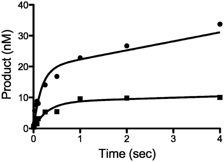
The Y-family DNA polymerase Rev1 is required for successful replication of G-quadruplex DNA (G4 DNA) in higher eukaryotes. Here we show that human Rev1 (hRev1) disrupts G4 DNA structures and prevents refolding in vitro. Nucleotidyl transfer by hRev1 is not necessary for mechanical unfolding to occur. hRev1 binds G4 DNA substrates with Kd,DNA values that are 4-15-fold lower than those of non-G4 DNA substrates. The pre-steady-state rate constant of deoxycytidine monophosphate (dCMP) insertion opposite the first tetrad-guanine by hRev1 is ∼56% as fast as that observed for non-G4 DNA substrates. Thus, hRev1 can promote fork progression by either dislodging tetrad guanines to unfold the G4 DNA, which could assist in extension by other DNA polymerases, or hRev1 can prevent refolding of G4 DNA structures. The hRev1 mechanism of action against G-quadruplexes helps explain why replication progress is impeded at G4 DNA sites in Rev1-deficient cells and illustrates another unique feature of this enzyme with important implications for genome maintenance.


Figures

Figure 1.
Figure 1.
The c-MYC promoter sequence forms G4 DNA in vitro. (a) The c-MYC promoter contains a 27-nt sequence (Pu27mer) with five runs of tandem guanines (1–5) that can form several G4 DNA structures. Tetrad-associated guanines are marked in bold, and the Pu27mer numbering scheme is in accordance with previous reports in the literature. A mutant of the wild-type Pu27mer, the Myc2345 G14/23T loop isomer, was chosen for our study of hRev1 activity on G4 DNA substrates. (b) DMS footprinting was performed with Myc2345 G14/23T 42-mer DNA in the presence or absence of an annealed 23-mer primer. Tetrad-associated guanines are protected from reaction with DMS on addition of potassium, indicative of G4 DNA folding. The addition of a primer does not alter the protection afforded to the G4 DNA-forming sequences. The nucleotide numbering scheme depicted to the left of each gel follows that of the Pu27mer (a). (c) Quantification of template base reactivity to DMS for experiments performed with each substrate either with KCl (100 mM, black bars) or without KCl (white bars) reveals a strong protection of guanines predicted to participate in the G4 DNA structure, indicating that both G4 DNA substrates (i.e. with or without 23-mer primer) behave as expected. A cartoon illustration of the DNA structures that depicts our interpretation of the experimental results is shown for clarity.
Figure 2.
Figure 2.
hRev1330-833 alters the pattern of DMS reactivity for G4 DNA-forming sequences. (a) Primer–template G4 DNA substrate (23/42mer, 5 pmoles) was incubated with hRev1330-833 (5 and 25 pmoles) either without salt (−) or in the presence of KCl (40 mM, +; 100 mM, ++), and DMS footprinting was performed. (b) Quantification of template base reactivity for experiments performed without salt (a) either in the absence of enzyme (‘white’ bars) or in the presence of hRev1330–833 (5 pmoles, ‘blue’ bars; 25 pmoles, ‘red’ bars) reveals that hRev1330–833 binds to unstructured ssDNA and attenuates reactivity with DMS. The mean ± SD is shown (n = 6). (c) Quantification of template base reactivity for experiments performed with KCl (40 mM, a) either in the absence of enzyme (‘white’ bars) or in the presence of hRev1330–833 (5 pmoles, ‘blue’ bars; 25 pmoles, ‘red’ bars) reveals that the tetrad-associated guanine (G22) at the extreme 3'′-terminus is deprotected on addition of hRev1330–833. The mean (± SD is shown (n = 3). (d) Quantification of template base reactivity for experiments performed with KCl (100 mM, a) either in the absence of enzyme (‘white’ bars) or in the presence of hRev1330–833 (5 pmoles, ‘blue’ bars; 25 pmoles, ‘red’ bars) reveals that the tetrad-associated guanines in the template strand are deprotected on addition of hRev1330–833. The mean ± SD is shown (n = 3). A cartoon illustration of the DNA structures that depicts our interpretation of the effect of hRev1330–833 binding is shown for clarity.
Figure 3.
Figure 3.
hRev1330–833 preferentially binds to G4 DNA-containing substrates. (a) hRev1330–833 was titrated into a solution containing either FAM-labeled ss-G4 DNA (square) or ss-non-G4 DNA (circle) oligonucleotides (1 nM). Changes in fluorescence polarization were measured, and the resulting data fit to a quadratic equation to yield the following equilibrium dissociation constants: ss-G4 DNA, Kd,DNA = 15.7 ± 8.7 nM; ss-non-G4 DNA, Kd,DNA = 65.7 ± 8.8 nM. (b) hRev1330–833 was titrated into a solution containing either FAM-labeled primer–template G4 DNA (square) or primer–template non-G4 DNA (circle) oligonucleotides (1 nM). Changes in fluorescence polarization were measured, and the resulting data fit to a quadratic equation to yield the following equilibrium dissociation constants: primer–template G4 DNA, Kd,DNA = 8.4 ± 2.5 nM; primer–template non-G4 DNA, Kd,DNA = 129 ± 20 nM. The reported values represent the mean ± SD (n = 3).
Figure 4.
Figure 4.
hRev1330–833 can disrupt G4 DNA structures. (a) Schematic illustration of the fluorescence-quenching assay used to monitor G4 DNA folding and unfolding. FAM fluorescence is quenched by dabcyl (Dab) moiety when G4 DNA folds and increases when G4 DNA unfolds. (b) Fluorescence quenching for FAM/Dab-42-mer ss-G4 DNA (20 nM) was monitored by stopped-flow following mixing with buffer without KCl (dashed line), 40 mM KCl (black trace), 40 mM KCl with either 5 nM (‘purple’ trace), 10 nM (‘dark blue’ trace) or 15 nM (‘light blue’ trace) hRev1330–833. The resulting curves were fit to Equation 3 to yield the following kinetic parameters: 40 mM KCl, no hRev1330–833: A = −58.2 ± 0.1 × 10⁻² V, kobs = 22.9 ± 0.1 × 10⁻³ s−1; 40 mM KCl, 5 nM hRev1330–833: A = −42.8 ± 0.1 × 10⁻² V, kobs = 23.1 ± 0.1 × 10⁻³ s−1; 40 mM KCl, 10 nM hRev1330–833: A = −10.6 ± 0.1 × 10⁻² V, kobs = 20.0 ± 0.5 × 10⁻³ s−1; 40 mM KCl, 15 nM hRev1330–833: A = −7.73 ± 0.05 × 10⁻² V, kobs = 14.7 ± 0.6 × 10⁻³ s−1. The fit of the data to Equation 3 is shown. (c) G4 DNA unfolding was monitored for a pre-formed FAM/Dab-42-mer ss-G4 DNA (20 nM) following addition of buffer (‘black’ trace) and either 20 nM (‘dark blue’ trace), 200 nM (‘light blue’ trace) hRev1330–833 or 200 nM Dpo4 (‘red’ trace). The data for experiments with hRev1330–833 were fit to Equation 4 to yield the following kinetic parameters: 20 nM hRev1330–833: n = 2, A = 6.6 ± 0.4 × 10⁻² V, kobs = 153.4 ± 0.1 × 10⁻⁴ s−1, k2 = 14.7 ± 0.1 × 10⁻⁵ V s−1; 200 nM hRev1330–833: n = 1, A = 23.1 ± 0.1 × 10⁻² V, kobs = 87.1 ± 0.4 × 10⁻⁴ s−1, k2 = 17.9 ± 0.6 × 10⁻⁵ V s−1. The fit of the data to Equation 4 is shown. (d) G4 DNA unfolding was monitored for FAM/Dab-18/42-mer G4 DNA (20 nM) following addition of either 200 nM hRev1330–833 alone (‘light blue’ trace), 200 nM hRev1330–833 with 2 mM MgCl2 (‘green’ trace), or 200 nM hRev1330–833 with 50 µM dCTP and 2 mM MgCl2 (‘purple’ trace). All experiments were performed in the presence of KCl (40 mM). The data for all three experiments were fit to Equation 4 to yield the following kinetic parameters: hRev1330–833 alone: n = 1, A = 15.2 ± 0.7 × 10⁻² V, kobs = 72.0 ± 0.7 × 10⁻⁴ s−1, k2 = 13.9 ± 0.1 × 10⁻⁵ V s−1; hRev1330–833 with MgCl2: n = 1, A = 9.9 ± 0.6 × 10⁻² V, kobs = 58.4 ± 0.8 × 10⁻⁴ s−1, k2 = 15.2 ± 0.1 × 10⁻⁵ V s−1; hRev1330–833 with dCTP/MgCl2: n = 1, A = 11.1 ± 0.6 × 10⁻² V, kobs = 71.4 ± 0.9 × 10⁻⁴ s−1, k2 = 14.6 ± 0.1 × 10⁻⁵ V s−1. The fit of the data to Equation 4 is shown. The data represent the average of two to four experiments, and the error represents the standard error of the fit. A cartoon schematic is shown to the left of each reaction trace to better illustrate the experimental design and our interpretation of the results.
Figure 5.
Figure 5.
Representative dCMP insertion results for 23/42-mer DNA substrates. (a) Results of a time-course experiment measuring hRev1330–833 (5 nM) insertion of dCTP (1 mM) on 23/42-mer non-G4 DNA or G4 DNA substrates (200 nM). hRev1330–833 can insert dCMP opposite the first three tetrad-guanines (G22–G20) but the rate of insertion is less than that observed for the non-G4 DNA substrate. Representative gels for steady-state kinetic analysis of hRev1 (10 nM) insertion of dCTP at the concentrations indicated on either (b) 23/42-mer non-G4 DNA or (c) 23/42-mer G4 DNA (200 nM) substrates. The reaction time for non-G4 DNA substrates was 5 min and that for G4 DNA substrates was 10 min.
Figure 6.
Figure 6.
Pre-steady-state kinetic analysis of hRev1330–833 activity on G4 DNA substrates. hRev1330–833 (400 nM) was pre-incubated with 23/42-mer DNA (100 nM), and the reaction initiated by addition of dCTP (250 µM) and MgCl2 (2 mM). Product formation was plotted as a function of time and the data fit to Equation 4 (n = 1), yielding the following kinetic parameters: non-G4 DNA (circle), A = 19.4 ± 1.6 nM, kobs = 5.9 ± 1.4 s−1, k2 = 2.9 ± 0.4 nM s−1; G4 DNA (square), A = 8.5 ± 0.8 nM, kobs = 3.3 ± 0.8 s−1 and k2 = 0.46 ± 0.17 nM s−1.

Similar articles

Cited by

References

    1. Huppert JL, Balasubramanian S. Prevalence of quadruplexes in the human genome. Nucleic Acids Res. 2005;33:2908–2916. - PMC - PubMed
    1. Henderson E, Hardin CC, Walk SK, Tinoco I, Jr, Blackburn EH. Telomeric DNA oligonucleotides form novel intramolecular structures containing guanine-guanine base pairs. Cell. 1987;51:899–908. - PubMed
    1. Zahler AM, Williamson JR, Cech TR, Prescott DM. Inhibition of telomerase by G-quartet DNA structures. Nature. 1991;350:718–720. - PubMed
    1. Lipps HJ, Rhodes D. G-quadruplex structures: in vivo evidence and function. Trends Cell Biol. 2009;19:414–422. - PubMed
    1. Biffi G, Tannahill D, McCafferty J, Balasubramanian S. Quantitative visualization of DNA G-quadruplex structures in human cells. Nat. Chem. 2013;5:182–186. - PMC - PubMed

Publication types