. 2014 Jan 21;111(3):966-71.
doi: 10.1073/pnas.1314997111. Epub 2013 Dec 23.

Crystal structures of a pentameric ligand-gated ion channel provide a mechanism for activation

Affiliations

Crystal structures of a pentameric ligand-gated ion channel provide a mechanism for activation

Ludovic Sauguet et al. Proc Natl Acad Sci U S A. .

Abstract

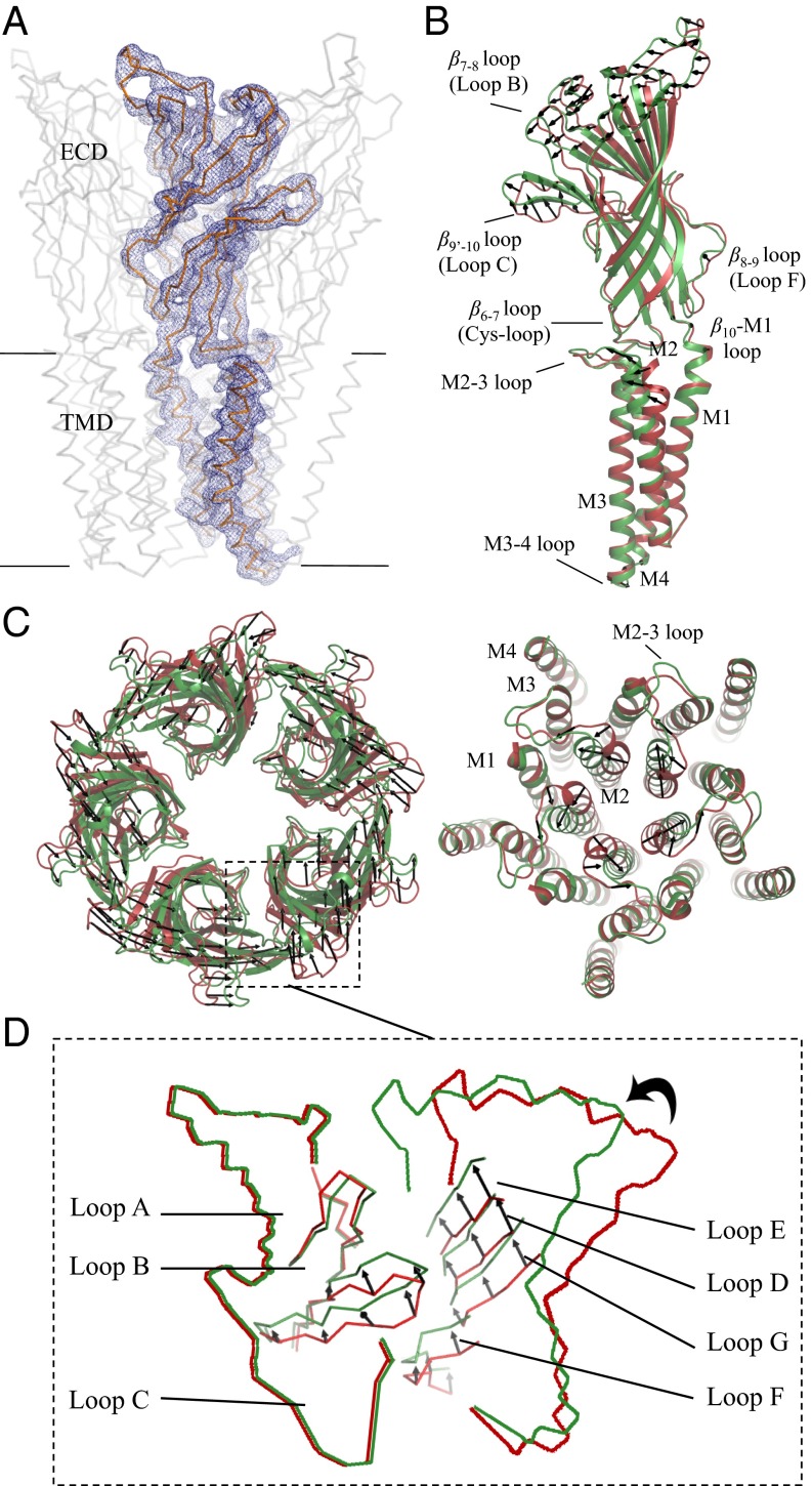
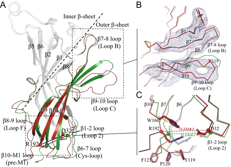
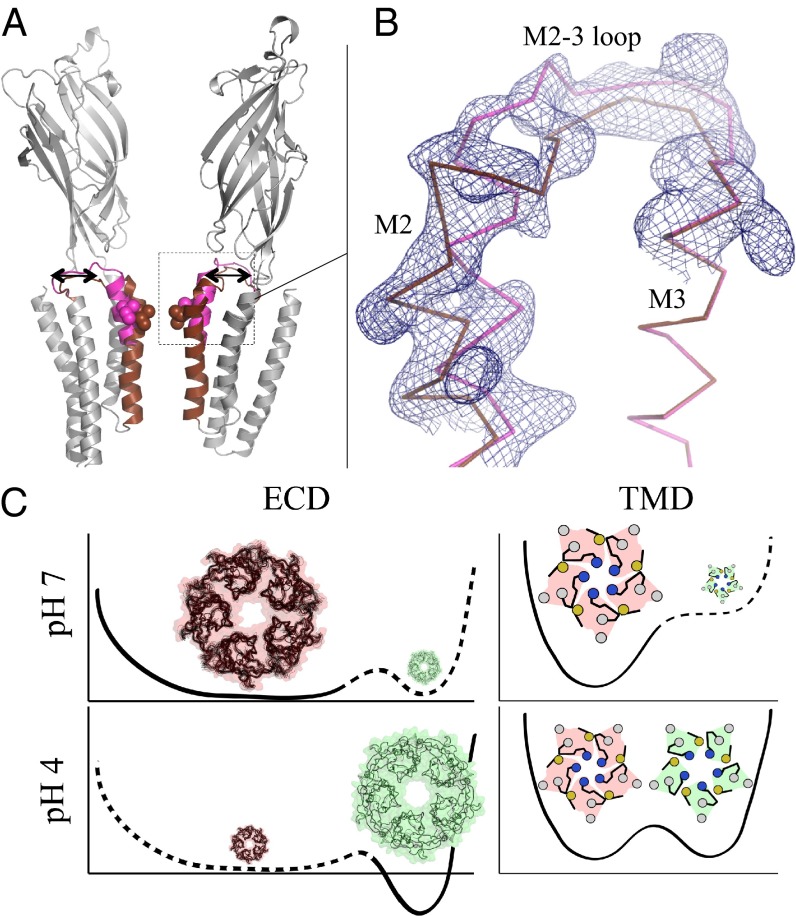
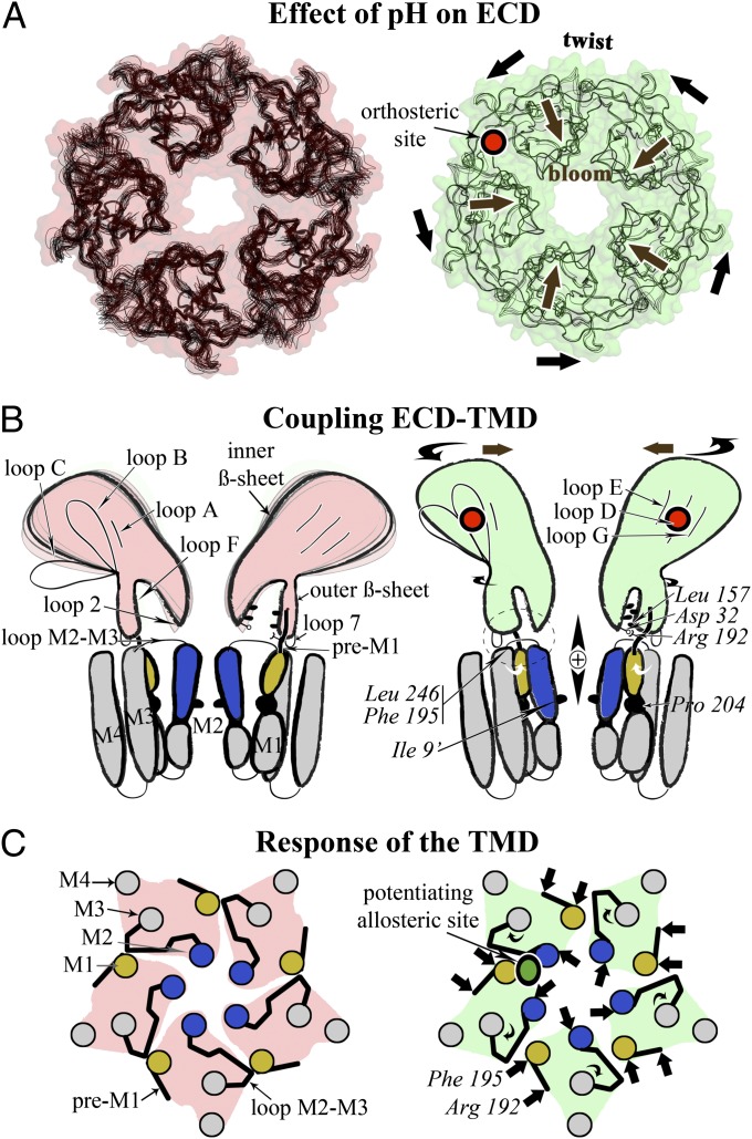
Pentameric ligand-gated ion channels mediate fast chemical transmission of nerve signals. The structure of a bacterial proton-gated homolog has been established in its open and locally closed conformations at acidic pH. Here we report its crystal structure at neutral pH, thereby providing the X-ray structures of the two end-points of the gating mechanism in the same pentameric ligand-gated ion channel. The large structural variability in the neutral pH structure observed in the four copies of the pentamer present in the asymmetric unit has been used to analyze the intrinsic fluctuations in this state, which are found to prefigure the transition to the open state. In the extracellular domain (ECD), a marked quaternary change is observed, involving both a twist and a blooming motion, and the pore in the transmembrane domain (TMD) is closed by an upper bend of helix M2 (as in locally closed form) and a kink of helix M1, both helices no longer interacting across adjacent subunits. On the tertiary level, detachment of inner and outer β sheets in the ECD reshapes two essential cavities at the ECD-ECD and ECD-TMD interfaces. The first one is the ligand-binding cavity; the other is close to a known divalent cation binding site in other pentameric ligand-gated ion channels. In addition, a different crystal form reveals that the locally closed and open conformations coexist as discrete ones at acidic pH. These structural results, together with site-directed mutagenesis, physiological recordings, and coarse-grained modeling, have been integrated to propose a model of the gating transition pathway.

Keywords: X-ray crystallography; allostery; cys-loop receptor; signal transduction.


Conflict of interest statement

The authors declare no conflict of interest.

Figures

Fig. 1.
Fig. 1.
Superimposition of the pH 4 and pH 7 GLIC structures. (A) The Cα trace of a full-length monomer of GLIC is represented in gray. The blue mesh is the 2mFo–DFc NCS-averaged electron density map contoured at a level of 1.5 σ. (B) Side view of a full-length monomer; superimposition of the open pH 4 (green) and closed pH 7 (red) GLIC structures; the black arrows show the direction of the motion observed from the closed form to the open form. (C) Upper view of ECDs. (D) Upper view of TMDs. (E) Enlarged view of the orthosteric agonist binding site at the interface.
Fig. 2.
Fig. 2.
Tertiary reorganization observed in an ECD monomer. (A) The structural elements that differ between the pH 4 and pH 7 ECD GLIC structures are shown in green for pH 4 and in red for pH 7 GLIC. Other parts of the structures are in gray. Residues Asp32 and Arg192 are shown as sticks. The dashed line delineates the inner and outer β sheets. (B) Enlarged view of the orthosteric agonist binding site illustrating the detachment of loop C from loop B. The blue mesh is the 2mFo–DFc NCS-averaged electron density map contoured at a level of 1.5 σ. (C) The interface between the ECD and the TMD at the level of the β1–β2, β6–β7, and β10–M1 loops. The dashed line indicates the distance between the Cα atoms of Asp32 and Arg192.
Fig. 3.
Fig. 3.
GLIC open and LC forms coexist at pH 4. (A) Ribbon representation of the GLIC receptor showing the equilibrium of the M2 helices and M2–M3 loops corresponding to the coexisting LC (brown) and open (magenta) conformations in GLICHis10 structure. The rest of the structure is colored in gray. The receptor is viewed from the side, and the two front subunits are removed for clarity. (B) Enlarged view showing the 2mFo–DFc electron density map surrounding the M2 helix and the M2–M3 loop contoured at a level of 1 σ (blue mesh). (C) Energy landscape inferred from the pH 7 crystal form (Upper, pink) and the pH 4 form (Lower, green) described here, for each domain (TMD and ECD).
Fig. 4.
Fig. 4.
A sequential model for the activation of pLGICs. (A) The reduced flexibility in the ECD when going from the pH 7 form (Left, pink) to the pH 4 form (Right, green) is illustrated with superimposed atomic models. The quaternary associated twist and bloom motions are indicated with black arrows. The binding site of the agonist is shown in red. (B) Changes of the ECD–TMD interface in both states are shown in a schematic way. Residues shown experimentally to be involved in these interactions are indicated. The top of M2 helix is in blue, and the N terminus of M1 is in yellow. (C) Schematic view from the top and down the C5 axis of the TMD conformation in both states. The binding site for a positive modulator (anesthetics) is shown in green.

Similar articles

Cited by

References

    1. Corringer PJ, et al. Structure and pharmacology of pentameric receptor channels: From bacteria to brain. Structure. 2012;20(6):941–956. - PubMed
    1. Hilf RJ, Dutzler R. X-ray structure of a prokaryotic pentameric ligand-gated ion channel. Nature. 2008;452(7185):375–379. - PubMed
    1. Hilf RJ, Dutzler R. Structure of a potentially open state of a proton-activated pentameric ligand-gated ion channel. Nature. 2009;457(7225):115–118. - PubMed
    1. Bocquet N, et al. X-ray structure of a pentameric ligand-gated ion channel in an apparently open conformation. Nature. 2009;457(7225):111–114. - PubMed
    1. Hibbs RE, Gouaux E. Principles of activation and permeation in an anion-selective Cys-loop receptor. Nature. 2011;474(7349):54–60. - PMC - PubMed

Publication types