. 2014 Jan 7;111(1):469-74.
doi: 10.1073/pnas.1321454111. Epub 2013 Dec 23.

Protein kinase LKB1 regulates polarized dendrite formation of adult hippocampal newborn neurons

Affiliations

Protein kinase LKB1 regulates polarized dendrite formation of adult hippocampal newborn neurons

Wei Huang et al. Proc Natl Acad Sci U S A. .

Abstract

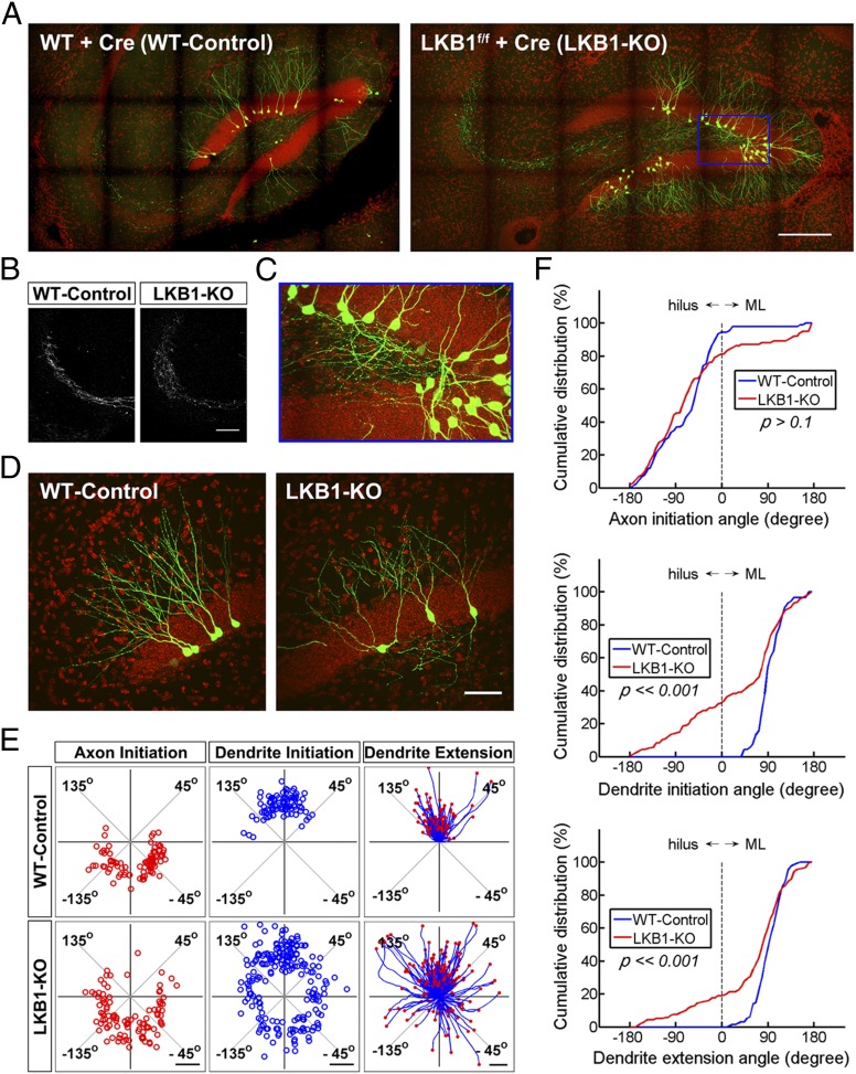
Adult-born granule cells in the dentate gyrus of the rodent hippocampus are important for memory formation and mood regulation, but the cellular mechanism underlying their polarized development, a process critical for their incorporation into functional circuits, remains unknown. We found that deletion of the serine-threonine protein kinase LKB1 or overexpression of dominant-negative LKB1 reduced the polarized initiation of the primary dendrite from the soma and disrupted its oriented growth toward the molecular layer. This abnormality correlated with the dispersion of Golgi apparatus that normally accumulated at the base and within the initial segment of the primary dendrite, and was mimicked by disrupting Golgi organization via altering the expression of Golgi structural proteins GM130 or GRASP65. Thus, besides its known function in axon formation in embryonic pyramidal neurons, LKB1 plays an additional role in regulating polarized dendrite morphogenesis in adult-born granule cells in the hippocampus.

Keywords: Golgi deployment; adult neurogenesis; neuronal polarization.


Conflict of interest statement

The authors declare no conflict of interest.

Figures

Fig. 1.
Fig. 1.
Expression pattern of LKB1 in the adult dentate gyrus. Sample confocal images of hippocampal slices in adult Nestin-CreER/Rosa26-YFP mice that were double-stained with antibodies against GFAP/LKB1, MCM2/LKB1, DCX/LKB1, and NeuN/LKB1. White dotted circles, the nuclei of colabeled cells. Observation time: 1-mo postinjection. (Scale bar, 10 μm.)
Fig. 2.
Fig. 2.
LKB1 deletion in adult-born granule cells affects oriented initiation and extension of the primary dendrite. (A) Large-scale montage images of hippocampal sections in wild-type and LKB1-flox mice that were infected with Ubi-Cre-E2A-GFP retroviruses. Red, DAPI; green, GFP. Observation time: 3-wk postinjection. (Scale bar, 200 μm.) (B) Sample confocal images of Mossy fibers in the CA3 region of WT and LKB1-flox mice infected with Ubi-Cre-E2A-GFP, respectively. (Scale bar, 100 μm.) (C) Confocal image of aberrant dendrites extending into the hilus, a higher magnification view (10:3) of the region marked by the blue box in A. (D) Sample confocal images of retrovirus-infected granule cells in WT and LKB1-flox mice at a higher resolution. (Scale bar, 50 μm.) (E) Angular distribution of the axon and dendrite initiation sites on the soma, and tracing of primary dendrites. [Scale bars, 5 μm (Left, Center) 20 μm (Right).] (n = 80–120 cells from four to six mice for each plot.) (F) Cumulative percentage plots for the angular distribution of axon and dendrite initiation sites, and end points of primary dendrites (P value as marked; K-S test).
Fig. 3.
Fig. 3.
LKB1 deletion leads to dispersion of Golgi apparatus. (A) Sample images of immunostained Golgi apparatus within retrovirus-labeled cells after removing signals within surrounding cells. Green, GFP; red, GRASP65. Observation time: 3-wk postinjection. (Scale bar, 10 μm.) (B) Summation of immunofluorescence intensities of GRASP65-labled Golgi apparatus for all retrovirus-infected neurons (n = 60–80 cells, from four mice each). (Scale bar, 5 μm.) (C) Average percentage of total fluorescence in each of four quadrants (marked by dashed lines, see B), calculated for each cell before averaging. The same dataset as in B. Values represent mean ± SEM (***P < < 0.001; t test).
Fig. 4.
Fig. 4.
Down-regulating LKB1 and manipulating of Golgi structural proteins led to similar impairment of dendrite morphognesis. (A and C) Sample confocal images of neurons infected with retroviruses that expressed scrambled-shRNA, LKB1-shRNA, or GM130-shRNA, as well as that expressed GFP or GRASP65. Observation time: 3-wk postinjection. (Scale bar, 50 μm.) (B and D) Angular distribution of dendrite initiation sites and tracing of primary dendrites. [Scale bars, 5 μm (Upper) 20 μm (Lower).] (n = 160–240 cells, from 8 to 12 mice each.) (E) Cumulative percentage plots for the angular distribution of dendrite initiation sites and end points of primary dendrites for all data in B and D. (P value as marked; K-S test).

Similar articles

Cited by

References

    1. Ming GL, Song H. Adult neurogenesis in the mammalian central nervous system. Annu Rev Neurosci. 2005;28:223–250. - PubMed
    1. Clelland CD, et al. A functional role for adult hippocampal neurogenesis in spatial pattern separation. Science. 2009;325(5937):210–213. - PMC - PubMed
    1. Sahay A, et al. Increasing adult hippocampal neurogenesis is sufficient to improve pattern separation. Nature. 2011;472(7344):466–470. - PMC - PubMed
    1. Nakashiba T, et al. Young dentate granule cells mediate pattern separation, whereas old granule cells facilitate pattern completion. Cell. 2012;149(1):188–201. - PMC - PubMed
    1. Santarelli L, et al. Requirement of hippocampal neurogenesis for the behavioral effects of antidepressants. Science. 2003;301(5634):805–809. - PubMed

Publication types

MeSH terms

LinkOut - more resources