Review
. 2007 Sep;83(6):164-74.
doi: 10.2183/pjab.83.164.

Development of mast cells

Affiliations
Review

Development of mast cells

Yukihiko Kitamura et al. Proc Jpn Acad Ser B Phys Biol Sci. 2007 Sep.

Abstract

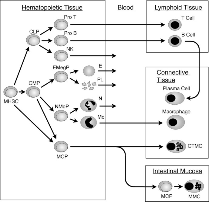
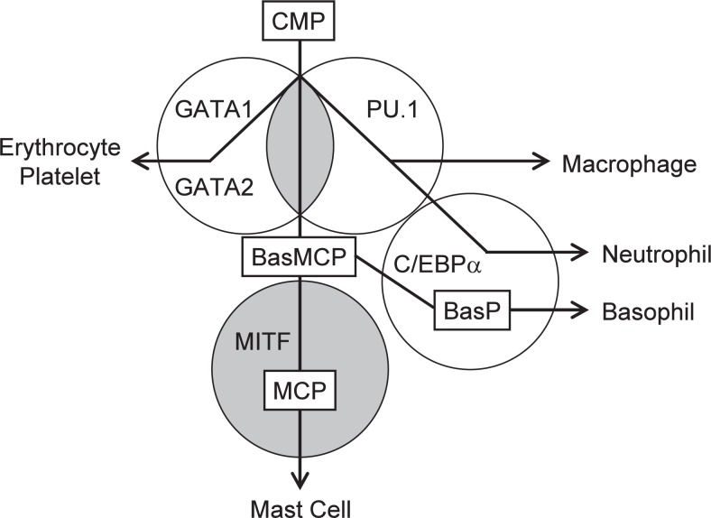
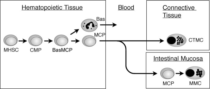
Mast cells are progeny of the multipotential hematopoietic stem cell (MHSC). Mast cell-committed progenitors (MCPs) leave hematopoietic tissues, migrate in peripheral blood, invade to connective or mucosal tissue, proliferate and differentiate to morphologically identifiable mast cells. Phenotype of mast cells (connective tissue-type or mucosal type) is determined by the site of lodgment of MCPs. Most progeny of the multipotential hematopoietic stem cell lose proliferation potential after maturation, but connective tissue-type mast cells (CTMCs) possess appreciable proliferation potential after maturation. Even after functioning by degranulation, CTMCs proliferate and restore the original morphology. The most important cytokine for development and survival of mast cells is KIT ligand, and the KIT receptor tyrosine kinase is expressed through the whole developmental process of mast cells from MHSC to mature mast cells. The loss-of-function mutation of KIT gene results in depletion of mast cells, whereas its gain-of-function mutation causes mast cell tumors. Since mast cells are involved in various disease processes, intervention in development of mast cells might be beneficial to the treatment.

Keywords: KIT; MITF; allergy; basophil; hematopoietic stem cell; mast cell.


Figures

Fig. 1.
Fig. 1.
Genetically mast cell-deficient mouse. (A) A wildtype (WB X C57BL/6)F1-+/+ (WBB6F1-+/+) mouse. (B) A mast cell-deficient WBB6F1-KitW/KitW-v mouse with white coat and black eyes. The WBB6F1-KitSl/KitSl-d mouse shows the undistinguishable appearance. (C) Skin section of the WBB6F1-+/+ mouse stained with toluidine blue. Many toluidine blue-positive mast cells are observed. (D) Skin section of WBB6F1-KitW/KitW-v mouse. No mast cells are detectable.
Fig. 2.
Fig. 2.
Development of mast cells from the multipotential hematopoietic stem cell. CLP, common lymphoid progenitor; CMP, common myeloid progenitor; CTMC, connective tissue-type mast cell; E, erythrocyte: EMegP, erythrocyte/megakaryocyte progenitor; MCP, mast cell-committed progenitor; MHSC, multipotential hematopoietic stem cell; MMC, mucosal mast cell; Mo, monocyte; N, neutrophil; NMoP, neutrophil/monocyte progenitor; PL, platelet.
Fig. 3.
Fig. 3.
Transcription factors that are involved in development of mast cells. GATA-1, GATA-2 and PU.1 are required in the early stage; C/EBPalpha in the intermediate stage; and MITF in the late stage. BasP, basophil progenitor; BasMCP, basophil/mast cell progenitor; CMP, common myeloid progenitor; MCP, mast cell-committed progenitor.
Fig. 4.
Fig. 4.
Differentiation of basophils and mast cells from the multipotential hematopoietic stem cell. The presence of bipotent progenitor committed to basophils and mast cells (BasMCP) was identified in the spleen of mice. Bas, basophil; BasMCP, basophil/mast cell progenitor; CMP, common myeloid progenitor; CTMC, connective tissue-type mast cell; MCP, mast cell-committed progenitor; MHSC, multipotential hematopoietic stem cell; MMC, mucosal mast cell.
None

Similar articles

Cited by

References

    1. Selye, H. (1965) The Mast Cells. Butterworths, Washington
    1. Ishizaka, T. and Ishizaka, K. (1984) Activation of mast cells for mediator release through IgE receptors. Prog. Allergy 34, 188–235 - PubMed
    1. Galli, S.J. and Kitamura, Y. (1987) Genetically mast-cell-deficient W/Wv and Sl/Sld mice. Their value for the analysis of the roles of mast cells in biologic responses in vivo. Am. J. Pathol. 127, 191–198 - PMC - PubMed
    1. Galli, S.J. and Wershil, B.K. (1996) The two faces of the mast cell. Nature 381, 21–22 - PubMed
    1. Ruitenberg, E.J. and Elgersma, A. (1976) Nature 264, 258–260 - PubMed

LinkOut - more resources