Review
. 2007 Dec;83(9-10):296-319.
doi: 10.2183/pjab/83.296.

Physiology and pathophysiology of prostanoid receptors

Affiliations
Review

Physiology and pathophysiology of prostanoid receptors

Shuh Narumiya. Proc Jpn Acad Ser B Phys Biol Sci. 2007 Dec.

Abstract

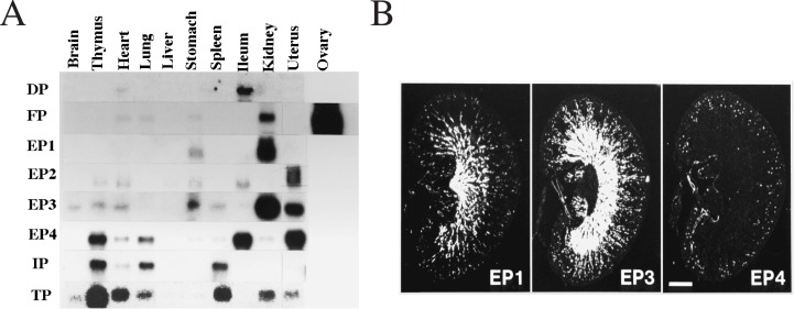
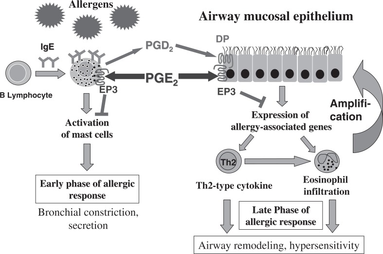
Prostanoids, consisting of prostaglandins (PGs) and thromboxanes (TXs), are oxygenated products of C20 unsaturated fatty acids. They include PGD2, PGE2, PGF2 α , PGI2, and TXA2. Given that aspirin-like nonsteroidal anti-inflammatory drugs exert their actions by suppressing prostanoid production, prostanoids have been implicated in processes inhibited by these drugs, including inflammation, fever, and pain. Prostanoids also contribute to vascular homeostasis, reproduction, and regulation of kidney and gastrointestinal functions. How prostanoids exert such a variety of actions had remained unclear, however. Prostanoids are released outside of cells immediately after their synthesis and exert their actions by binding to receptors on target cells. We have identified a family of eight types or subtypes of G protein-coupled receptors that mediate prostanoid actions. Another G protein-coupled receptor was also identified as an additional receptor for PGD2. Genes for these receptors have been individually disrupted in mice, and analyses of these knockout mice have not only elucidated the molecular and cellular mechanisms of known prostanoid actions but also revealed previously unknown actions. In this article, I review the physiological and pathophysiological roles of prostanoids and their receptors revealed by these studies.

Keywords: G protein–coupled receptor; cyclooxygenase; prostaglandin; thromboxane.


Figures

Fig. 1
Fig. 1
Pathway of prostanoid biosynthesis. Arachidonic acid is metabolized by the action of cyclooxygenase (COX) first to prostaglandin endoperoxide (PGG2) and then to PGH2, which is subsequently converted to various PGs and TXA2 by respective synthases. Aspirin-like nonsteroidal anti-inflammatory drugs (NSAIDs) exert their effects by inhibiting COX and thereby suppressing prostanoid biosynthesis.
Fig. 2
Fig. 2
(A) Membrane topology model for the human TXA2 receptor (modified from ref. with permission). Each amino acid is represented by the single-letter code within a circle. White letters in black circles indicate residues that are conserved among members of the prostanoid receptor family. The model is based on hydrophilicity analysis of the amino acid sequence. Asparagine residues at positions 4 and 16 are glycosylated. (B) Phylogenetic tree and functional grouping of prostanoid receptors (modified from ref. with permission).
Fig. 3
Fig. 3
(A) Northern blot analysis of transcripts derived from the eight types or subtypes of prostanoid receptor in mouse tissues (from ref. with permission). (B) In situ hybridization of mRNAs for the EP1, EP3, and EP4 subtypes of the PGE receptor in mouse kidney (from ref. with permission). Scale bar, 1 mm.
Fig. 4
Fig. 4
(A) Impaired febrile response to LPS in mice deficient in EP3 (from ref. with permission). Wild-type (closed circles), EP1−/− (open circles), or EP3−/− (open triangles) mice were injected with LPS at time zero; control wild-type mice were injected with vehicle (closed triangles). (B) Current model for the neural pathway that underlies fever generation. OVLT, organosum vasculatum lamina terminalis; POA, preoptic area. (C) Impaired ACTH response to LPS in mice deficient in EP1 or EP3 (from ref. with permission). Wild-type (BL6) or the indicated EP knockout mice were injected with LPS or vehicle (−) and the plasma concentration of ACTH was measured 1 h thereafter. **P < 0.01.
Fig. 5
Fig. 5
Roles and mechanisms of action of the PGE2-EP2 pathway in expansion of the cumulus oophorus and fertilization (A) and of the PGF2α-FP pathway in induction of parturition (B).
Fig. 6
Fig. 6
Atherosclerosis in the innominate artery of ApoE−/−, ApoE−/−TP−/−, and ApoE−/−IP−/− mice (from ref. with permission). Arterial cross-sections prepared from 45-week-old mice were stained with hematoxylin-eosin.
Fig. 7
Fig. 7
A model for functional antagonism between the PGD2-DP pathway and the PGE2-EP3 pathway in development of allergic inflammation associated with asthma. PGD2 is released in response to mast cell activation and facilitates allergic inflammation at least in part by acting at DP in the airway epithelium. On the other hand, PGE2 is produced in the surrounding tissues during mast cell activation and suppresses development of allergic inflammation by inhibiting induction of various asthma-related genes in the epithelium.
Fig. 8
Fig. 8
Sites of prostanoid actions in skin immune responses. The PGE2-EP4 pathway and the PGD2-DP pathway regulate dendritic cell migration and maturation in mutually antagonistic manners, whereas the TXA2-TP pathway regulates the interaction between dendritic cells and naïve T cells in draining lymph nodes. LC, Langerhans cell.
Fig. 9
Fig. 9
Exacerbation of DSS-induced colitis in EP4-deficient mice (from ref. with permission). EP4−/− mice exhibit severe hemorrhagic colitis in response to the administration of 3% DSS in drinking water, which induces only marginal colitis in wild-type mice. The gross appearance (A) and hematoxylin-eosin staining (B) of the intestine from wild-type (EP4+/+) or EP4−/− mice are shown.
Fig. 10
Fig. 10
Ovariectomy-induced bone loss and its prevention by administration of an EP4-selective agonist in rats (from ref. with permission). Control rats as well as ovariectomized rats treated with vehicle (Ovx) or with a low or high dose of ONO-4819 before and after surgery were examined for bone loss by X-ray computed tomography.
None

Similar articles

Cited by

References

    1. Schaloske, R. H. and Dennis, E. A. (2006) Biochim. Biophys. Acta 1761, 1246–1259 - PubMed
    1. Smith, W. L., DeWitt, D. L. and Garavito, R. M. (2000) Annu. Rev. Biochem. 69, 145–182 - PubMed
    1. Murakami, M. and Kudo, I. (2004) Prog. Lipid Res. 43, 3–35 - PubMed
    1. Bito, L.Z. (1972) J. Physiol. (Lond.) 221, 371–387 - PMC - PubMed
    1. Jones, R.L., Peesapati, V. and Wilson, N.H. (1982) Br. J. Pharmacol. 76, 423–438 - PMC - PubMed

LinkOut - more resources