. 2009 Dec 7:1:39-52.
doi: 10.2147/bctt.s6255. eCollection 2009.

p130Cas substrate domain signaling promotes migration, invasion, and survival of estrogen receptor-negative breast cancer cells

Affiliations

p130Cas substrate domain signaling promotes migration, invasion, and survival of estrogen receptor-negative breast cancer cells

Anna C Cunningham-Edmondson et al. Breast Cancer (Dove Med Press). .

Abstract

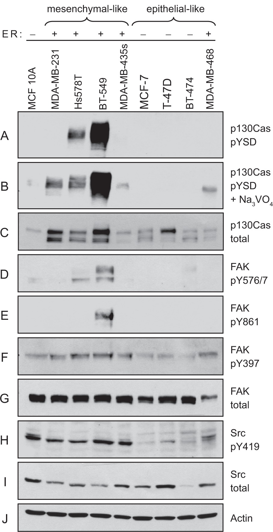
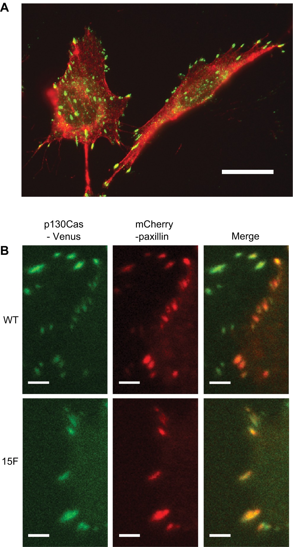
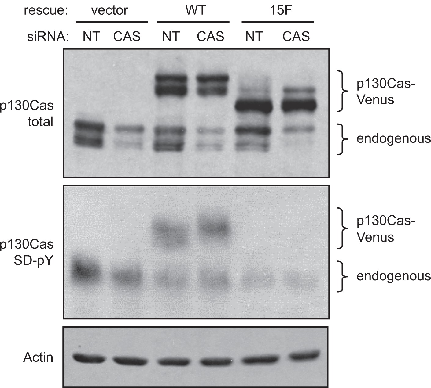
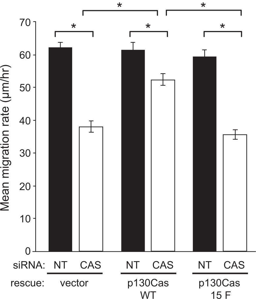
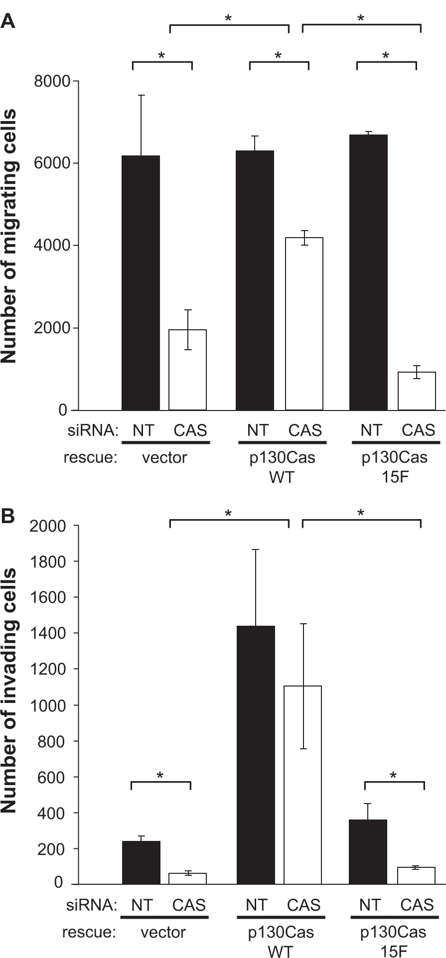
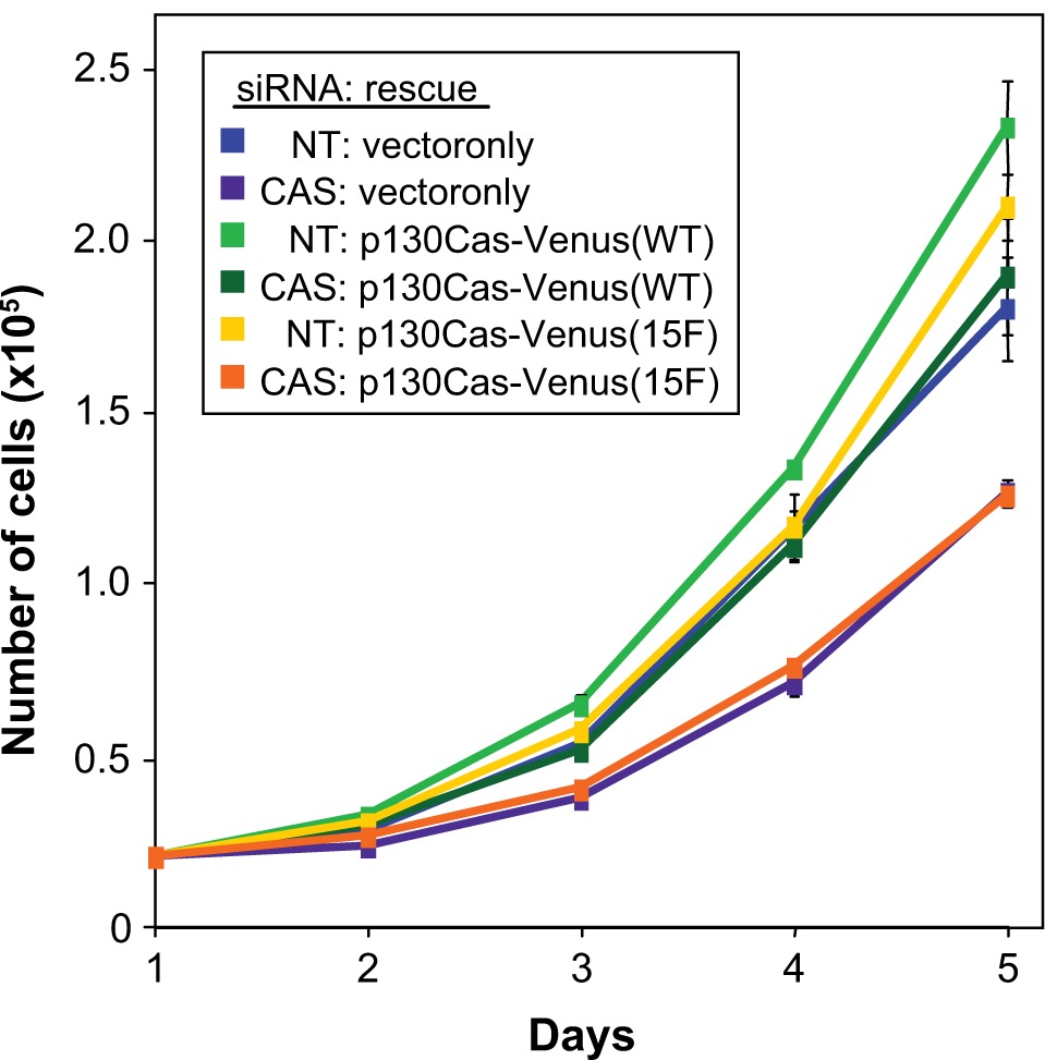
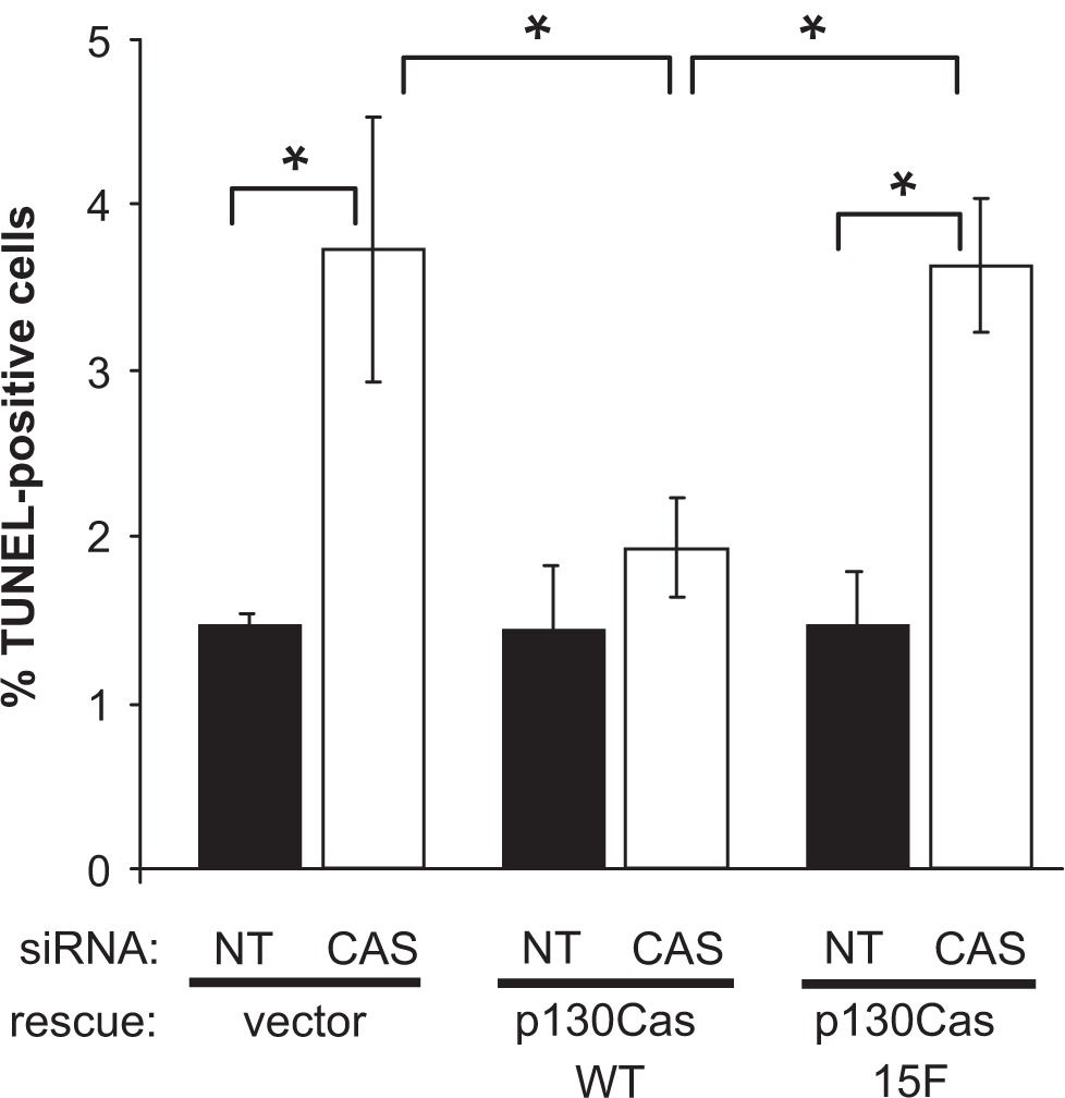
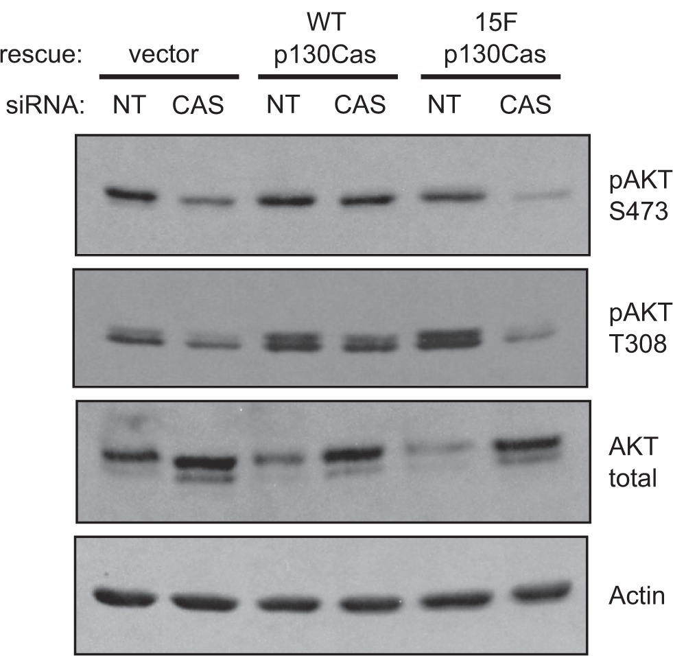
Elevated Src tyrosine kinase activity is commonly observed in breast cancer and likely contributes to neoplasia and malignancy. p130Cas ("Crk-associated substrate") is a major Src substrate found at the sites where integrins mediate cell adhesion to the extracellular matrix. Src phosphorylates multiple tyrosines in the p130Cas "substrate domain" (SD) and this signaling event has been implicated in the promotion of cell motility, primarily from studies on fibroblasts. In breast cancer, studies on p130Cas have focused on its role in conferring antiestrogen resistance to cells that express the estrogen receptor (ER+). However, little is known regarding the role of p130Cas in the more aggressive estrogen receptor negative (ER-) breast cancers for which there is a need for development of effective targeted therapies. We found high levels of p130Cas SD tyrosine phosphorylation to be a common characteristic of ER- breast cancer cell lines, with particularly high levels observed for the BT-549 cell line. Using RNA interference to knock down p130Cas expression in BT-549 cells, combined with rescue by WT p130Cas versus a signaling-deficient control, we provide evidence that p130Cas SD tyrosine phosphorylation is an important signaling event in the migration, invasion, proliferation, and survival of this ER-breast cancer cell line.

Keywords: BCAR1; FAK; Src; adhesion; integrins; tyrosine phosphorylation.


Figures

Figure 1
Figure 1
Elevated p130Cas SD tyrosine phosphorylation and Src activity in ER-breast cancer cell lines. Total cell lysates prepared from four representative mesenchymal-like breast cancer cell lines (MDA-MB-231, Hs578T, BT-549, and MDA-MB-435s) and four representative epithelial-like breast cancer cell lines (MCF-7, T-47D, BT-474, and MDA-MB-468) were analyzed by immunoblotting. MCF-10A was also included as a control representative of noncancerous breast epithelial cells. A and B) P130Cas SD tyrosine phosphorylation (pYSD) was detected using equimolar amounts of phosphospecific antibodies pCAS-165, pCAS-249, pCAS-410. In B) the cells were pretreated with sodium vanadate (Na3VO4) to enhance detection by the pCas antibodies. C) Total p130Cas protein was detected using the CAS-TL antibody. DG) FAK tyrosine phosphorylation levels were evaluated using phosphospecific antibodies against known Src targets (Tyr-576/Tyr577 and Tyr-861), and the FAK autophosphorylation site Tyr-397. H and I) Activated Src was detected using a phosphospecific antibody against the autophosphorylation site Tyr-419 (H). J) Actin was detected as a loading control.
Figure 2
Figure 2
Localization of p130Cas SD tyrosine phosphorylation and p130Cas-Venus variants to focal adhesions. A) Cell immunostaining was performed on BT-549 cells grown on fibronectin. Phosphorylated p130Cas SD was detected using pCAS-165 antibody followed by the FITC-conjugated anti-rabbit IgG (green). F-actin, including stress fibers associated with focal adhesions, was visualized using Alexa 594-phalloidin (red). Bar indicates 20 μm. B) TIRF microscopy was used to show the localization of WT and 15F p130Cas-Venus variants to focal adhesions within BT-549 cells. Cells expressing p130Cas-Venus variants (green) were transiently transfected with plasmid mCherry-C1-paxillin (red), an established focal adhesion marker. The images show focal adhesions located near the cell peripheries. Bars indicate 5 μm.
Figure 3
Figure 3
Knockdown of p130Cas in BT-549 cells and expression of siRNA-resistant mouse p130Cas-Venus variants. BT-549 cells transfected with either the CAS siRNA or NT control siRNA were lysed 48 hours post-transfection, and total cell lysates were assessed by immunoblot analysis of replicate blots. The p130Cas knockdown was performed on BT-549 cells that stably express either WT mouse p130Cas (resistant to the siRNA) or the signaling-deficient 15F mouse p130Cas variant, both carrying the Venus fluorophore tag. Cells stably expressing Venus only (empty rescue vector) were also included in the analysis. (Top panel) Both endogenous p130Cas and the p130Cas-Venus variants were detected by immunoblotting with the CAS-TL antibody. (Middle panel) Tyrosine phosphorylation of the p130Cas SD was assessed using a mixture of phosphospecific antibodies (pCAS-165, pCAS-249, pCAS-410). Prior to lysis, cells were treated with vanadate to enhance the detection of p130Cas SD tyrosine phosphorylation. (Bottom panel) Actin was detected as a loading control.
Figure 4
Figure 4
p130Cas SD tyrosine phosphorylation is required for efficient BT-549 cell migration. BT-549 cells stably expressing mouse p130Cas-Venus variants (WT vs 15F) or Venus only (rescue vector) were transfected with either the CAS or NT siRNA and analyzed for migration by scratch wound assay. After wounding, cell movement into the denuded area was recorded over a 10-hour period and mean migration rates (distance in μm/hour) were determined from measurements of the distance moved by 120 individual cells from each experimental condition. Error bars represent standard error of the mean. Statistical significance was determined by Student’s t-test (*P < 0.001).
Figure 5
Figure 5
p130Cas SD tyrosine phosphorylation promotes BT549 cell invasion. BT549 cells stably expressing mouse p130Cas-Venus variants (WT vs 15F) or Venus only (rescue vector) were transfected with either the CAS or NT siRNA and analyzed for migration and invasion by modified Boyden chamber assays. For each experimental condition, triplicate assays were performed wherein 100,000 cells suspended in serum-free media were placed in the top of the chamber and allowed to migrate, over 24 hours, through porous filters towards media containing 10% serum. A) Migration through uncoated filters. B) Invasion through Matrigel-coated filters. Mean numbers of migrating or invading cells per chamber are shown for a representative experiment. Error bars represent the standard error of the mean. Statistical significance was determined by Student’s t-test (*P < 0.05).
Figure 6
Figure 6
p130Cas SD tyrosine phosphorylation is required for efficient BT-549 cell proliferation. BT-549 cells stably expressing mouse p130Cas-Venus variants (WT vs 15F) or Venus only (rescue vector) were transfected with either the CAS or NT siRNA and analyzed for proliferation rate by determining growth curves. For each experimental condition, 20,000 cells were plated in replicate dishes in media containing 10% FBS and cells were counted, in triplicate, at daily intervals over the subsequent five-day period. Shown are resulting adherent growth curves from a representative experiment, obtained by plotting the mean number of cells counted at each time point Error bars represent standard deviations. Statistical significance was assessed on days 3–5 by Student’s t-test. Proliferation rates of two populations, CAS siRNA:vector only (purple line) and CAS siRNA:p130Cas-Venus (15F) (orange line), were significantly less than the others (P < 0.002).
Figure 7
Figure 7
p130Cas SD tyrosine phosphorylation enhances BT-549 cell survival. BT-549 cells stably expressing mouse p130Cas-Venus variants (WT vs 15F) or Venus only (rescue vector) were transfected with either the CAS or NT siRNA and analyzed for apoptosis using the TUNEL assay. For each experimental condition, the percentage of TUNEL-positive cells was determined by scoring at least 1200 cells. The plots show average values obtained from three independent experiments. Error bars represent standard error of the mean. Significant differences were determined by Student’s t-test (*P < 0.05).
Figure 8
Figure 8
p130Cas SD tyrosine phosphorylation promotes AKT activation in BT-549 cells. BT-549 cells stably expressing mouse p130Cas-Venus variants (WT vs 15F) or Venus only (rescue vector) were transfected with either the CAS or NT siRNA. Forty-eight hours post-transfection, total cell lysates were prepared and analyzed by immunoblotting. (Top three panels) The activation state of AKT was assessed using phosphospecific antibodies against either the Ser-473 or Thr-308 sites, relative to the total AKT protein levels. (Bottom panel) Actin was detected as a loading control. Note: This analysis was performed on replicate blots of the same samples used in Figure 3 to assess p130Cas.

Similar articles

Cited by

References

    1. Frame MC. Src in cancer: deregulation and consequences for cell behaviour. Biochim Biophys Acta. 2002;1602(2):114–130. - PubMed
    1. Summy JM, Gallick GE. Src family kinases in tumor progression and metastasis. Cancer Metastasis Rev. 2003;22(4):337–358. - PubMed
    1. Playford MP, Schaller MD. The interplay between Src and integrins in normal and tumor biology. Oncogene. 2004;23(48):7928–7946. - PubMed
    1. Mayer BJ, Hamaguchi M, Hanafusa H. A novel viral oncogene with structural similarity to phospholipase C. Nature. 1988;332(6161):272–275. - PubMed
    1. Reynolds AB, Kanner SB, Wang HC, Parsons JT. Stable association of activated pp60src with two tyrosine-phosphorylated cellular proteins. Mol Cell Biol. 1989;9(9):3951–3958. - PMC - PubMed