. 2012 Jul 30:4:115-24.
doi: 10.2147/BCTT.S34655. eCollection 2012.

Sonomammographic characteristics of invasive lobular carcinoma

Affiliations

Sonomammographic characteristics of invasive lobular carcinoma

Osama R Kombar et al. Breast Cancer (Dove Med Press). .

Abstract

Objective: The objective of our study was to identify characteristic features of invasive lobular carcinoma on mammography and ultrasound examinations.

Materials and methods: This is a retrospective multicenter study of women with biopsy-proven invasive lobular carcinoma. All patients had undergone diagnostic sonomammography. The imaging findings were identified by experienced breast imagers. Final surgical pathology results were used as the reference standard.

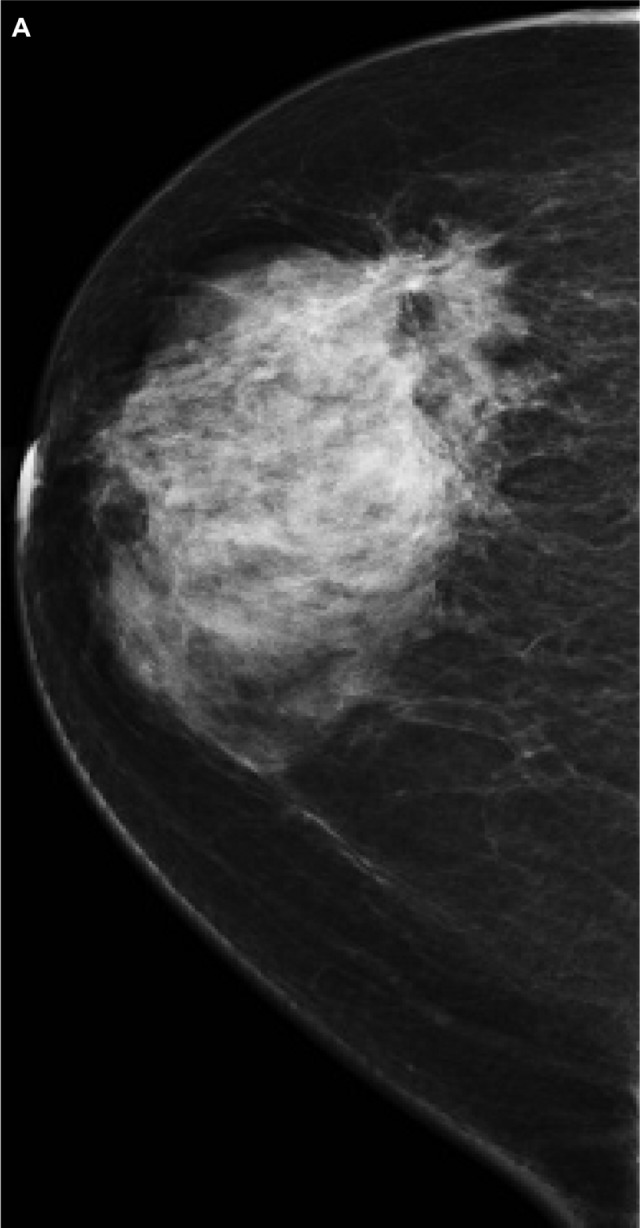
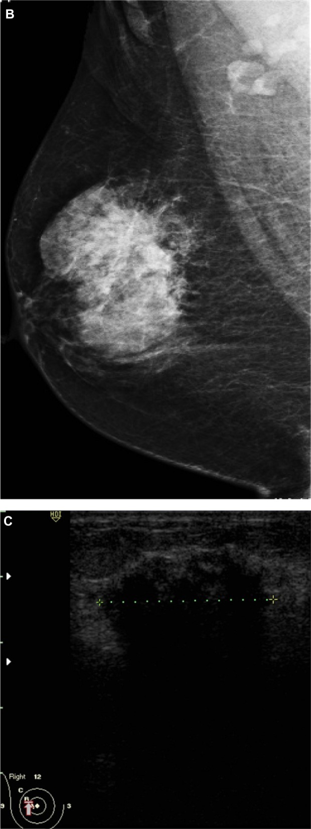
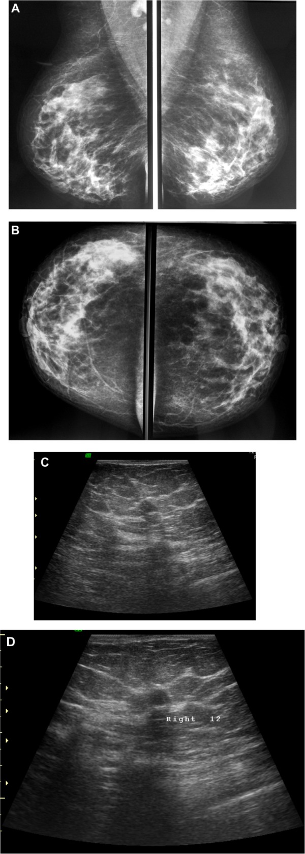
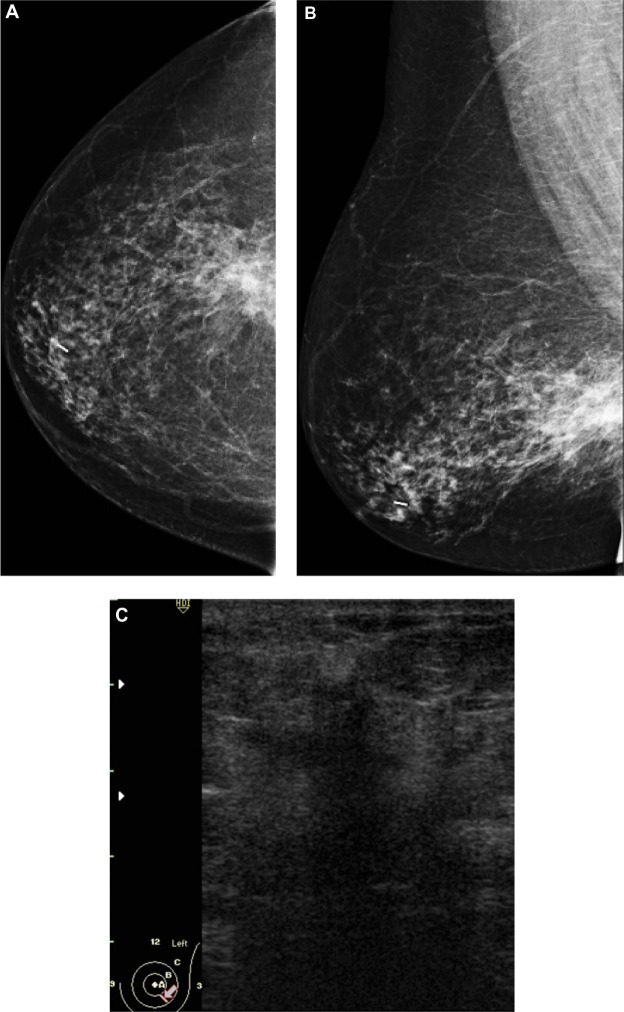
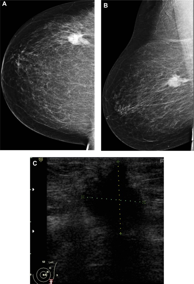
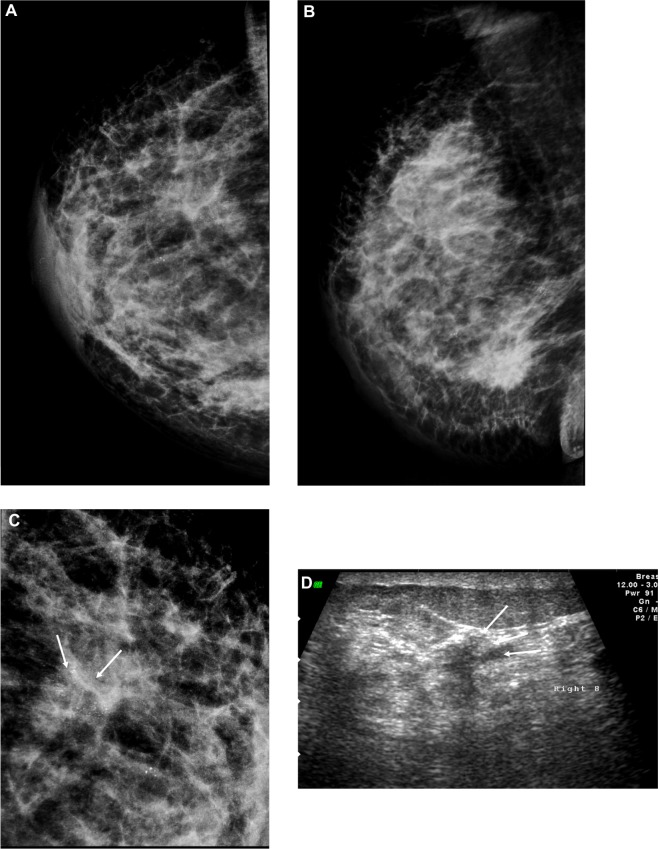
Results: Thirty-two women ranging in age from 42 to 63 years old (mean age, 53 years), All had biopsy-proven invasive lobular carcinomas. Common features on mammogram included dense mass followed by architectural distortion; three cases showed breast asymmetry and one case was reported as normal. On ultrasound, common features included solid mass with spiculated margins, posterior shadowing, and perpendicular to the skin.

Conclusion: Although no specific features could be linked to invasive lobular carcinoma, care should be directed to subtle signs such as architectural distortion and breast asymmetry in order not to miss any lesions. The combination of mammographic and sonographic helps to decrease the relatively high false negative diagnosis of this type of breast cancer.

Keywords: breast; cancer; mammography; ultrasound.


Figures

Figure 1
Figure 1
A 51-year-old female presented with hard right breast after suffering from right breast trauma 2 months previously. (A and B) Craniocaudal and mediolateral mammogram of the right breast revealed a dense breast with architectural distortion noted at upper outer quadrant (more evident on craniocaudal view). (C) Ultrasound revealed an ill-defined irregular hypoechoic mass with posterior acoustic shadowing at 9 o’clock of the right breast. Note: The longitudinal axis of the mass was parallel to that of the skin.
Figure 1
Figure 1
A 51-year-old female presented with hard right breast after suffering from right breast trauma 2 months previously. (A and B) Craniocaudal and mediolateral mammogram of the right breast revealed a dense breast with architectural distortion noted at upper outer quadrant (more evident on craniocaudal view). (C) Ultrasound revealed an ill-defined irregular hypoechoic mass with posterior acoustic shadowing at 9 o’clock of the right breast. Note: The longitudinal axis of the mass was parallel to that of the skin.
Figure 2
Figure 2
A 48-year-old female patient presented with palpable right axillary lymph node. (A and B) Craniocaudal and mediolateral mammogram revealed asymmetric breast density with no definite masses. (C and D) Ultrasound revealed three small hypoechoic masses at 1–11 o’clock of the right breast (only two are displayed here). Note: The masses have smooth margins, posterior acoustic shadowing, and their long axis was parallel to that of the skin.
Figure 3
Figure 3
A 58-year-old female with palpable mass in the left breast. (A and B) CC and ML mammogram films revealed ill-defined dense opacity with spiculated margins surrounded by architectural distortion at the lower central area of the breast, which looks to be attached to the pectoralis muscle posteriorly. (C) On ultrasound, ill-defined, spiculated, hypoechoic, solid mass with posterior shadowing measuring 16 × 13 mm is seen at 5 o’clock of the breast. Notes: The mass was inseparable from the underlying muscle. Another smaller, similar hypoechoic mass is seen close by measuring 6 × 5 mm. Abbreviations: CC, craniocaudal; ML, mediolateral.
Figure 4
Figure 4
A 72-year-old female with left breast mass. (A and B) Craniocaudal and mediolateral mammogram of left breast revealed an irregular spiculated, dense mass (35 × 25 mm) in the upper outer quadrant close to the pectoral muscle. Single benign macrocalcification is noted in the central area. (C) Ultrasound of the left breast revealed a spiculated, hypoechoic, solid mass (30 × 23 mm) with strong acoustic shadow at 3–4 o’clock. Note: The mass extends to the pectoralis muscle with its long axis perpendicular to the skin. Abbreviations: CC, craniocaudal; ML, mediolateral.
Figure 5
Figure 5
A 48-year-old female patient presented with hard right breast mass of 2 months duration. (AC) CC, ML, and magnified mammogram views of the right breast revealed an irregular spiculated dense mass at the lower outer quadrant of the right breast. A magnified view shows clusters of pleomorphic microcalcifications within the lesion (arrows). Four foci of calcifications are seen away from the lesion. (D) Ultrasound revealed an ill-defined mass of heterogeneous echopattern at 8 o’clock of left breast. Note: The mass shows foci of microcalcifications (arrows). Abbreviations: CC, craniocaudal; ML, mediolateral.

Similar articles

Cited by

References

    1. Li CI, Daling JR. Changes in breast cancer incidence rates in the United States by histologic subtype and race/ethnicity, 1995 to 2004. Cancer Epidemiol Biomarkers Prev. 2007;16:2773–2780. - PubMed
    1. Kopans DB. Malignant lesions of the lobule. In: Kopans DB, editor. Breast Imaging. Baltimore, MD: Lippincott Williams & Wilkins; 2007. pp. 866–870.
    1. Rosenberg RD, Hunt WC, Williamson MR, et al. Effects of age, breast density, ethnicity, and estrogen replacement therapy on screening mammographic sensitivity and cancer stage at diagnosis: review of 183,134 screening mammograms in Albuquerque, New Mexico. Radiology. 1998;209:511–518. - PubMed
    1. Krecke KN, Gisvold JJ. Invasive lobular carcinoma of the breast: mammographic findings and extent of disease at diagnosis in 184 patients. AJR Am J Roentgenol. 1993;161:957–960. - PubMed
    1. Adejolu M, Krishnamurthy S, Whitman GJ. Ultrasound of invasive lobular carcinoma. Ultrasound Clin. 2011;6:313–325.