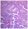
Association between Pituitary Langerhans Cell Histiocytosis and Papillary Thyroid Carcinoma
- PMID: 24367234
- PMCID: PMC3869627
- DOI: 10.4137/CCRep.S13021
Association between Pituitary Langerhans Cell Histiocytosis and Papillary Thyroid Carcinoma
Abstract
Here we report a case of panhypopituitarism caused by pituitary Langerhans cell hystocitosis (LCH) in a 22-year-old woman affected by papillary thyroid carcinoma (PTC). Although several cases of the coexistence of PTC and LCH within thyroid tissue have been described in relative literature, in this case, the patient presented a unique suprasellar retrochiasmatic histocytosis localization which, to the best of our knowledge, had never been described before in association with PTC. Even if this aspect is not addressed in the present case report, it is worth noting that about 50% of the patients affected either by LCH or PTC are characterized by activating mutations of the proto-oncogene BRAF. This, along with other clinical studies, may warrant further biomolecular large-scale case study investigations in order to evaluate a possible connection between the 2 conditions and shed light on the etiology of these diseases, which are still largely unknown.
Keywords: Langherans cell hystiocytosis; hypopituitarism; papillary thyroid carcinoma.
Figures


Similar articles
-
BRAF gene mutations in synchronous papillary thyroid carcinoma and Langerhans cell histiocytosis co-existing in the thyroid gland: a case report and literature review.BMC Cancer. 2019 Feb 22;19(1):170. doi: 10.1186/s12885-019-5372-3. BMC Cancer. 2019. PMID: 30795755 Free PMC article. Review.
-
Concomitant Langerhans cell histiocytosis of cervical lymph nodes in adult patients with papillary thyroid carcinoma: A report of two cases and review of the literature.Autops Case Rep. 2021 Mar 12;11:e2021253. doi: 10.4322/acr.2021.253. eCollection 2021. Autops Case Rep. 2021. PMID: 34307217 Free PMC article.
-
Thyroid Langerhans cell histiocytosis concurrent with papillary thyroid carcinoma: A case report and literature review.Front Med (Lausanne). 2023 Jan 19;9:1105152. doi: 10.3389/fmed.2022.1105152. eCollection 2022. Front Med (Lausanne). 2023. PMID: 36743683 Free PMC article.
-
Langerhans cell histiocytosis and Erdheim-Chester disease, both with cutaneous presentations, and papillary thyroid carcinoma all harboring the BRAF(V600E) mutation.J Cutan Pathol. 2016 Mar;43(3):270-5. doi: 10.1111/cup.12636. Epub 2015 Nov 20. J Cutan Pathol. 2016. PMID: 26454140
-
Langerhans cell histiocytosis of the thyroid complicated by papillary thyroid carcinoma: A case report and brief literature review.Medicine (Baltimore). 2017 Sep;96(35):e7954. doi: 10.1097/MD.0000000000007954. Medicine (Baltimore). 2017. PMID: 28858125 Free PMC article. Review.
Cited by
-
Langerhans Cell Histiocytosis, Non-Langerhans histiocytosis and concurrent Papillary Thyroid Carcinoma with BRAF V600E mutations: A case report and literature review.Hum Pathol (N Y). 2019 Sep;17:200302. doi: 10.1016/j.hpcr.2019.200302. Epub 2019 Jun 15. Hum Pathol (N Y). 2019. PMID: 31592429 Free PMC article. No abstract available.
-
Occult Langerhans Cell Histiocytosis Presenting with Papillary Thyroid Carcinoma, a Thickened Pituitary Stalk and Diabetes Insipidus.Case Rep Endocrinol. 2016;2016:5191903. doi: 10.1155/2016/5191903. Epub 2016 Aug 30. Case Rep Endocrinol. 2016. PMID: 27656301 Free PMC article.
References
-
- Kinder BK. Well differentiated thyroid cancer. Curr Opin Oncol. 2003;15:71–7. - PubMed
-
- Jemal A, Siegel R, Ward E, Hao Y, Xu J, Thun MJ. Cancer Statistics, 2009. Ca Cancer J Clin. 2009;59:225–49. - PubMed
-
- Nikiforov YE, Biddinger PW, Thompson LDR, editors. Diagnostic Pathology and Molecular Genetics of the Thyroid. Philadelphia, PA: Lippincott Williams & Wilkins; 2009.
-
- American Thyroid Association (ATA) Guidelines Taskforce on Thyroid Nodules and Differentiated Thyroid Cancer. Cooper DS, Doherty GM, et al. Revised American Thyroid Association management guidelines for patients with thyroid nodules and differentiated thyroid cancer. Thyroid. 2009;19:1167–214. - PubMed
-
- Pacini F, Schlumberger M, Dralle H, Elisei R, Smit JW, Wiersinga W European Thyroid Cancer Taskforce. European consensus for the management of patients with differentiated thyroid carcinoma of the follicular epithelium. Eur J Endocrinol. 2006;154:787–803. - PubMed
Publication types
LinkOut - more resources
Full Text Sources
Other Literature Sources
Research Materials

