. 2013;9(12):e1003801.
doi: 10.1371/journal.ppat.1003801. Epub 2013 Dec 19.

Intestinal CD103+ dendritic cells are key players in the innate immune control of Cryptosporidium parvum infection in neonatal mice

Affiliations

Intestinal CD103+ dendritic cells are key players in the innate immune control of Cryptosporidium parvum infection in neonatal mice

Louis Lantier et al. PLoS Pathog. 2013.

Abstract

Cryptosporidium parvum is a zoonotic protozoan parasite found worldwide, that develops only in the gastrointestinal epithelium and causes profuse diarrhea. Using a mouse model of C. parvum infection, we demonstrated by conditional depletion of CD11c+ cells that these cells are essential for the control of the infection both in neonates and adults. Neonates are highly susceptible to C. parvum but the infection is self-limited, whereas adults are resistant unless immunocompromised. We investigated the contribution of DC to the age-dependent susceptibility to infection. We found that neonates presented a marked deficit in intestinal CD103+ DC during the first weeks of life, before weaning, due to weak production of chemokines by neonatal intestinal epithelial cells (IEC). Increasing the number of intestinal CD103+ DC in neonates by administering FLT3-L significantly reduced susceptibility to the infection. During infections in neonates, the clearance of the parasite was preceded by a rapid recruitment of CD103+ DC mediated by CXCR3-binding chemokines produced by IEC in response to IFNγ. In addition to this key role in CD103+ DC recruitment, IFNγ is known to inhibit intracellular parasite development. We demonstrated that during neonatal infection CD103+ DC produce IL-12 and IFNγ in the lamina propria and the draining lymph nodes. Thus, CD103+DC are key players in the innate immune control of C. parvum infection in the intestinal epithelium. The relative paucity of CD103+ DC in the neonatal intestine contributes to the high susceptibility to intestinal infection.


Conflict of interest statement

The authors have declared that no competing interests exist.

Figures
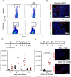
Figure 1
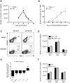
Figure 1. CD11c+ cells are necessary for the control of C. parvum infection.
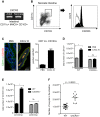
(A) Seven day-old heterozygous CD11c-DTR neonates were infected with 5.105 C. parvum oocysts and some animals were treated with DT 4 dpi. Spleen cells and intestinal cells were collected separately 24 hours later and CD11c+ cell depletion was analyzed by flow cytometry. (B) Neonates were infected and treated as in (A), and sections of the small intestine from CD11c-DTR neonates collected 5 dpi were processed for histology: C. parvum is stained in red, and CD11c is stained in green (Original magnification ×200). (C) Neonates were infected and treated as in (A). The parasite load was assessed daily from 5 dpi to 8 dpi in CD11c-DTR neonates treated or not treated with DT 4 dpi (n = 7–12 neonatal mice per group). (D) Adult CD11c-DTR were infected with 106 C. parvum oocysts and treated twice (d−1; d+2) or not treated with DT. Parasite load was evaluated 4 dpi in the whole intestine of adult mice (p<0.001, n = 8 mice per group). In the same experiment, histological sections of small intestine were collected 4 dpi (C. parvum staining in red; original magnification ×200).
Figure 2
Figure 2. Rapid recruitment of CD11c+ CD103+ in the infected mucosa.
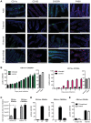
(A) Sections of the small intestine of mice at different ages: 7 days old, 13 days old, 13 days old inoculated with C. parvum at 7 days of age, and uninfected adult mice were stained with Hoechst dye and antibodies against CD11c, CD103 and F4/80. CX3CR1GFP/WT mice were used for CX3CR1 detection. Original magnification ×200, scale bars indicate 100 µm. Double staining for CD11c+ CD103+, CD11c+ CX3CR1+ and F4/80+ CX3CR1+ are provided in Figure S2. (B) Number of CD11c+CX3CR1+ and CD11c+ CD103+ double-positive cells in the intestine of infected animals and controls. (C) CD11c+ CX3CR1+ and CD11c+ CD103+ double-positive cells were quantified by flow cytometry for infected animals 6 dpi and for age matched controls (n = 5 animals per group). Gating strategies are provided in Figure S5, A. (D) As in (B) but the location in the mucosa of CD11c+ CX3CR1+ and CD11c+ CD103+ double-positive cells was evaluated in the intestine of infected animals 6 dpi. IE: intraepithelial; LP: Lamina propria; MM: Muscularis mucosae. (E) As in (B), CD11c+ CD103+ double-positive cells were enumerated in the intestine of axenic or conventional neonates at age 13 days and post weaning (22 days old). For (B, D, E) the values reported were obtained by counting double-positive cells in sections of the small intestine of mice and for each point are the means ± SEM of at least 30 optical fields from two animals from different litters.
Figure 3
Figure 3. In vivo amplification of CD11c+ CD103+ DC by FLT3-L enhances neonatal resistance to C. parvum infection.
(A) WT and CX3CR1GFP/WT neonates were injected daily for 6 consecutive days from birth with 1 µg FLT3-L. The first three injections were subcutaneous and the next three intraperitoneal. Histological sections of the ilea were collected from FLT3-L-treated or untreated mice at 7 days of age. Sections were stained with Hoechst dye and antibodies against CD11c and CD103. CX3CR1GFP/WT mice were used for CX3CR1 detection. Single staining is shown, to provide a clearer overview of the distribution of the positive cells in the villi (scale bars indicate 100 µm). Double positive CD11c+CX3CR1+ cells and CD11c+ CD103+ DC in ileal sections were counted. The graph reports means ± SEM of at least 20 optical fields from two different animals giving similar results; the values are fold amplification (treated/untreated animals). (B) Control neonates and neonates treated with FLT3-L for 6 days were orally infected with 5.105 oocysts of C. parvum at 7 days of age and the parasite load was evaluated 6 dpi. Values are means ± SEM (p<0.001, n = 8 neonatal mice per group). (C) Seven day-old WT and IL-12p40−/− neonates were orally infected with 5.105 oocysts of C. parvum and the parasite load was evaluated at various times post infection. Values are means ± SEM (n = 5–8 neonatal mice per group at each time point). (D) Same experiment as in (B) but with IL12p40−/− neonates. Values are means ± SEM (ns: not significant, n = 10–11 neonatal mice per group). The Mann-Whitney non-parametric analyses were considered significant when p values were less than <0.05. Figure 3D, p>0.05 is non-significant (ns).
Figure 4
Figure 4. Weak chemokine expression by epithelial cells of neonates is upregulated by the infection.
RNA was extracted from intestinal epithelial cells isolated from 13 day-old control neonates, infected (13 day-old, 6 dpi) neonates and control adult mice. Chemokine mRNAs were assayed by qRT-PCR. Values were all normalized to IEC isolated from control neonates (*p<0.05, **p<0.01; n = 4–6 in each group).
Figure 5
Figure 5. The chemokine receptor CXCR3 plays a major role in the recruitment of CD103+ DC during C. parvum infection.
(A) CD11c+ MHCII+ CD103+ DC isolated from the intestines of uninfected adults and infected neonates were sorted by flow cytometry. CXCR3 expression in each sample was evaluated by RT-PCR (amplicons on a 2% agarose gel). (B) Surface expression of CXCR3 on intestinal CD11c+CD103+ DC (previously gated on MHCII+ cells) isolated from infected neonates (6 dpi) was analyzed by flow cytometry. Gating strategies are provided in Figure S5, B. The grey line histogram represents the isotype control and the black filled histogram the staining with anti-CXCR3 monoclonal antibody. (C) Recombinant CXCL10 was administered (1 µg/per os) to 7 day-old neonates for 3 consecutive days starting from day 7. At 9 days of age, CD11c+ CD103+ labeling and counting were performed on intestinal sections. CD11c+ cells are stained in green, CD103+ cells in red and nuclei in blue with Hoechst dye (scale bars indicate 20 µm). Data in the right-hand panel were obtained by counting double-positive cells per field in sections of the small intestine. Reported values are means ± SEM of at least 30 optical fields from two neonates from different litters. (D) CD11c+ MHCII+ CD103+ DC recruitment after CXCL10 treatment in WT and CXCR3−/− neonates was also analyzed by flow cytometry. Numbers of CD11c+MHCII+CD103+ cells in the small intestine in treated and untreated neonates (*p<0.05, n = 4 neonates for each group) are shown. (E) Seven day-old WT and CXCR3−/− neonates (*p<0.05, n = 4 neonates for each group) were infected with C. parvum and CD103+ DC recruitment was analyzed by flow cytometry 3 dpi. Gating strategies are provided in Figure S5, B. (F) Seven day-old WT and CXCR3−/− neonates were infected with C. parvum and the parasite load was evaluated 6 dpi. (p<0.0001, n = 12–16 mice per group).
Figure 6
Figure 6. IFNγ plays a key role in the recruitment of CD103+DC during infection.
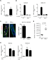
(A) Basal mRNA level of IFNγ in the intestine of neonate and adult mice (p<0.001, n = 8 mice per group). (B) Seven day-old C57BL/6J WT and IFNγ−/− neonates were infected with C. parvum. mRNAs for CXCL9 and CXCL10 in isolated IEC were assayed by qRT-PCR in infected (6 dpi) and in uninfected age-matched control neonates (n = 6 neonatal mice for each group,** p<0.005). (C) The presence of CD11c+CD103+ DC was evaluated in the ileal villi of WT and IFNγ−/− mice 6 dpi. Sections of small intestine were immunostained and CD11c+ CD103+ double-positive cells (white arrow) were counted. CD11c+ cells are stained in green, CD103+ cells in red and nuclei in blue with Hoechst dye (scale bars indicate 20 µm). Data in the right-hand panel were obtained by counting double-positive cells per field in sections of the small intestine of C57BL/6J WT and IFNγ−/− mice at 6 dpi. Reported values are means ± SEM of at least 30 optical fields from two neonates from different litters. (D) Seven day-old WT and IFNγ−/− neonates were infected with C. parvum. The parasite loads 6 dpi in WT and IFNγ−/− neonatal mice are reported. Values represent means ± SEM (n = 6 neonatal mice per group). (E) CMT-93 cells were left unstimulated or stimulated with IFNγ (10 ng/ml) or infected with C. parvum at a ratio of three oocysts/cell. Expression of CXCL9 and CXCL10 mRNAs was analyzed by qRT-PCR 24 h later (*p<0.01 relative to control CMT-93 cells).
Figure 7
Figure 7. CD103+ dendritic cell subsets contribute to IL-12p40 and IFNγ production.
(A) Seven day-old neonates were infected with C. parvum and ilea were collected 6 dpi for mRNA extraction and subsequent qRT-PCR. For each gene, mRNA expression is represented as a fold increase with respect to uninfected neonates. The values are means ± SEM (n = 6 neonatal mice per group). (B) Gene expression after CD11c+ cell depletion was analyzed by injecting DT into CD11c-DTR neonates at 4 dpi. RNA was extracted from the ileum 6 dpi. For each gene, mRNA abundance is represented as fold decrease relative to untreated neonates. The values reported are means ± SEM (n = 6 neonatal mice per group). (C) Total numbers of CD11c+MHCII+CD103+ in the MLN of infected (6 dpi) and age matched control neonates (13-day-old) in the left panel. Data from the same experiment, in the right panel, show the total numbers of CD8α+ and CD8α− CD103+ DC subsets (n = 4 pools, 2–3 neonates/pool). (D) isolation of CD103+ cell subsets from the MLN of neonates was first based on selection of CD11chi MHC IIhi. Double-positive cells were further separated into two subsets based on CD8α expression. Gating strategies are provided in Figure S5, C. CD103+ cells, CD103+ CD8α− and CD103+ CD8α+ subsets were isolated from pooled MLN obtained 6 dpi from numerous neonates (2–4 pools, 6–44 neonates per pool). CD103+ DC were also isolated from their age-matched controls (2 pools, 72 neonates per pool) for normalization. Isolated cells were used for qRT-PCR analysis. Gene expression in pools of samples was assessed by qRT-PCR. For each gene, mRNA expression in the different subsets isolated from infected animals is represented as fold differences to that in CD103+ DC from control neonates. The reported values are means ± SEM. (E) MLN were isolated from infected neonates 6 dpi, and CD11c+ MHCII+ CD103+ DC were sorted by FACS and cultured in vitro. After 24 h of culture, supernatants were assayed by ELISA for IFNγ and IL-12p40 (n = 5 pools, 2 neonates/pool). Some cells were cultured in the presence of 10 ng/ml of IL-12 and IFNγ production assayed. (F) CD103+ CD11c+ MHC II+ DC were isolated from the intestine of infected neonates. Due to the low frequency of CD11c+CD103+ DC in the intestine of uninfected neonates at 13 days of age, CD11c+CD103+ DC were isolated from their MLN for normalization. RNA extraction and qRT-PCR were performed immediately after cell sorting. Gating strategies are provided in Figure S5, B. The reported values are means ± SEM (6 pools of neonatal mice per group; between 8–21 neonates per pool). (G) Intracellular staining of IFNγ in samples from infected (6 dpi) and uninfected animals (13 day-old). Panels show intracellular staining of IFNγ in CD11c+MHCII+CD103+ DC (panels are representative of two independent experiments).
Figure 8
Figure 8. NKp46+NK1.1+ Natural killer cells and conventional T cells are not essential for the control of the acute phase of C. parvum infection.
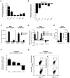
Seven day-old neonates were orally infected with 5.105 C. parvum oocysts and the parasite load in the whole intestine was evaluated at various times post infection. (A) Parasite load was evaluated in CD3ε−/− and WT neonates. Data are means ± SEM (n = 6 neonatal mice per group). (B) Four weeks after infection of CD3ε−/− and WT neonates, and apparent recovery from the infection, mice were injected with 1 µg of dexamethasone, by the IP route, daily for 3 days. Fecal smears were used to quantify oocyst excretion every day. Data are means ± SEM (n = 5 mice per group). (C) Flow cytometry analysis of NKp46+NK1.1+ cells in the intestine and spleen of infected heterozygous NKp46-DTR neonates and infected wild-type littermates, all treated with DT on d−1; d+1 and d+3 post infection. The results shown are for representative animals from the same litter. (D) Same experiment as in (C). The parasite load was evaluated at 4, 6 and 8 dpi. Data are means ± SEM (n = 6–15 neonatal mice per group). (E) Similar experiment as described in (C) and cytokine mRNAs in the ilea of neonates 6 dpi were analyzed by qRT-PCR. The bars represent the mean values ± SEM of the ratios of the relative expression value for mRNA in the intestine of NKp46-depleted neonatal mice to that in non-depleted wild type littermates (n = 6). (F) Parasite load was evaluated 4, 6 and 8 dpi in IL15−/− and WT neonates. Data are means ± SEM (n = 6 neonatal mice per group).

Similar articles

Cited by

References

    1. Scallan E, Hoekstra RM, Angulo FJ, Tauxe RV, Widdowson MA, et al. (2011) Foodborne illness acquired in the United States−major pathogens. Emerg Infect Dis 17: 7–15. - PMC - PubMed
    1. Chen XM, Keithly JS, Paya CV, LaRusso NF (2002) Cryptosporidiosis. N Engl J Med 346: 1723–1731. - PubMed
    1. Stockinger S, Hornef MW, Chassin C (2011) Establishment of intestinal homeostasis during the neonatal period. Cell Mol Life Sci 68: 3699–3712. - PMC - PubMed
    1. Adkins B, Leclerc C, Marshall-Clarke S (2004) Neonatal adaptive immunity comes of age. Nat Rev Immunol 4: 553–564. - PubMed
    1. Renz H, Brandtzaeg P, Hornef M (2012) The impact of perinatal immune development on mucosal homeostasis and chronic inflammation. Nat Rev Immunol 12: 9–23. - PubMed

Publication types