. 2013;9(12):e1003813.
doi: 10.1371/journal.ppat.1003813. Epub 2013 Dec 19.

Biphasic euchromatin-to-heterochromatin transition on the KSHV genome following de novo infection

Affiliations

Biphasic euchromatin-to-heterochromatin transition on the KSHV genome following de novo infection

Zsolt Toth et al. PLoS Pathog. 2013.

Abstract

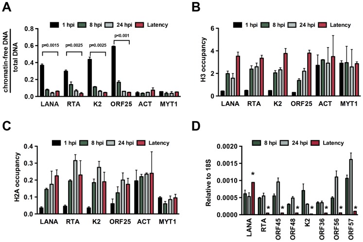
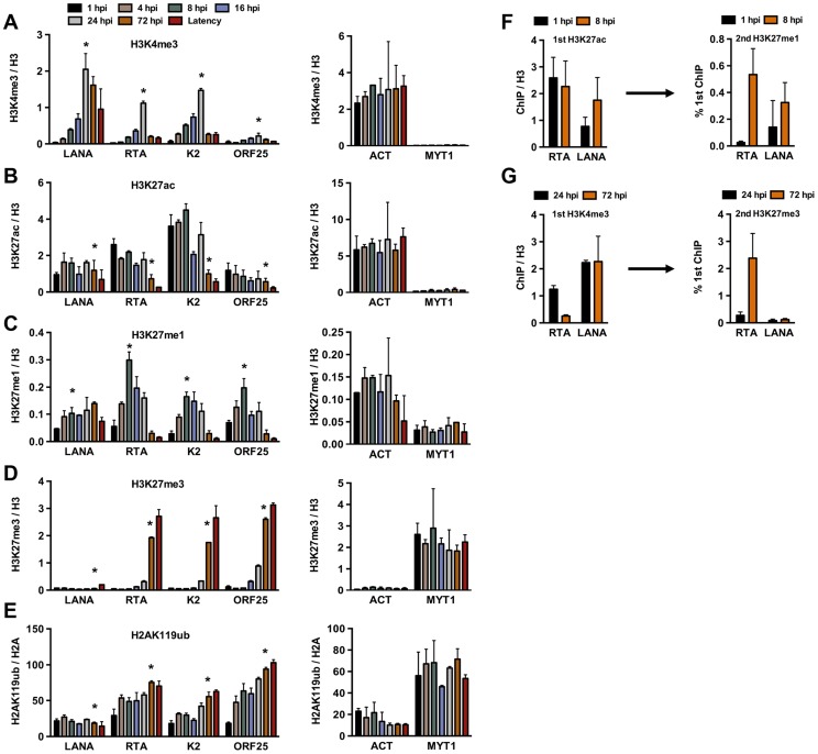
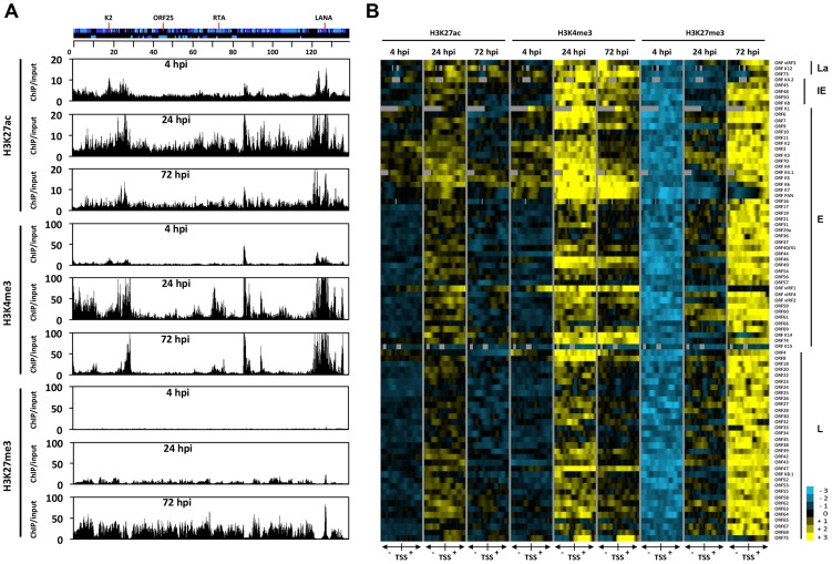
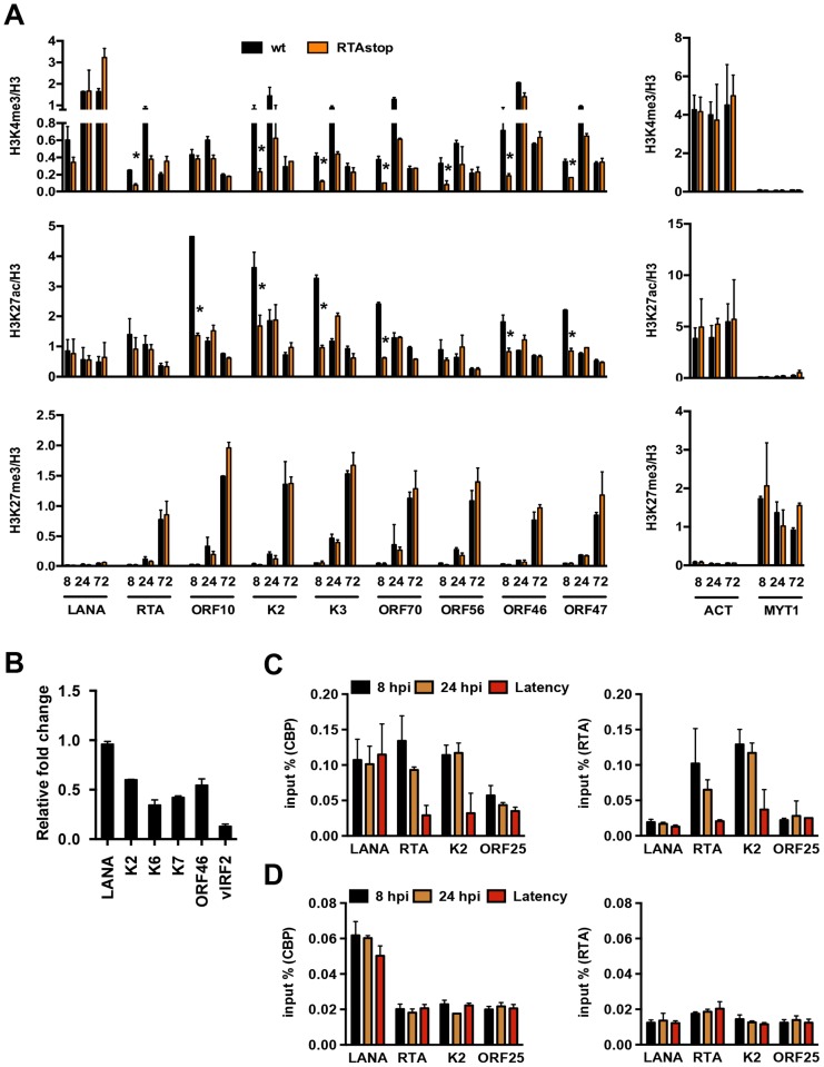
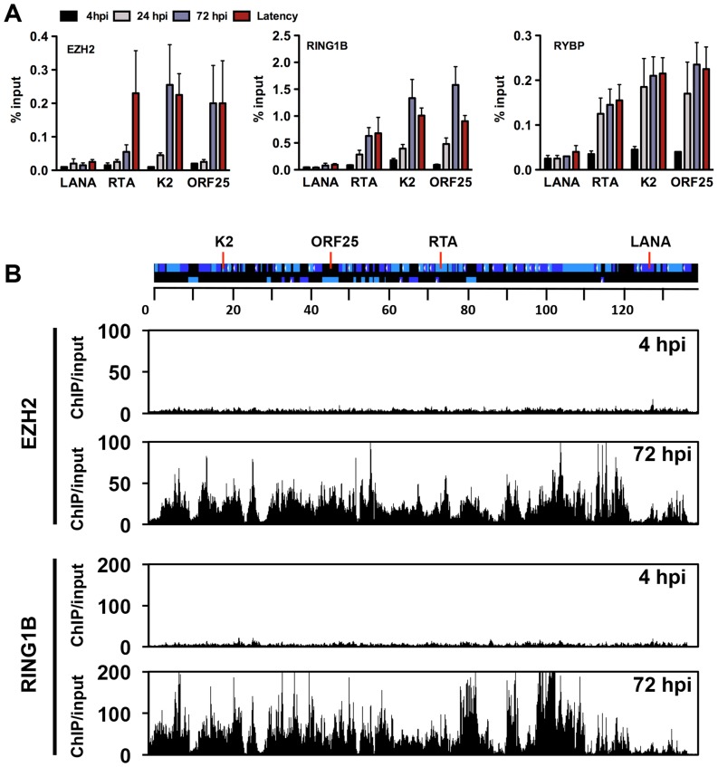
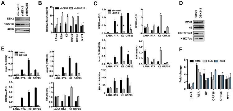
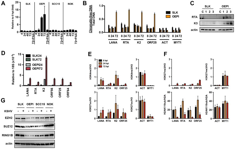
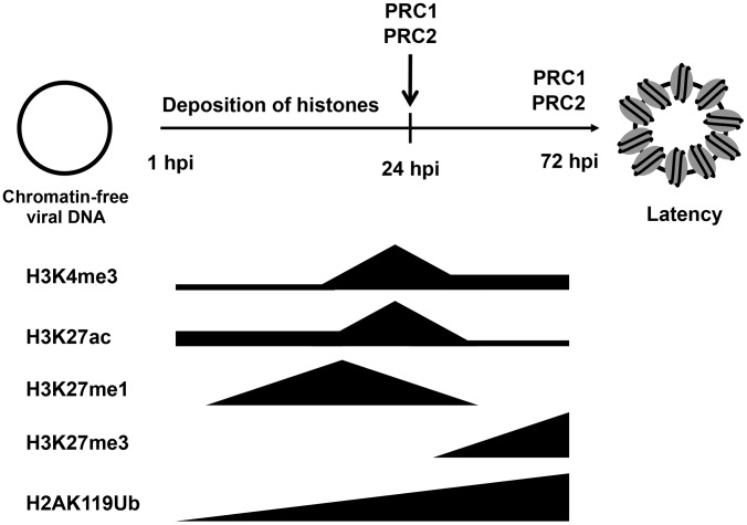
The establishment of latency is an essential step for the life-long persistent infection and pathogenesis of Kaposi's sarcoma-associated herpesvirus (KSHV). While the KSHV genome is chromatin-free in the virions, the viral DNA in latently infected cells has a chromatin structure with activating and repressive histone modifications that promote latent gene expression but suppress lytic gene expression. Here, we report a comprehensive epigenetic study of the recruitment of chromatin regulatory factors onto the KSHV genome during the pre-latency phase of KSHV infection. This demonstrates that the KSHV genome undergoes a biphasic chromatinization following de novo infection. Initially, a transcriptionally active chromatin (euchromatin), characterized by high levels of the H3K4me3 and acetylated H3K27 (H3K27ac) activating histone marks, was deposited on the viral episome and accompanied by the transient induction of a limited number of lytic genes. Interestingly, temporary expression of the RTA protein facilitated the increase of H3K4me3 and H3K27ac occupancy on the KSHV episome during de novo infection. Between 24-72 hours post-infection, as the levels of these activating histone marks declined on the KSHV genome, the levels of the repressive H3K27me3 and H2AK119ub histone marks increased concomitantly with the decline of lytic gene expression. Importantly, this transition to heterochromatin was dependent on both Polycomb Repressive Complex 1 and 2. In contrast, upon infection of human gingiva-derived epithelial cells, the KSHV genome underwent a transcription-active euchromatinization, resulting in efficient lytic gene expression. Our data demonstrate that the KSHV genome undergoes a temporally-ordered biphasic euchromatin-to-heterochromatin transition in endothelial cells, leading to latent infection, whereas KSHV preferentially adopts a transcriptionally active euchromatin in oral epithelial cells, resulting in lytic gene expression. Our results suggest that the differential epigenetic modification of the KSHV genome in distinct cell types is a potential determining factor for latent infection versus lytic replication of KSHV.


Conflict of interest statement

The authors have declared that no competing interests exist.

Figures

Figure 1
Figure 1. De novo chromatinization of the KSHV genome following infection.
(A) FAIRE assay showing the degree of chromatinization of the indicated viral and cellular promoters in SLK cells infected for 1, 8 or 24 hours or in latently infected SLK cells. (B) and (C) The enrichment of histones H3 and H2A was calculated as ChIP/input (% Input or occupancy) at the indicated viral and cellular promoters following de novo infection at 1, 8 and 24 hpi and in latently infected SLK cells. (D) Real time RT-PCR analysis was performed to determine the expression of the indicated viral genes in SLK cells infected by KSHV for 8 and 24 hours and in latently infected SLK cells. Expression levels of viral genes are shown relative to those of 18S. A 2-tailed student's t-test was performed between 8 hpi and latency for each tested lytic gene and all p values were less than 0.05.
Figure 2
Figure 2. Analysis of the deposition of histone modifications on KSHV promoters during de novo infection using time-course ChIP assays.
ChIPs were performed using latently infected SLK cells or SLK cells following de novo infection for the indicated hours post infection (hpi) and the enrichment of histone modifications was measured by qPCR using primers specific for the indicated viral and cellular promoters. ChIPs for each histone modification were normalized for the amount of the relevant histones at each promoter. T-test was applied to compare the values between the indicated time points (*). (A) H3K4me3 ChIP (for LANA, RTA and K2 p<0.05, for ORF25 p = 0.1 between 1 and 24 hpi). (B) H3K27ac ChIP (for LANA p = 0.6, RTA and K2 p<0.05, for ORF25 p = 0.18 between 1 and 72 hpi). (C) H3K27me1 ChIP (for LANA p = 0.063, for RTA, K2 and ORF25 p<0.05 between 1 and 8 hpi). (D) H3K27me3 ChIP (for LANA p = 0.33, for RTA, K2 and ORF25 p<0.005 between 1 and 72 hpi). (E) H2AK119ub ChIP (for LANA p = 0.3, for RTA, K2 and ORF25 p<0.02 between 1 and 72 hpi). (F) Sequential deposition of H3K27ac and H3K27me1 on viral promoters during infection was confirmed by sequential ChIP assays. The first ChIP was performed with H3K27ac-specific antibody at 1 hpi and 8 hpi, followed by elution of the immunoprecipitated DNA, The eluted DNA was used as the input for a second ChIP performed with H3K27me1 antibody. (G) Sequential deposition and colocalization of H3K4me3 and H3K27me3 on the RTA promoter following de novo infection were confirmed by sequential ChIP assays. The first ChIP was performed with H3K4me3-specific antibody at 24 hpi and 72 hpi followed by the second ChIP for H3K27me3 using the eluted first ChIPs as the input.
Figure 3
Figure 3. Genome-wide view of the deposition of histone modifications on the KSHV genome following de novo infection.
(A) ChIP-on-chip was performed for H3K27ac, H3K4me3 and H3K27me3 at 4, 24 and 72 hpi using SLK infected cells. The alternating dark and light blue squares represent viral ORFs. (B) Heat map representation of changes in histone modifications at the gene regulatory regions of KSHV genes grouped by expression class (latent, La, immediate-early, IE, early, E, late, L). The rows display the relative abundance of the indicated histone modification within the −1 kb to +1 kb genomic regions flanking the translational start site (TSS) of each viral gene. The blue and yellow colors denote lower-than-average and higher-than-average enrichment, respectively, whereas gray represents missing values for enrichment due to lack of probes in those genomic regions.
Figure 4
Figure 4. RTA is involved in the deposition of activating histone marks on the KSHV genome following de novo infection.
(A) Comparative ChIP analysis of H3K4me3, H3K27ac and H3K27me3 at the indicated viral promoters of wild type (wt) or RTA knockout (RTAstop) KSHV-infected SLK cells at 8, 24 and 72 hpi. The promoters of the cellular actin (ACT) and MYT1 genes were used as controls. ChIPs were normalized for the amount of histone H3 at each promoter. The asterisk denotes p<0.05 between wt and RTAstop at 8 hpi. (B) RT-qPCR analysis of viral gene expression in RTAstop KSHV-infected SLK cells at 24 hpi. The expression of the indicated viral genes was calculated relative to wt KSHV-infected cells. (C) ChIP experiments showing the binding of RTA and CBP on the RTA-responsive RTA and K2 promoters as well as the LANA promoter in latently-infected cells and naïve cells infected for 8 or 24 hours by wild type KSHV. ORF25 promoter was used as a negative control. (D) RTA and CBP binding on viral promoters in RTAstop KSHV-infected SLK cells.
Figure 5
Figure 5. Recruitment of components of the PRC2 and PRC1 complexes onto the KSHV genome during de novo infection.
(A) Time-course ChIP analysis of the binding of EZH2 (PRC2), RING1B and RYBP (PRC1) onto KSHV promoters at 4, 24, and 72 hpi and during latency. (B) Genome-wide recruitment of EZH2 and RING1B to the KSHV genome at 4 and 72 hpi. The Pearson correlation between the binding of EZH2 and RING1B is 0.6 at 72 hpi. Labels are the same as in Figure 3.
Figure 6
Figure 6. Both PRC2 and PRC1 are involved in the inhibition of lytic gene expression following de novo infection.
(A) Immunoblot analysis of EZH2 and RING1B expression in shRNA-treated SLK cells. (B) RT-qPCR analysis of viral gene expression of KSHV infected cells upon depletion of EZH2 and RING1B expression. The expression of viral genes and MYT1 cellular gene is shown relative to that of KSHV infected SLK cells treated with scrambled shRNA. (C) ChIP assays show that depletion of the EZH2 expression during de novo infection altered the deposition profiles of histone modifications and led to the reduction of RING1B recruitment on the KSHV genome. The asterisk denotes p<0.02. (D) Following 2 days of treatment with either DMSO or the EZH2 inhibitor, GSK343, SLK cell lysates were used for immunoblot analysis with the indicated antibodies. (E) ChIP analysis of the recruitment of PcG proteins and the deposition of histone modifications on KSHV promoters in KSHV infected cells pretreated with GSK343. (F) GSK343-treated TIME, SLK and 293T cells were infected by KSHV for 72 hours and RT-PCR was performed to test the expression of viral genes and the cellular gene MYT1. The fold change represents the induction of viral gene expression in GSK343-treated cells compared to DMSO-treated cells.
Figure 7
Figure 7. Euchromatinization of the KSHV genome in gingival oral epithelial cells following de novo infection.
(A) Measurement of viral DNA replication in SLK, OEPI, SCC15 and NOK cells infected by KSHV for 4, 24, 48 and 72 hours. The viral DNA polymerase inhibitor, PAA, was applied to block the replication of KSHV. (B) FAIRE assay showing the degree of chromatinization of the indicated viral and cellular promoters in SLK and OEPI cells infected for 8, 24 or 72 hours. (C) SLK and OEPI cells were infected with KSHV for 1, 2 and 3 days and immunoblots were performed to test the expression of RTA and K3 viral proteins. Actin served as a loading control. The “C” indicates immunoblot analysis of uninfected cells. (D) Quantitative RT-PCR analysis of viral gene expression in KSHV infected SLK and OPEI cells. (E) and (F) ChIP analysis of the indicated histone modifications on a selection of KSHV promoters in OEPI cells at 8, 24 and 72 hpi. The cellular promoters (ACT and MYT1) were used as controls. (G) Comparative immunoblot analysis of the indicated cellular proteins between SLK and oral epithelial cells.
Figure 8
Figure 8. Biphasic euchromatin-to-heterochromatin transition on the KSHV genome following de novo infection.
As soon as the viral genome enters the nucleus, histones are recruited on the viral DNA, resulting in the chromatinization of the KSHV genome. The viral genome initially adopts a transcriptionally permissive chromatin, characterized by high levels of the H3K27ac and H3K4me3 and this is accompanied by the transient expression of lytic genes. Subsequently, the PRC2 and PRC1 are recruited to viral genome, where they are responsible for the enrichment of H3K27me3 and H2Ak119ub on the viral chromatin as well as the repression of lytic genes. During latency, both PRC2 and PRC1 remain on the KSHV genome for the maintenance of the repression of lytic genes.

References

    1. Cesarman E, Moore PS, Rao PH, Inghirami G, Knowles DM, et al. (1995) In vitro establishment and characterization of two acquired immunodeficiency syndrome-related lymphoma cell lines (BC-1 and BC-2) containing Kaposi's sarcoma-associated herpesvirus-like (KSHV) DNA sequences. Blood 86: 2708–2714. - PubMed
    1. Chang Y, Cesarman E, Pessin MS, Lee F, Culpepper J, et al. (1994) Identification of herpesvirus-like DNA sequences in AIDS-associated Kaposi's sarcoma. Science 266: 1865–1869. - PubMed
    1. Greene W, Kuhne K, Ye F, Chen J, Zhou F, et al. (2007) Molecular biology of KSHV in relation to AIDS-associated oncogenesis. Cancer treatment and research 133: 69–127. - PMC - PubMed
    1. Ye F, Zhou F, Bedolla RG, Jones T, Lei X, et al. (2011) Reactive oxygen species hydrogen peroxide mediates Kaposi's sarcoma-associated herpesvirus reactivation from latency. PLoS Pathog 7: e1002054. - PMC - PubMed
    1. Chang J, Renne R, Dittmer D, Ganem D (2000) Inflammatory cytokines and the reactivation of Kaposi's sarcoma-associated herpesvirus lytic replication. Virology 266: 17–25. - PubMed

Publication types

MeSH terms