. 2013;9(12):e1004001.
doi: 10.1371/journal.pgen.1004001. Epub 2013 Dec 19.

Base pairing interaction between 5'- and 3'-UTRs controls icaR mRNA translation in Staphylococcus aureus

Affiliations

Base pairing interaction between 5'- and 3'-UTRs controls icaR mRNA translation in Staphylococcus aureus

Igor Ruiz de los Mozos et al. PLoS Genet. 2013.

Abstract

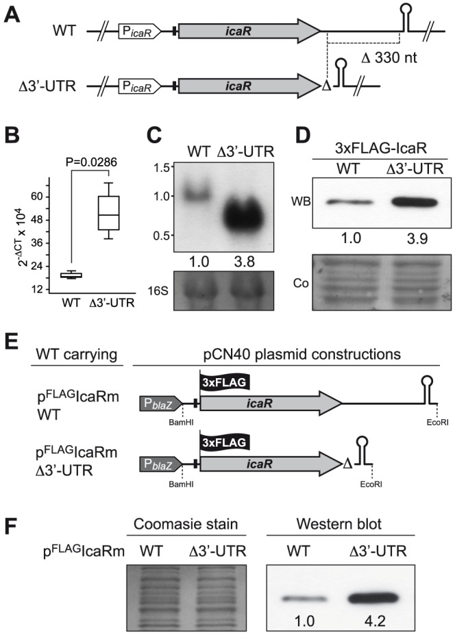
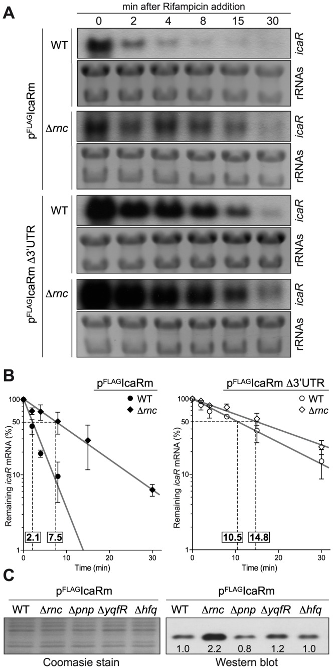
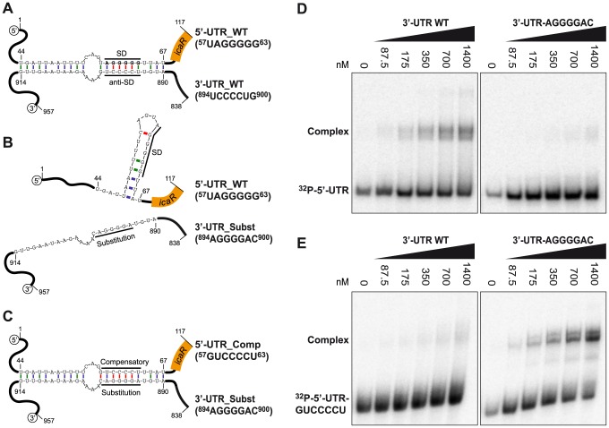
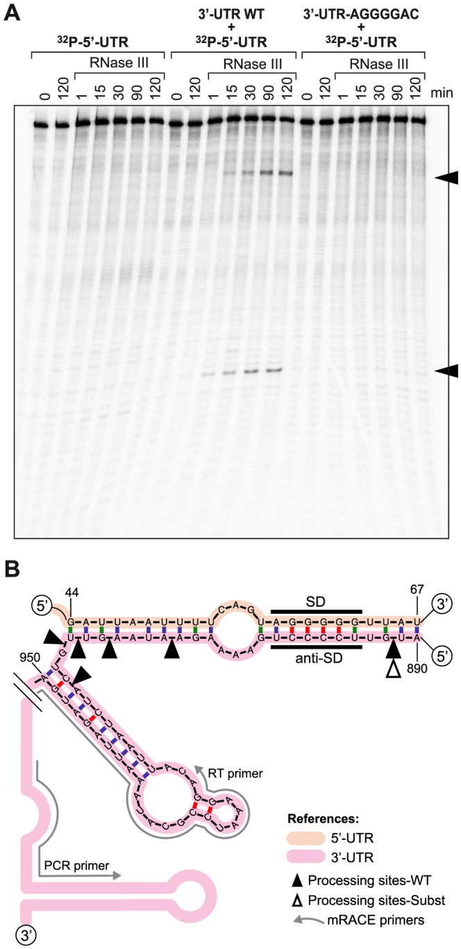
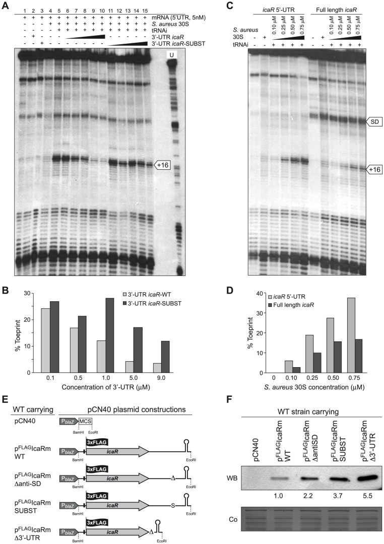
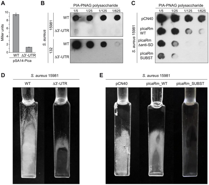
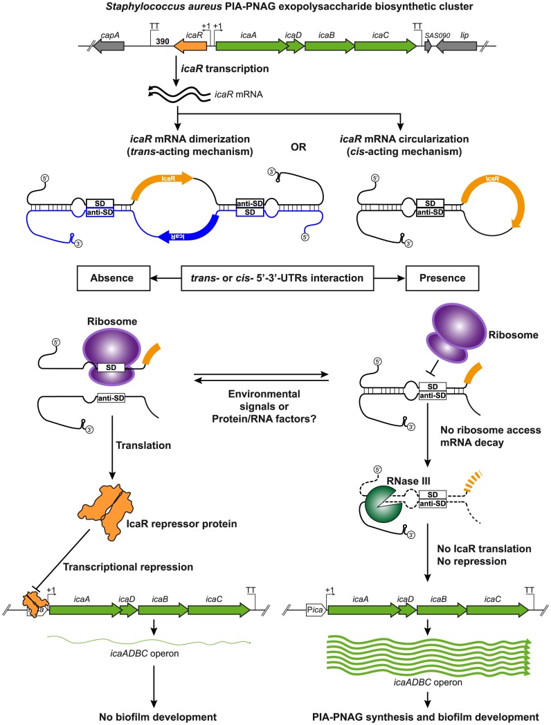
The presence of regulatory sequences in the 3' untranslated region (3'-UTR) of eukaryotic mRNAs controlling RNA stability and translation efficiency is widely recognized. In contrast, the relevance of 3'-UTRs in bacterial mRNA functionality has been disregarded. Here, we report evidences showing that around one-third of the mapped mRNAs of the major human pathogen Staphylococcus aureus carry 3'-UTRs longer than 100-nt and thus, potential regulatory functions. We selected the long 3'-UTR of icaR, which codes for the repressor of the main exopolysaccharidic compound of the S. aureus biofilm matrix, to evaluate the role that 3'-UTRs may play in controlling mRNA expression. We showed that base pairing between the 3'-UTR and the Shine-Dalgarno (SD) region of icaR mRNA interferes with the translation initiation complex and generates a double-stranded substrate for RNase III. Deletion or substitution of the motif (UCCCCUG) within icaR 3'-UTR was sufficient to abolish this interaction and resulted in the accumulation of IcaR repressor and inhibition of biofilm development. Our findings provide a singular example of a new potential post-transcriptional regulatory mechanism to modulate bacterial gene expression through the interaction of a 3'-UTR with the 5'-UTR of the same mRNA.


Conflict of interest statement

The authors have declared that no competing interests exist.

Figures

Figure 1
Figure 1. Schematic representation of how long bacterial 3′-UTRs can be generated.
A transcript ending in a transcriptional terminator located far away from the corresponding protein stop codon generates a bona fide long 3′-UTR. In other cases, despite the presence of a transcriptional terminator (TT) close to the end of the protein stop codon, transcription may continue downstream the predicted TT, generating a terminating-read-through-dependent long 3′-UTR. In addition, several transcripts end at a TT that is part of the expression platform of a riboswitch. In this case the long 3′-UTRs will be generated only when the riboswitch is in an OFF configuration. Otherwise, if the riboswitch is in an ON configuration, a polycistronic transcript is generated.
Figure 2
Figure 2. icaR mRNA contains a conserved long 3′-UTR.
(A) Schematic representation of the icaR mRNA molecule, mapped by RACE, showing the length of the 5′-UTR, ORF and 3′-UTR. The nucleotide sequence of the 3′-UTR is shown. The sequence of the transcriptional terminator is underlined and its secondary structure, predicted by Mfold, is shown. (B) Northern blots carried out with two different riboprobes, one targeting the icaR ORF region and the other, the icaR mRNA 3′-UTR.
Figure 3
Figure 3. icaR 3′-UTR post-transcriptionally modulates IcaR expression.
(A) Schematic representation of chromosomal 3′-UTR deletion. Note that the transcriptional terminator is not affected by the deletion. (B) qRT-PCR analysis of icaR mRNA levels in S. aureus 15981 wild type and Δ3′-UTR strains grown in TSB-gluc at 37°C until exponential phase (OD600 nm = 0.8). The gyrB transcript was used as an endogenous control, and the results were expressed as the n-fold difference relative to the control gene (2−ΔCt, where ΔCt represents the difference in threshold cycle between the target and control genes). (C) A representative Northern blot showing icaR mRNA of wild type and Δ3′UTR strains grown in TSB-gluc at 37°C until exponential phase (OD600 nm = 0.8). Lower panel shows 16S ribosome band stained with ethidium bromide as loading control. (D) A representative Western blot showing IcaR protein levels expressed from strains shown in panel A. The 3XFLAG tagged IcaR protein was detected with commercial anti-3XFLAG antibodies. Numbers below the image show relative band quantification according to densitometry analysis performed with ImageJ (http://rsbweb.nih.gov/ij/). A Coomassie stained gel portion is shown as loading control. (E) Schematic representation of plasmid constructions constitutively expressing the 3XFLAG tagged IcaR protein from the whole icaR mRNA or the mRNA carrying the 3′-UTR deletion. (F) A representative Western blot showing IcaR protein levels of strains shown in panel E. The 3XFLAG tagged IcaR protein was detected with commercial anti-3XFLAG antibodies. Densitometry analysis is also shown. On the left, a Coomassie stained gel portion is shown as loading control.
Figure 4
Figure 4. Deletion of rnc gene, which encodes the double stranded endoribonuclease RNase III, affects icaR mRNA stability and IcaR protein levels.
(A) Half-life measurement of icaR wild type and Δ3′-UTR mRNAs constitutively expressed in the wild type and Δrnc mutant strains. These strains were grown in TSB-gluc at 37°C until exponential phase (OD600 nm = 0.8) and then rifampicin (300 µg/ml) was added. Samples for RNA extraction were taken at the indicated time points (min). The experiment was repeated three times and representative images are shown. (B) Levels of icaR mRNA were quantified by densitometry of Northern blot autoradiographies using ImageJ (http://rsbweb.nih.gov/ij/). Each of mRNA levels was relativized to mRNA levels at time 0. The logarithm values of relative mRNA levels were subjected to linear regression analysis and plotted as a function of time. Error bars indicate the standard deviation of mRNA levels from three independent experiments. Dashed lines indicate the time at which 50% of mRNA remained. The half-life of mRNAs is shown above of X-axis. (C) Representative Western blot showing IcaR protein levels in different mutant strains constitutively expressing the 3XFLAG tagged IcaR from the PblaZ promoter. Tagged IcaR protein was detected with commercial anti-3XFLAG antibodies. On the left, a Coomassie stained gel portion is shown as loading control. rnc, double-stranded endoribonuclease RNase III; pnp, polynucleotide phosphorylase PNPase; yqfR, (SAOUHSC_01659), ATP-dependent RNA helicase containing a DEAD box domain; hfq, RNA chaperone, host factor-1 protein.
Figure 5
Figure 5. A UCCCCUG motif is necessary for the interaction between the 3′-UTR and the Shine-Dalgarno region of icaR mRNA in vitro.
(A) Schematic representation of the 5′-3′-UTRs interaction. A UCCCCUG motif located at the 3′-UTR pairs the UAGGGGG Shine-Dalgarno region located at the 5′-UTR. The numbers indicate the relative position of the nucleotides in the full-length icaR mRNA (B) Substitution of the 894 UCCCCUG 900 motif by 894 AGGGGAC 900, disrupts the pairing predicted by Mfold program. (C) Introduction of a compensatory mutation sequence (57 GUCCCCU 63) in the 5′-UTR, complementary to the substituted 894 AGGGGAC 900 motif, restores complex formation. (D) Gel shift analysis of the 5′ and 3′-UTR icaR mRNA interaction. The 32P-labeled 5′-UTR fragment (1–117-nt) was incubated with increasing concentrations of unlabeled 3′-UTR (3′-UTR WT) or substituted 3′-UTR (3′-UTR-AGGGGAC) (838–957-nt). (E) Similarly, the 32P-labeled compensatory-5′-UTR fragment (32P-5′-UTR-GUCCCCU) was incubated with increasing concentrations of unlabeled 3′-UTR (3′-UTR WT) or substituted 3′-UTR (3′-UTR-AGGGGAC). After 30 min of incubation at 37°C, the mixture was analysed by electrophoresis in a native 5% polyacrylamide gel and PhosphorImager (Molecular Dynamics).
Figure 6
Figure 6. In vitro and in vivo RNase III-mediated processing of the double stranded region generated by icaR 5′-3′-UTRs interaction.
(A) In vitro RNase III activity assay. A 32P-labelled 5′-UTR fragment was incubated with purified recombinant S. aureus RNase III during different times in the absence or presence of either the 3′-UTR fragment or the substituted 3′-UTR fragment. The two RNA bands that are generated by the presence of the wild type 3′-UTR are indicated with arrows. (B) Schematic representation showing in vivo mRACE results. Mapping of icaR mRNA fragments naturally generated in vivo was carried out with circularized RNAs and two outward primers (RT and PCR) that pair next to the transcriptional terminator. Black and white triangles indicate in vivo processing sites identified in the icaR mRNA wild type and the icaR mRNA with the UCCCC substitution respectively.
Figure 7
Figure 7. The 5′-3′-UTRs interaction interferes with the translational initiation complex.
(A, B) Formation of the ternary complex between icaR 5′-UTR fragment (5 nM), S. aureus 30S ribosomal subunit, and initiator tRNA was monitored in the absence or in the presence of increasing concentrations of wild-type 3′-UTR fragment and substituted 3′-UTR fragment. The toeprint at position +16 is indicated. The quantification of the toeprint (B) was first normalized according to the full-length extension product bands using the SAFA software , and the toeprint signal (given in %) represents the yield of the toeprint obtained in the presence of the competitor RNA versus the yield of the toeprint obtained in the absence of the competitor RNA. (C, D) Formation of the ternary complex with the 5′-UTR fragment and the full-length icaR mRNA molecule was monitored using different S. aureus 30S concentrations. A reverse transcriptase pause at the Shine-Dalgarno (SD) sequence occurring in the full-length icaR mRNA molecule is indicated with an arrow. The quantification of toeprint experiment (D) is described above in B. (E) Schematic representation of plasmid constructions constitutively expressing the 3XFLAG tagged IcaR protein from the different icaR mRNA alleles. (F) A representative Western blot showing IcaR protein levels in strains shown in panel E. The 3XFLAG tagged IcaR protein was detected with commercial anti-3XFLAG antibodies. Band quantification according to densitometry analysis is shown. A Coomassie stained gel portion is shown as loading control.
Figure 8
Figure 8. In vivo relevance of the interaction between the 3′-UTR and the Shine-Dalgarno region of icaR mRNA.
(A) β-galatosidase assays measuring icaA promoter activity in the wild type and Δ3′-UTR strains grown in TSB-gluc at 37°C until exponential phase (OD600 nm = 0.8). Each bar represents the average of three independent assays. (B) Consequences of icaR mRNA 3′-UTR deletion on PIA-PNAG exopolysaccharide synthesis and biofilm production of S. aureus 15981 and 132 strains. (C) In vivo effects of either the mutation or the substitution of the 894 UCCCCUG 900 motif on PIA-PNAG synthesis. Quantification of PIA-PNAG exopolysaccharide biosynthesis by dot-blot. Serial dilutions (1/5) of the samples were spotted onto nitrocellulose membranes and PIA-PNAG production was detected with specific anti-PIA-PNAG antibodies. (D) Biofilm development of the wild type and Δ3′-UTR strains grown in microfermentors under continuous flow for 8 h at 37°C. The glass slides where bacteria form the biofilm are shown. (E) Biofilm development of the S. aureus 15981 with the pCN40, pIcaRm_WT and pIcaRm_SUBST plasmids grown in microfermentors under continuous flow for 8 h at 37°C.
Figure 9
Figure 9. Modulation of IcaR expression by 5′-3′-UTRs interaction.
A model of the potential post-transcriptional regulatory mechanism controlling IcaR expression mediated by the 3′-UTR interaction with the Shine-Dalgarno region is shown. Once icaR gene is transcribed, the 3′-UTR interacts either in trans or cis with the 5′-UTR through the anti-SD UCCCCUG motif. This interaction has two main consequences: i) it interferes with ribosome access to the SD region to inhibit the formation of the translational initiation complex and ii) it promotes RNase III-dependent mRNA decay. In consequence, IcaR repressor is less expressed and thus icaADBC transcription occurs, favouring PIA-PNAG biosynthesis and biofilm development. When the interaction between icaR 3′- and 5′-UTR regions does not happen, ribosome binds the SD and proceeds with IcaR protein translation. The resulting IcaR protein binds to icaADBC operon promoter inhibiting its transcription and consequently biofilm formation.

Similar articles

Cited by

References

    1. Mazumder B, Seshadri V, Fox PL (2003) Translational control by the 3′-UTR: the ends specify the means. Trends Biochem Sci 28: 91–98 doi:10.1016/S0968-0004(03)00002-1 - DOI - PubMed
    1. Brodersen P, Voinnet O (2009) Revisiting the principles of microRNA target recognition and mode of action. Nat Rev Mol Cell Biol 10: 141–148 doi:10.1038/nrm2619 - DOI - PubMed
    1. Mangus DA, Evans MC, Jacobson A (2003) Poly(A)-binding proteins: multifunctional scaffolds for the post-transcriptional control of gene expression. Genome Biol 4: 223 doi:10.1186/gb-2003-4-7-223 - DOI - PMC - PubMed
    1. Garneau NL, Wilusz J, Wilusz CJ (2007) The highways and byways of mRNA decay. Nat Rev Mol Cell Biol 8: 113–126 doi:10.1038/nrm2104 - DOI - PubMed
    1. Matoulkova E, Michalova E, Vojtesek B, Hrstka R (2012) The role of the 3′ untranslated region in post-transcriptional regulation of protein expression in mammalian cells. RNA Biol 9: 563–576 doi:10.4161/rna.20231 - DOI - PubMed

Publication types

MeSH terms

Substances