. 2013;9(12):e1004033.
doi: 10.1371/journal.pgen.1004033. Epub 2013 Dec 19.

Reactivation of chromosomally integrated human herpesvirus-6 by telomeric circle formation

Affiliations

Reactivation of chromosomally integrated human herpesvirus-6 by telomeric circle formation

Bhupesh K Prusty et al. PLoS Genet. 2013.

Abstract

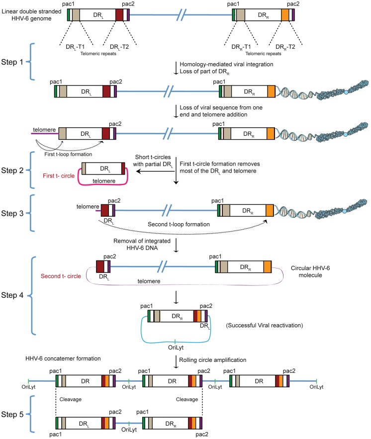
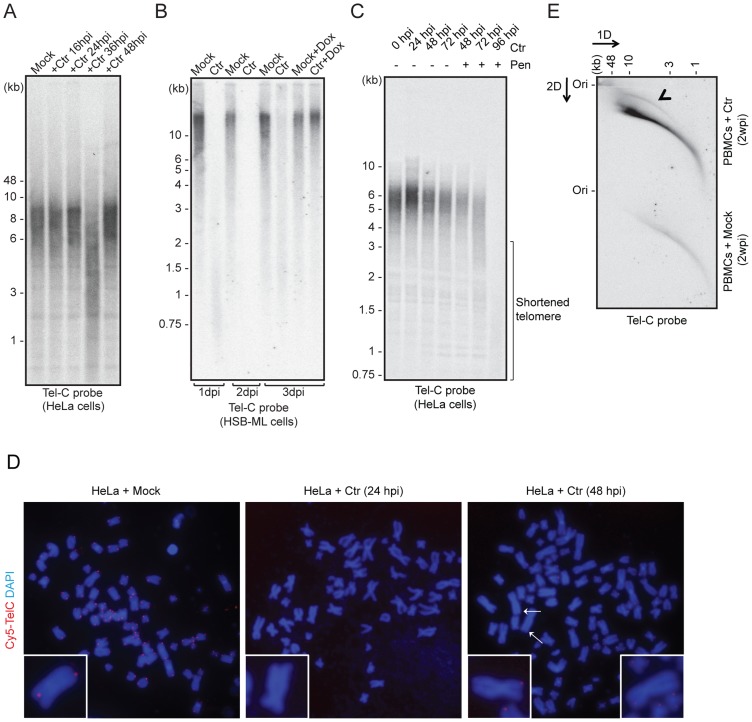
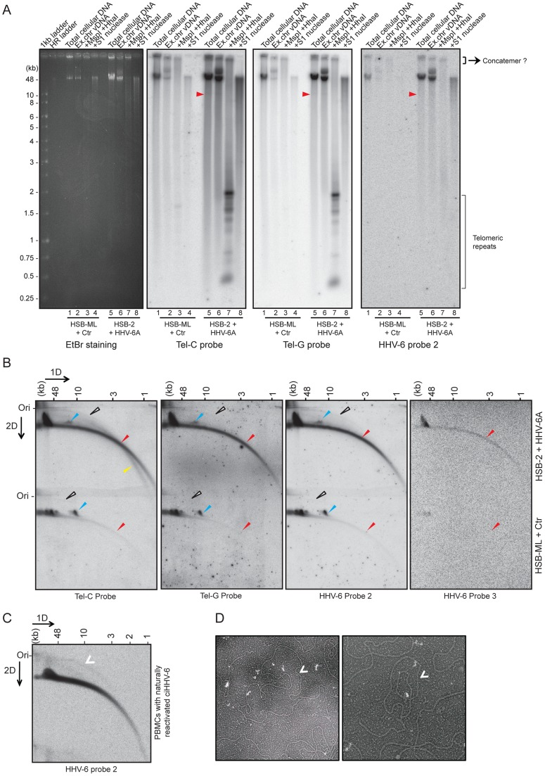
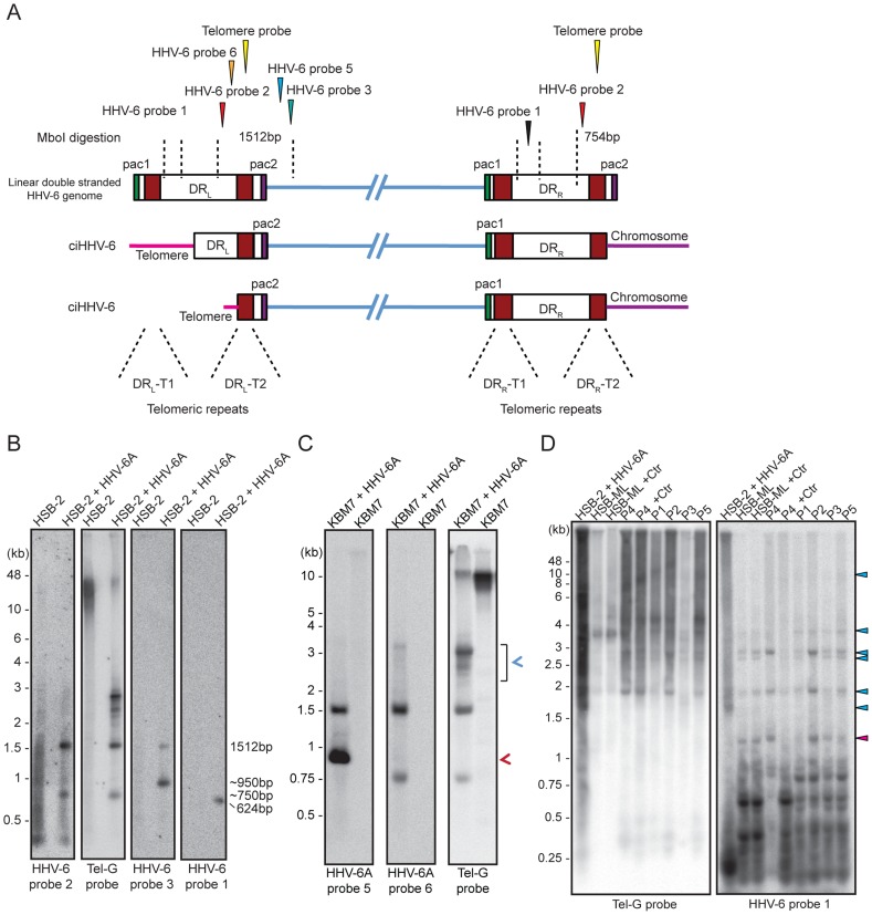
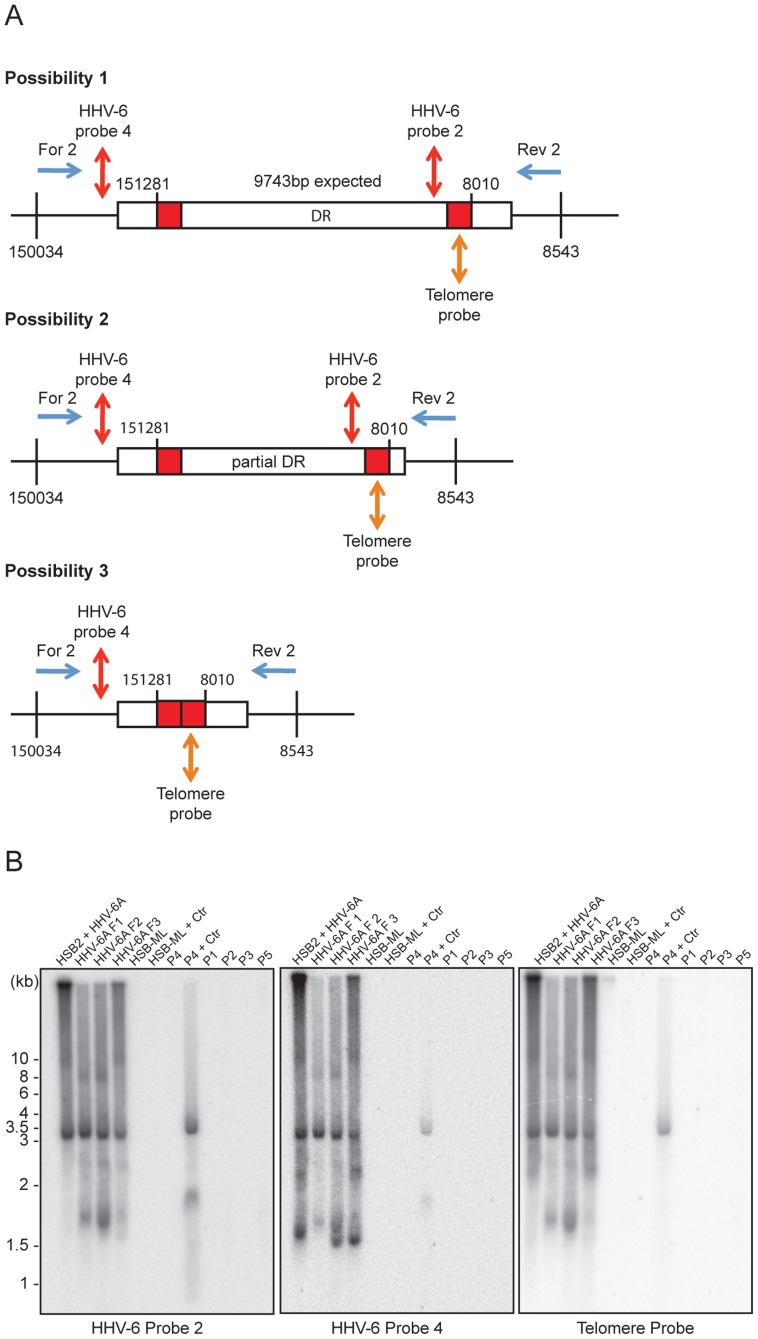
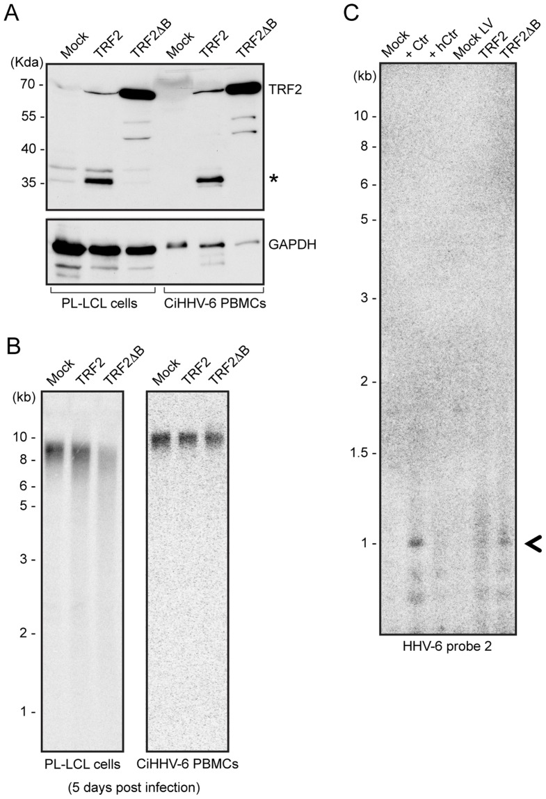
More than 95% of the human population is infected with human herpesvirus-6 (HHV-6) during early childhood and maintains latent HHV-6 genomes either in an extra-chromosomal form or as a chromosomally integrated HHV-6 (ciHHV-6). In addition, approximately 1% of humans are born with an inheritable form of ciHHV-6 integrated into the telomeres of chromosomes. Immunosuppression and stress conditions can reactivate latent HHV-6 replication, which is associated with clinical complications and even death. We have previously shown that Chlamydia trachomatis infection reactivates ciHHV-6 and induces the formation of extra-chromosomal viral DNA in ciHHV-6 cells. Here, we propose a model and provide experimental evidence for the mechanism of ciHHV-6 reactivation. Infection with Chlamydia induced a transient shortening of telomeric ends, which subsequently led to increased telomeric circle (t-circle) formation and incomplete reconstitution of circular viral genomes containing single viral direct repeat (DR). Correspondingly, short t-circles containing parts of the HHV-6 DR were detected in cells from individuals with genetically inherited ciHHV-6. Furthermore, telomere shortening induced in the absence of Chlamydia infection also caused circularization of ciHHV-6, supporting a t-circle based mechanism for ciHHV-6 reactivation.


Conflict of interest statement

The authors have declared that no competing interests exist.

Figures

Figure 1
Figure 1. Proposed model for the reactivation of ciHHV-6.
ciHHV-6 DNA has to be processed two times in order to form a t-circle containing a single DR, which can then undergo rolling circle amplification to form linear HHV-6 DNA. DR, direct repeats; DRL, left direct repeat; DRR, right direct repeat; T1, heterogeneous telomeric repeats; T2, homogeneous telomeric repeats; pac1 and pac2, packaging sequence 1 and 2; OriLyt, origin of lytic replication.
Figure 2
Figure 2. C. trachomatis (Ctr) infection induces telomere alterations.
(A) HeLa (wild type) (B) HSB-ML cells were infected with C. trachomatis for different time intervals. Mock-infected or 1 µg/ml doxycycline (Dox)-treated cells were used as control. Total genomic DNA (10 µg) was digested with MspI and HhaI and telomeric sequences were detected by Southern hybridization using an end-labeled (CCCTAA)4 probe (Tel-C probe). The positions of molecular weight markers are shown on the left. Dox was added to cells wherever mentioned (+Dox) after 24 h post Ctr infection and the cells were grown for further time intervals. Mock, cells with no Chlamydia infection. (C) Penicillin (Pen)-induced Chlamydia persistence prevents telomere repairing. Where indicated, 100 U of penicillin was added 24 h post Chlamydia infection. (D) FISH analysis of metaphase chromosomes showing telomere shortening in HeLa cells infected with Chlamydia. Telomeres were hybridized with a Cy5-tagged telomeric C-rich (Tel-C) probe and are shown in the red channel; DNA was stained with 4′,6-diamidino-2-phenylindole (DAPI) and is shown in the blue channel. Arrows indicate missing telomeric staining from single sister chromatids. Representative individual chromosomes are enlarged to show telomere staining. (E) Detection of circular telomeric DNA in PBMCs after Chlamydia infection. Freshly isolated PBMCs from healthy individuals were either mock infected or with Ctr for 2 days. Subsequently, cells were cultured for 2 more weeks in presence of doxycycline. Genomic DNA from each sample was digested with HinfI and RsaI and was processed for 2D-DNA electrophoresis and subsequent Southern hybridization with Tel-C probe. Black arrowhead indicates circular telomeric DNA. hpi, hour post infection; dpi, days post infection; wpi, week post infection.
Figure 3
Figure 3. Extra-chromosomal HHV-6 DNA exists in different conformations and contains telomeric repeats of different sizes.
(A) Purified viral DNA was either digested with MspI and HhaI or with S1 nuclease. All DNA samples were subjected to Southern hybridization using end-labeled (CCCTAA)4 probe (Tel-C probe), (TTAGGG)4 probe (Tel-G probe) or a HHV-6 DR specific probe (HHV-6 probe 2). HSB-ML+Ctr, Chlamydia-infected HSB-ML cells; HR ladder, high-range DNA ladder. The position of molecular weight markers is shown on the left. Smaller viral DNAs (possible short t-circles) are marked with red arrowhead. Possible concatemeric viral DNA are indicated. (B) Total genomic DNA from each cell type was processed for 2D-DNA electrophoresis and subsequent Southern hybridization with telomere-specific probes as well as with HHV-6 probes 2 and 3. Colored arrowheads mark different forms of viral DNA. White arrowheads: double-stranded circular HHV-6 DNA; Red arrowheads: double-stranded linear DNA; Blue arrowheads: short t-circles containing HHV-6 DR; Yellow arrowheads: single-stranded viral DNA (G-strand). DRL: left direct repeat. The positions of the size markers for the first-dimension gel electrophoresis are shown at the top, the origin of the second-dimension gel electrophoresis is indicated (Ori). (C) Detection of double-stranded full length circular HHV-6 DNA in PBMCs of ciHHV-6 patients with natural viral reactivation and with in vitro HHV-6 reactivation. Total genomic DNA from each cell type was processed for neutral-neutral 2D-DNA electrophoresis and subsequent Southern hybridization with HHV-6 probe 2. White arrowheads indicate double-stranded circular HHV-6 DNA. (D) Defined DNA fragments of ∼10 kb were gel eluted and prepared for EM by surface spreading. Shown is a reverse contrast image. Arrows mark circular t-circles of different sizes.
Figure 4
Figure 4. Detection of differentially processed ciHHV-6 ends.
(A) Diagram of possible processing of the ciHHV-6 DRL-T2 end after viral integration. Approximate location of MboI digestion sites within and around viral DR are marked with dotted lines. The location of probes used to detect specific regions of viral DNA and their expected sizes are indicated. Different positions marked are based on HHV-6A U1102 genome (X83413.1). (B) Total genomic DNA from HSB-2 cells, with or without HHV-6A infection, were digested with MboI and processed for Southern hybridization. The membrane was stripped and re-probed sequentially with HHV-6 probe 2, Tel-G probe, HHV-6 probe 3 and finally with HHV-6 probe 1 (See table S1). (C) Similar experiment was performed in KBM-7 cells using HHV-6A probe 5, 6 and Tel-G probe. Chromosomal ends having DRL-T2 overhang is marked with red arrowhead. Short t-circles, which can be detected using Tel-G probe and HHV-6A probe 5 but not with HHV-6A probe 6 are marked with blue arrowhead. The positions of molecular weight markers are shown on the left. (D) Inverse PCR using forward and reverse primer directed out of the DRL (see Figure S4B for orientation). Equal amount of the PCR products were subjected to Southern hybridization using probes against telomeric repeats (Tel-G probe). The membrane was rehybridized with a second probe against viral DR (HHV-6 probe 1). P1–P5 represents 5 different DNA samples from ciHHV-6 individuals. P4+Ctr represents PBMCs from P4 after 2 weeks of Chlamydia infection. The positions of molecular weight markers are shown on the left. The bands, which hybridize with both the probes, represent the extra-chromosomal short t-circles (blue arrow head). The band marked with red arrowhead represents short t-circles with extremely short telomeric repeats, which do not hybridize with telomeric probe.
Figure 5
Figure 5. PCR amplification of circular HHV-6 genomes having a single direct repeat (DR).
(A) Location of primers and southern hybridization probes to detect each possible combination of circular viral DNA (see table S1). Different positions marked are based on the HHV-6A U1102 genome (X83413.1). (B) Total DNA was used for PCR using a primer pair facing against each other but located outside the viral DR (see table S1). PCR products were used for Southern hybridization using HHV-6 probe 2. The membrane was re-hybridized with HHV-6 probe 4 and later on with telomere (Tel-G) probe. P1–P5 represents 5 different DNA samples from ciHHV-6 individuals. P4+Ctr represents PBMCs from P4 after 2 weeks of Chlamydia infection. The position of molecular weight markers run is shown on the left.
Figure 6
Figure 6. TRF2ΔB overexpression induces formation of circular HHV-6 genome in ciHHV-6 cells.
(A) TRF2 and TRF2ΔB was over-expressed in an EBV immortalized cell line (PL-LCL) derived from blood cells of a ciHHV-6 person and in freshly isolated PBMCs of another ciHHV-6 person using lentiviral vectors. Protein expression was checked after 3 days of lentivirus infection by Western blot analysis. GAPDH served as loading control. *, Uncharacterized protein. (B) Telomere restriction fragment assay was performed in the same cells after 5 days of lentiviral infection using telomere specific probe. (C) Formation of circular extra-chromosomal HHV-6 DNA was detected in the same cells by inverse PCR. Data represents the PCR amplifications from PL-LCL cells. As a control PL-LCL cells were infected with C. trachomatis or heat inactivated Ctr (hCtr). Amplified PCR product, which was validated by sequencing, is marked with a black arrowhead.

Similar articles

Cited by

References

    1. Tanaka-Taya K, Sashihara J, Kurahashi H, Amo K, Miyagawa H, et al. (2004) Human herpesvirus 6 (HHV-6) is transmitted from parent to child in an integrated form and characterization of cases with chromosomally integrated HHV-6 DNA. J Med Virol 73: 465–473. - PubMed
    1. Arbuckle JH, Medveczky MM, Luka J, Hadley SH, Luegmayr A, et al. (2010) The latent human herpesvirus-6A genome specifically integrates in telomeres of human chromosomes in vivo and in vitro. Proc Natl Acad Sci USA 107: 5563–5568. - PMC - PubMed
    1. Daibata M, Taguchi T, Nemoto Y, Taguchi H, Miyoshi I (1999) Inheritance of chromosomally integrated human herpesvirus 6 DNA. Blood 94: 1545. - PubMed
    1. Clark DA (2000) Human herpesvirus 6. Rev Med Virol 10: 155–173. - PubMed
    1. Campadelli-Fiume G, Mirandola P, Menotti L (1999) Human herpesvirus 6: An emerging pathogen. Emerg Infect Dis 5: 353–366. - PMC - PubMed

Publication types

MeSH terms

LinkOut - more resources