

Desensitization properties of P2X3 receptors shaping pain signaling
- PMID: 24367291
- PMCID: PMC3854565
- DOI: 10.3389/fncel.2013.00245
Desensitization properties of P2X3 receptors shaping pain signaling
Abstract
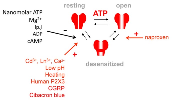
ATP-gated P2X3 receptors are mostly expressed by nociceptive sensory neurons and participate in transduction of pain signals. P2X3 receptors show a combination of fast desensitization onset and slow recovery. Moreover, even low nanomolar agonist concentrations unable to evoke a response, can induce desensitization via a phenomenon called "high affinity desensitization." We have also observed that recovery from desensitization is agonist-specific and can range from seconds to minutes. The recovery process displays unusually high temperature dependence. Likewise, recycling of P2X3 receptors in peri-membrane regions shows unexpectedly large temperature sensitivity. By applying kinetic modeling, we have previously shown that desensitization characteristics of P2X3 receptor are best explained with a cyclic model of receptor operation involving three agonist molecules binding a single receptor and that desensitization is primarily developing from the open receptor state. Mutagenesis experiments suggested that desensitization depends on a certain conformation of the ATP binding pocket and on the structure of the transmembrane domains forming the ion pore. Further molecular determinants of desensitization have been identified by mutating the intracellular N- and C-termini of P2X3 receptor. Unlike other P2X receptors, the P2X3 subtype is facilitated by extracellular calcium that acts via specific sites in the ectodomain neighboring the ATP binding pocket. Thus, substitution of serine275 in this region (called "left flipper") converts the natural facilitation induced by extracellular calcium to receptor inhibition. Given their strategic location in nociceptive neurons and unique desensitization properties, P2X3 receptors represent an attractive target for development of new analgesic drugs via promotion of desensitization aimed at suppressing chronic pain.
Keywords: P2X3 receptor; desensitization; extracellular ATP; pain; sensory neuron.
Figures
References
-
- Alexander K., Niforatos W., Bianchi B., Burgard E. C., Lynch K. J., Kowaluk E. A., et al. (1999). Allosteric modulation and accelerated resensitization of human P2X(3) receptors by cibacron blue. J. Pharmacol. Exp. Ther. 291 1135–1142 - PubMed
Publication types
LinkOut - more resources
Full Text Sources
Other Literature Sources